Automatic Tuberculosis Detection Using Chest X-Ray Analysis with Position Enhanced Structural Information

Hermann Jepdjio Nkouanga, Szilard Vajda

Responsive image

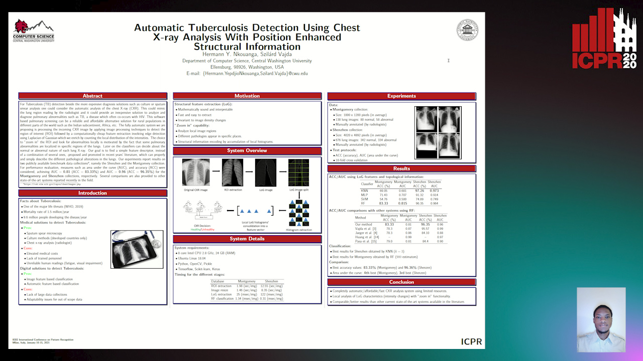
Auto-TLDR; Automatic Chest X-ray Screening for Tuberculosis in Rural Population using Localized Region on Interest

Slides Poster

For Tuberculosis (TB) detection beside the more expensive diagnosis solutions such as culture or sputum smear analysis one could consider the automatic analysis of the chest X-ray (CXR). This could mimic the lung region reading by the radiologist and it could provide a cheap solution to analyze and diagnose pulmonary abnormalities such as TB which often co- occurs with HIV. This software based pulmonary screening can be a reliable and affordable solution for rural population in different parts of the world such as India, Africa, etc. Our fully automatic system is processing the incoming CXR image by applying image processing techniques to detect the region on interest (ROI) followed by a computationally cheap feature extraction involving edge detection using Laplacian of Gaussian which we enrich by counting the local distribution of the intensities. The choice to ”zoom in” the ROI and look for abnormalities locally is motivated by the fact that some pulmonary abnormalities are localized in specific regions of the lungs. Later on the classifiers can decide about the normal or abnormal nature of each lung X-ray. Our goal is to find a simple feature, instead of a combination of several ones, -proposed and promoted in recent years’ literature, which can properly describe the different pathological alterations in the lungs. Our experiments report results on two publicly available data collections1, namely the Shenzhen and the Montgomery collection. For performance evaluation, measures such as area under the curve (AUC), and accuracy (ACC) were considered, achieving AUC = 0.81 (ACC = 83.33%) and AUC = 0.96 (ACC = 96.35%) for the Montgomery and Schenzen collections, respectively. Several comparisons are also provided to other state- of-the-art systems reported recently in the field.

Similar papers

Inception Based Deep Learning Architecture for Tuberculosis Screening of Chest X-Rays

Dipayan Das, K.C. Santosh, Umapada Pal

Responsive image

Auto-TLDR; End to End CNN-based Chest X-ray Screening for Tuberculosis positive patients in the severely resource constrained regions of the world

Slides Poster Similar

The motivation for this work is the primary need of screening Tuberculosis (TB) positive patients in the severely resource constrained regions of the world. Chest X-ray (CXR) is considered to be a promising indicator for the onset of TB, but the lack of skilled radiologists in such regions degrades the situation. Therefore, several computer aided diagnosis (CAD) systems have been proposed to solve the decision making problem, which includes hand engineered feature extraction methods to deep learning or Convolutional Neural Network (CNN) based methods. Feature extraction, being a time and resource intensive process, often delays the process of mass screening. Hence an end to end CNN architecture is proposed in this work to solve the problem. Two benchmark CXR datasets have been used in this work, collected from Shenzhen (China) and Montgomery County (USA), on which the proposed methodology achieved a maximum abnormality detection accuracy (ACC) of 91.7\% (0.96 AUC) and 87.47\% (0.92 AUC) respectively. To the greatest of our knowledge, the obtained results are marginally superior to the state of the art results that have solely used deep learning methodologies on the aforementioned datasets.

Unsupervised Detection of Pulmonary Opacities for Computer-Aided Diagnosis of COVID-19 on CT Images

Rui Xu, Xiao Cao, Yufeng Wang, Yen-Wei Chen, Xinchen Ye, Lin Lin, Wenchao Zhu, Chao Chen, Fangyi Xu, Yong Zhou, Hongjie Hu, Shoji Kido, Noriyuki Tomiyama

Responsive image

Auto-TLDR; A computer-aided diagnosis of COVID-19 from CT images using unsupervised pulmonary opacity detection

Slides Poster Similar

COVID-19 emerged towards the end of 2019 which was identified as a global pandemic by the world heath organization (WHO). With the rapid spread of COVID-19, the number of infected and suspected patients has increased dramatically. Chest computed tomography (CT) has been recognized as an efficient tool for the diagnosis of COVID-19. However, the huge CT data make it difficult for radiologist to fully exploit them on the diagnosis. In this paper, we propose a computer-aided diagnosis system that can automatically analyze CT images to distinguish the COVID-19 against to community-acquired pneumonia (CAP). The proposed system is based on an unsupervised pulmonary opacity detection method that locates opacity regions by a detector unsupervisedly trained from CT images with normal lung tissues. Radiomics based features are extracted insides the opacity regions, and fed into classifiers for classification. We evaluate the proposed CAD system by using 200 CT images collected from different patients in several hospitals. The accuracy, precision, recall, f1-score and AUC achieved are 95.5%, 100%, 91%, 95.1% and 95.9% respectively, exhibiting the promising capacity on the differential diagnosis of COVID-19 from CT images.

Automatic Classification of Human Granulosa Cells in Assisted Reproductive Technology Using Vibrational Spectroscopy Imaging

Marina Paolanti, Emanuele Frontoni, Giorgia Gioacchini, Giorgini Elisabetta, Notarstefano Valentina, Zacà Carlotta, Carnevali Oliana, Andrea Borini, Marco Mameli

Responsive image

Auto-TLDR; Predicting Oocyte Quality in Assisted Reproductive Technology Using Machine Learning Techniques

Slides Poster Similar

In the field of reproductive technology, the biochemical composition of female gametes has been successfully investigated with the use of vibrational spectroscopy. Currently, in assistive reproductive technology (ART), there are no shared criteria for the choice of oocyte, and automatic classification methods for the best quality oocytes have not yet been applied. In this paper, considering the lack of criteria in Assisted Reproductive Technology (ART), we use Machine Learning (ML) techniques to predict oocyte quality for a successful pregnancy. To improve the chances of successful implantation and minimize any complications during the pregnancy, Fourier transform infrared microspectroscopy (FTIRM) analysis has been applied on granulosa cells (GCs) collected along with the oocytes during oocyte aspiration, as it is routinely done in ART, and specific spectral biomarkers were selected by multivariate statistical analysis. A proprietary biological reference dataset (BRD) was successfully collected to predict the best oocyte for a successful pregnancy. Personal health information are stored, maintained and backed up using a cloud computing service. Using a user-friendly interface, the user will evaluate whether or not the selected oocyte will have a positive result. This interface includes a dashboard for retrospective analysis, reporting, real-time processing, and statistical analysis. The experimental results are promising and confirm the efficiency of the method in terms of classification metrics: precision, recall, and F1-score (F1) measures.

Dealing with Scarce Labelled Data: Semi-Supervised Deep Learning with Mix Match for Covid-19 Detection Using Chest X-Ray Images

Saúl Calderón Ramirez, Raghvendra Giri, Shengxiang Yang, Armaghan Moemeni, Mario Umaña, David Elizondo, Jordina Torrents-Barrena, Miguel A. Molina-Cabello

Responsive image

Auto-TLDR; Semi-supervised Deep Learning for Covid-19 Detection using Chest X-rays

Slides Poster Similar

Coronavirus (Covid-19) is spreading fast, infecting people through contact in various forms including droplets from sneezing and coughing. Therefore, the detection of infected subjects in an early, quick and cheap manner is urgent. Currently available tests are scarce and limited to people in danger of serious illness. The application of deep learning to chest X-ray images for Covid-19 detection is an attractive approach. However, this technology usually relies on the availability of large labelled datasets, a requirement hard to meet in the context of a virus outbreak. To overcome this challenge, a semi-supervised deep learning model using both labelled and unlabelled data is proposed. We developed and tested a semi-supervised deep learning framework based on the Mix Match architecture to classify chest X-rays into Covid-19, pneumonia and healthy cases. The presented approach was calibrated using two publicly available datasets. The results show an accuracy increase of around $15\%$ under low labelled / unlabelled data ratio. This indicates that our semi-supervised framework can help improve performance levels towards Covid-19 detection when the amount of high-quality labelled data is scarce. Also, we introduce a semi-supervised deep learning boost coefficient which is meant to ease the scalability of our approach and performance comparison.

A Novel Computer-Aided Diagnostic System for Early Assessment of Hepatocellular Carcinoma

Ahmed Alksas, Mohamed Shehata, Gehad Saleh, Ahmed Shaffie, Ahmed Soliman, Mohammed Ghazal, Hadil Abukhalifeh, Abdel Razek Ahmed, Ayman El-Baz

Responsive image

Auto-TLDR; Classification of Liver Tumor Lesions from CE-MRI Using Structured Structural Features and Functional Features

Slides Poster Similar

Early assessment of liver cancer patients with hepatocellular carcinoma (HCC) is of immense importance to provide the proper treatment plan. In this paper, we have developed a two-stage classification computer-aided diagnostic (CAD) system that has the ability to detect and grade the liver observations from multiphase contrast enhanced magnetic resonance imaging (CE-MRI). The proposed approach consists of three main steps. First, a pre-processing is applied to the CE-MRI scans to delineate the tumor lesions that will be used as an ROI across the four different phases of the CE-MRI, (namely, the pre-contrast, late-arterial, portal-venous, and delayed-contrast). Second, a group of three features are modeled to provide a quantitative discrimination between the tumor lesions; namely: i) the tumor appearance that is modeled using a set of texture features, (namely; the first-order histogram, second-order gray-level co-occurrence matrix, and second-order gray-level run-length matrix), to capture any discrimination that may appear in the lesion texture, ii) the spherical harmonics (SH) based shape features that have the ability to describe the shape complexity of the liver tumors, and iii) the functional features that are based on the calculation of the wash-in/wash-out through that evaluate the intensity changes across the post-contrast phases. Finally, the aforementioned individual features were then integrated together to obtain the combined features to be fed to a machine learning classifier towards getting the final diagnostic decision. The proposed CAD system has been tested using hepatic observations that was obtained from 85 participating patients, 34 patients with benign tumors, 34 patients with intermediate tumors and 34 with malignant tumors. Using a random forests based classifier with a leave-one-subject-out (LOSO) cross-validation, the developed CAD system achieved an 87.1% accuracy in distinguishing the malignant, intermediate and benign tumors. The classification performance is then evaluated using k-fold (5/10-fold) cross-validation approach to examine the robustness of the system. The LR-1 lesions were classified from LR-2 benign lesions with 91.2% accuracy, while 85.3% accuracy was achieved differentiating between LR-4 and LR-5 malignant tumors. The obtained results hold a promise of the proposed framework to be reliably used as a noninvasive diagnostic tool for the early detection and grading of liver cancer tumors.

A Comparison of Neural Network Approaches for Melanoma Classification

Maria Frasca, Michele Nappi, Michele Risi, Genoveffa Tortora, Alessia Auriemma Citarella

Responsive image

Auto-TLDR; Classification of Melanoma Using Deep Neural Network Methodologies

Slides Poster Similar

Melanoma is the deadliest form of skin cancer and it is diagnosed mainly visually, starting from initial clinical screening and followed by dermoscopic analysis, biopsy and histopathological examination. A dermatologist’s recognition of melanoma may be subject to errors and may take some time to diagnose it. In this regard, deep learning can be useful in the study and classification of skin cancer. In particular, by classifying images with Deep Neural Network methodologies, it is possible to obtain comparable or even superior results compared to those of dermatologists. In this paper, we propose a methodology for the classification of melanoma by adopting different deep learning techniques applied to a common dataset, composed of images from the ISIC dataset and consisting of different types of skin diseases, including melanoma on which we applied a specific pre-processing phase. In particular, a comparison of the results is performed in order to select the best effective neural network to be applied to the problem of recognition and classification of melanoma. Moreover, we also evaluate the impact of the pre- processing phase on the final classification. Different metrics such as accuracy, sensitivity, and specificity have been selected to assess the goodness of the adopted neural networks and compare them also with the manual classification of dermatologists.

Appliance Identification Using a Histogram Post-Processing of 2D Local Binary Patterns for Smart Grid Applications

Yassine Himeur, Abdullah Alsalemi, Faycal Bensaali, Abbes Amira

Responsive image

Auto-TLDR; LBP-BEVM based Local Binary Patterns for Appliances Identification in the Smart Grid

Similar

Identifying domestic appliances in the smart grid leads to a better power usage management and further helps in detecting appliance-level abnormalities. An efficient identification can be achieved only if a robust feature extraction scheme is developed with a high ability to discriminate between different appliances on the smart grid. Accordingly, we propose in this paper a novel method to extract electrical power signatures after transforming the power signal to 2D space, which has more encoding possibilities. Following, an improved local binary patterns (LBP) is proposed that relies on improving the discriminative ability of conventional LBP using a post-processing stage. A binarized eigenvalue map (BEVM) is extracted from the 2D power matrix and then used to post-process the generated LBP representation. Next, two histograms are constructed, namely up and down histograms, and are then concatenated to form the global histogram. A comprehensive performance evaluation is performed on two different datasets, namely the GREEND and WITHED, in which power data were collected at 1 Hz and 44000 Hz sampling rates, respectively. The obtained results revealed the superiority of the proposed LBP-BEVM based system in terms of the identification performance versus other 2D descriptors and existing identification frameworks.

Influence of Event Duration on Automatic Wheeze Classification

Bruno M Rocha, Diogo Pessoa, Alda Marques, Paulo Carvalho, Rui Pedro Paiva

Responsive image

Auto-TLDR; Experimental Design of the Non-wheeze Class for Wheeze Classification

Slides Poster Similar

Patients with respiratory conditions typically exhibit adventitious respiratory sounds, such as wheezes. Wheeze events have variable duration. In this work we studied the influence of event duration on wheeze classification, namely how the creation of the non-wheeze class affected the classifiers' performance. First, we evaluated several classifiers on an open access respiratory sound database, with the best one reaching sensitivity and specificity values of 98% and 95%, respectively. Then, by changing one parameter in the design of the non-wheeze class, i.e., event duration, the best classifier only reached sensitivity and specificity values of 53% and 75%, respectively. These results demonstrate the importance of experimental design on the assessment of wheeze classification algorithms' performance.

Attack-Agnostic Adversarial Detection on Medical Data Using Explainable Machine Learning

Matthew Watson, Noura Al Moubayed

Responsive image

Auto-TLDR; Explainability-based Detection of Adversarial Samples on EHR and Chest X-Ray Data

Slides Poster Similar

Explainable machine learning has become increasingly prevalent, especially in healthcare where explainable models are vital for ethical and trusted automated decision making. Work on the susceptibility of deep learning models to adversarial attacks has shown the ease of designing samples to mislead a model into making incorrect predictions. In this work, we propose an explainability-based method for the accurate detection of adversarial samples on two datasets with different complexity and properties: Electronic Health Record (EHR) and chest X-ray (CXR) data. On the MIMIC-III and Henan-Renmin EHR datasets, we report a detection accuracy of 77% against the Longitudinal Adversarial Attack. On the MIMIC-CXR dataset, we achieve an accuracy of 88%; significantly improving on the state of the art of adversarial detection in both datasets by over 10% in all settings. We propose an anomaly detection based method using explainability techniques to detect adversarial samples which is able to generalise to different attack methods without a need for retraining.

Classify Breast Histopathology Images with Ductal Instance-Oriented Pipeline

Beibin Li, Ezgi Mercan, Sachin Mehta, Stevan Knezevich, Corey Arnold, Donald Weaver, Joann Elmore, Linda Shapiro

Responsive image

Auto-TLDR; DIOP: Ductal Instance-Oriented Pipeline for Diagnostic Classification

Slides Poster Similar

In this study, we propose the Ductal Instance-Oriented Pipeline (DIOP) that contains a duct-level instance segmentation model, a tissue-level semantic segmentation model, and three-levels of features for diagnostic classification. Based on recent advancements in instance segmentation and the Mask R-CNN model, our duct-level segmenter tries to identify each ductal individual inside a microscopic image; then, it extracts tissue-level information from the identified ductal instances. Leveraging three levels of information obtained from these ductal instances and also the histopathology image, the proposed DIOP outperforms previous approaches (both feature-based and CNN-based) in all diagnostic tasks; for the four-way classification task, the DIOP achieves comparable performance to general pathologists in this unique dataset. The proposed DIOP only takes a few seconds to run in the inference time, which could be used interactively on most modern computers. More clinical explorations are needed to study the robustness and generalizability of this system in the future.

Bridging the Gap between Natural and Medical Images through Deep Colorization

Lia Morra, Luca Piano, Fabrizio Lamberti, Tatiana Tommasi

Responsive image

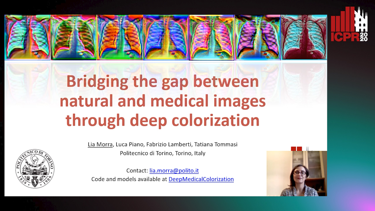
Auto-TLDR; Transfer Learning for Diagnosis on X-ray Images Using Color Adaptation

Slides Poster Similar

Deep learning has thrived by training on large-scale datasets. However, in many applications, as for medical image diagnosis, getting massive amount of data is still prohibitive due to privacy, lack of acquisition homogeneity and annotation cost. In this scenario transfer learning from natural image collections is a standard practice that attempts to tackle shape, texture and color discrepancy all at once through pretrained model fine-tuning. In this work we propose to disentangle those challenges and design a dedicated network module that focuses on color adaptation. We combine learning from scratch of the color module with transfer learning of different classification backbones obtaining an end-to-end, easy-to-train architecture for diagnostic image recognition on X-ray images. Extensive experiments show how our approach is particularly efficient in case of data scarcity and provides a new path for further transferring the learned color information across multiple medical datasets.

Deep Transfer Learning for Alzheimer’s Disease Detection

Nicole Cilia, Claudio De Stefano, Francesco Fontanella, Claudio Marrocco, Mario Molinara, Alessandra Scotto Di Freca

Responsive image

Auto-TLDR; Automatic Detection of Handwriting Alterations for Alzheimer's Disease Diagnosis using Dynamic Features

Slides Poster Similar

Early detection of Alzheimer’s Disease (AD) is essential in order to initiate therapies that can reduce the effects of such a disease, improving both life quality and life expectancy of patients. Among all the activities carried out in our daily life, handwriting seems one of the first to be influenced by the arise of neurodegenerative diseases. For this reason, the analysis of handwriting and the study of its alterations has become of great interest in this research field in order to make a diagnosis as early as possible. In recent years, many studies have tried to use classification algorithms applied to handwritings to implement decision support systems for AD diagnosis. A key issue for the use of these techniques is the detection of effective features, that allow the system to distinguish the natural handwriting alterations due to age, from those caused by neurodegenerative disorders. In this context, many interesting results have been published in the literature in which the features have been typically selected by hand, generally considering the dynamics of the handwriting process in order to detect motor disorders closely related to AD. Features directly derived from handwriting generation models can be also very helpful for AD diagnosis. It should be remarked, however, that the above features do not consider changes in the shape of handwritten traces, which may occur as a consequence of neurodegenerative diseases, as well as the correlation among shape alterations and changes in the dynamics of the handwriting process. Moving from these considerations, the aim of this study is to verify if the combined use of both shape and dynamic features allows a decision support system to improve performance for AD diagnosis. To this purpose, starting from a database of on-line handwriting samples, we generated for each of them a synthetic off-line colour image, where the colour of each elementary trait encodes, in the three RGB channels, the dynamic information associated to that trait. Finally, we exploited the capability of Deep Neural Networks (DNN) to automatically extract features from raw images. The experimental comparison of the results obtained by using standard features and features extracted according the above procedure, confirmed the effectiveness of our approach.

A Systematic Investigation on Deep Architectures for Automatic Skin Lesions Classification

Pierluigi Carcagni, Marco Leo, Andrea Cuna, Giuseppe Celeste, Cosimo Distante

Responsive image

Auto-TLDR; RegNet: Deep Investigation of Convolutional Neural Networks for Automatic Classification of Skin Lesions

Slides Poster Similar

Computer vision-based techniques are more and more employed in healthcare and medical fields nowadays in order, principally, to be as a support to the experienced medical staff to help them to make a quick and correct diagnosis. One of the hot topics in this arena concerns the automatic classification of skin lesions. Several promising works exist about it, mainly leveraging Convolutional Neural Networks (CNN), but proposed pipeline mainly rely on complex data preprocessing and there is no systematic investigation about how available deep models can actually reach the accuracy needed for real applications. In order to overcome these drawbacks, in this work, an end-to-end pipeline is introduced and some of the most recent Convolutional Neural Networks (CNNs) architectures are included in it and compared on the largest common benchmark dataset recently introduced. To this aim, for the first time in this application context, a new network design paradigm, namely RegNet, has been exploited to get the best models among a population of configurations. The paper introduces a threefold level of contribution and novelty with respect the previous literature: the deep investigation of several CNN architectures driving to a consistent improvement of the lesions recognition accuracy, the exploitation of a new network design paradigm able to study the behavior of populations of models and a deep discussion about pro and cons of each analyzed method paving the path towards new research lines.

Fine-Tuning Convolutional Neural Networks: A Comprehensive Guide and Benchmark Analysis for Glaucoma Screening

Amed Mvoulana, Rostom Kachouri, Mohamed Akil

Responsive image

Auto-TLDR; Fine-tuning Convolutional Neural Networks for Glaucoma Screening

Slides Poster Similar

This work aimed at giving a comprehensive and in-detailed guide on the route to fine-tuning Convolutional Neural Networks (CNNs) for glaucoma screening. Transfer learning consists in a promising alternative to train CNNs from stratch, to avoid the huge data and resources requirements. After a thorough study of five state-of-the-art CNNs architectures, a complete and well-explained strategy for fine-tuning these networks is proposed, using hyperparameter grid-searching and two-phase training approach. Excellent performance is reached on model evaluation, with a 0.9772 AUROC validation rate, giving arise to reliable glaucoma diagosis-help systems. Also, a benchmark analysis is conducted across all fine-tuned models, studying them according to performance indices such as model complexity and size, AUROC density and inference time. This in-depth analysis allows a rigorous comparison between model characteristics, and is useful for giving practioners important trademarks for prospective applications and deployments.

Confidence Calibration for Deep Renal Biopsy Immunofluorescence Image Classification

Federico Pollastri, Juan Maroñas, Federico Bolelli, Giulia Ligabue, Roberto Paredes, Riccardo Magistroni, Costantino Grana

Responsive image

Auto-TLDR; A Probabilistic Convolutional Neural Network for Immunofluorescence Classification in Renal Biopsy

Slides Poster Similar

With this work we tackle immunofluorescence classification in renal biopsy, employing state-of-the-art Convolutional Neural Networks. In this setting, the aim of the probabilistic model is to assist an expert practitioner towards identifying the location pattern of antibody deposits within a glomerulus. Since modern neural networks often provide overconfident outputs, we stress the importance of having a reliable prediction, demonstrating that Temperature Scaling, a recently introduced re-calibration technique, can be successfully applied to immunofluorescence classification in renal biopsy. Experimental results demonstrate that the designed model yields good accuracy on the specific task, and that Temperature Scaling is able to provide reliable probabilities, which are highly valuable for such a task given the low inter-rater agreement.

Automatic Semantic Segmentation of Structural Elements related to the Spinal Cord in the Lumbar Region by Using Convolutional Neural Networks

Jhon Jairo Sáenz Gamboa, Maria De La Iglesia-Vaya, Jon Ander Gómez

Responsive image

Auto-TLDR; Semantic Segmentation of Lumbar Spine Using Convolutional Neural Networks

Slides Poster Similar

This work addresses the problem of automatically segmenting the MR images corresponding to the lumbar spine. The purpose is to detect and delimit the different structural elements like vertebrae, intervertebral discs, nerves, blood vessels, etc. This task is known as semantic segmentation. The approach proposed in this work is based on convolutional neural networks whose output is a mask where each pixel from the input image is classified into one of the possible classes. Classes were defined by radiologists and correspond to structural elements and tissues. The proposed network architectures are variants of the U-Net. Several complementary blocks were used to define the variants: spatial attention models, deep supervision and multi-kernels at input, this last block type is based on the idea of inception. Those architectures which got the best results are described in this paper, and their results are discussed. Two of the proposed architectures outperform the standard U-Net used as baseline.

Magnifying Spontaneous Facial Micro Expressions for Improved Recognition

Pratikshya Sharma, Sonya Coleman, Pratheepan Yogarajah, Laurence Taggart, Pradeepa Samarasinghe

Responsive image

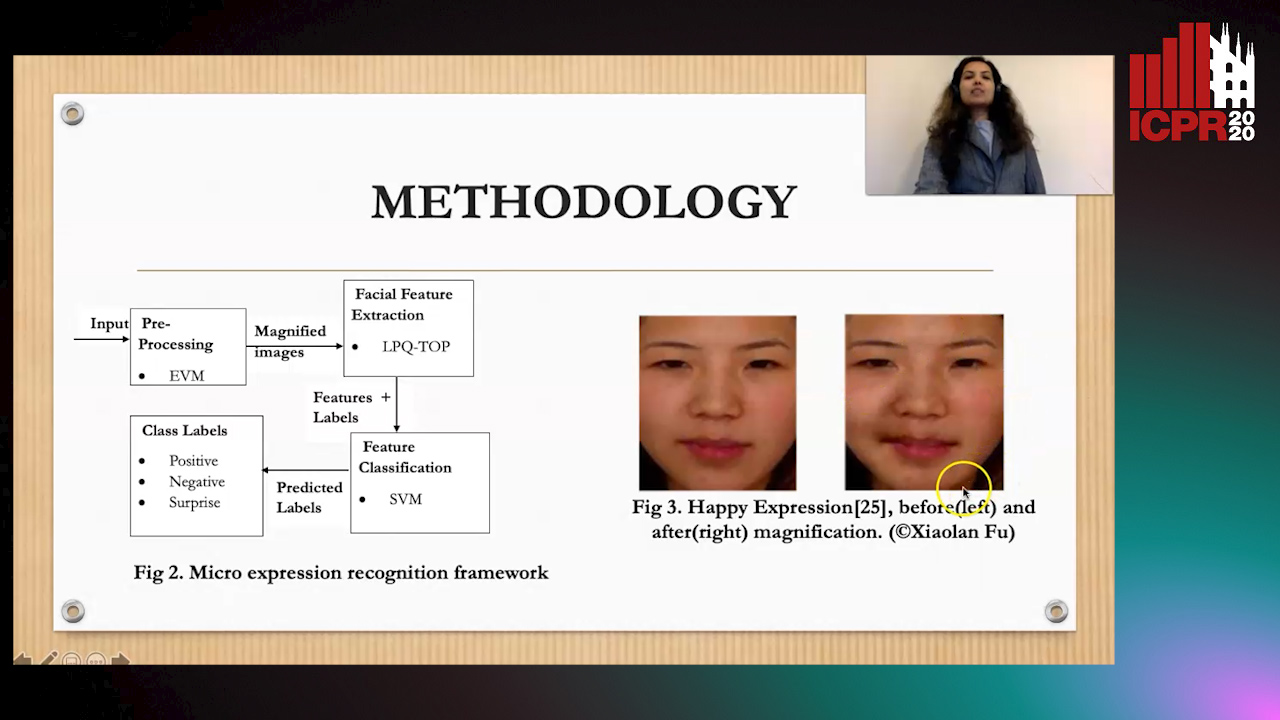
Auto-TLDR; Eulerian Video Magnification for Micro Expression Recognition

Slides Poster Similar

Building an effective automatic micro expression recognition (MER) system is becoming increasingly desirable in computer vision applications. However, it is also very challenging given the fine-grained nature of the expressions to be recognized. Hence, we investigate if amplifying micro facial muscle movements as a pre-processing phase, by employing Eulerian Video Magnification (EVM), can boost performance of Local Phase Quantization with Three Orthogonal Planes (LPQ-TOP) to achieve improved facial MER across various datasets. In addition, we examine the rate of increase for recognition to determine if it is uniform across datasets using EVM. Ultimately, we classify the extracted features using Support Vector Machines (SVM). We evaluate and compare the performance with various methods on seven different datasets namely CASME, CAS(ME)2, CASME2, SMIC-HS, SMIC-VIS, SMIC-NIR and SAMM. The results obtained demonstrate that EVM can enhance LPQ-TOP to achieve improved recognition accuracy on the majority of the datasets.

Documents Counterfeit Detection through a Deep Learning Approach

Darwin Danilo Saire Pilco, Salvatore Tabbone

Responsive image

Auto-TLDR; End-to-End Learning for Counterfeit Documents Detection using Deep Neural Network

Slides Poster Similar

The main topic of this work is on the detection of counterfeit documents and especially banknotes. We propose an end-to-end learning model using a deep learning approach based on Adapnet++ which manages feature extraction at multiple scale levels using several residual units. Unlike previous models based on regions of interest (ROI) and high-resolution documents, our network is feed with simple input images (i.e., a single patch) and we do not need high resolution images. Besides, discriminative regions can be visualized at different scales. Our network learns by itself which regions of interest predict the better results. Experimental results show that we are competitive compared with the state-of-the-art and our deep neural network has good ability to generalize and can be applied to other kind of documents like identity or administrative one.

Detecting Anomalies from Video-Sequences: A Novel Descriptor

Giulia Orrù, Davide Ghiani, Maura Pintor, Gian Luca Marcialis, Fabio Roli

Responsive image

Auto-TLDR; Trit-based Measurement of Group Dynamics for Crowd Behavior Analysis and Anomaly Detection

Slides Poster Similar

We present a novel descriptor for crowd behavior analysis and anomaly detection. The goal is to measure by appropriate patterns the speed of formation and disintegration of groups in the crowd. This descriptor is inspired by the concept of one-dimensional local binary patterns: in our case, such patterns depend on the number of group observed in a time window. An appropriate measurement unit, named "trit" (trinary digit), represents three possible dynamic states of groups on a certain frame. Our hypothesis is that abrupt variations of the groups' number may be due to an anomalous event that can be accordingly detected, by translating these variations on temporal trit-based sequence of strings which are significantly different from the one describing the "no-anomaly" one. Due to the peculiarity of the rationale behind this work, relying on the number of groups, three different methods of people group's extraction are compared. Experiments are carried out on the Motion-Emotion benchmark data set. Reported results point out in which cases the trit-based measurement of group dynamics allows us to detect the anomaly. Besides the promising performance of our approach, we show how it is correlated with the anomaly typology and the camera's perspective to the crowd's flow (frontal, lateral).

Prediction of Obstructive Coronary Artery Disease from Myocardial Perfusion Scintigraphy using Deep Neural Networks

Ida Arvidsson, Niels Christian Overgaard, Miguel Ochoa Figueroa, Jeronimo Rose, Anette Davidsson, Kalle Åström, Anders Heyden

Responsive image

Auto-TLDR; A Deep Learning Algorithm for Multi-label Classification of Myocardial Perfusion Scintigraphy for Stable Ischemic Heart Disease

Slides Poster Similar

For diagnosis and risk assessment in patients with stable ischemic heart disease, myocardial perfusion scintigraphy is one of the most common cardiological examinations performed today. There are however many motivations for why an artificial intelligence algorithm would provide useful input to this task. For example to reduce the subjectiveness and save time for the nuclear medicine physicians working with this time consuming task. In this work we have developed a deep learning algorithm for multi-label classification based on a modified convolutional neural network to estimate probability of obstructive coronary artery disease in the left anterior artery, left circumflex artery and right coronary artery. The prediction is based on data from myocardial perfusion scintigraphy studies conducted in a dedicated Cadmium-Zinc-Telluride cardio camera (D-SPECT Spectrum Dynamics). Data from 588 patients was available, with stress images in both upright and supine position, as well as a number of auxiliary parameters such as angina symptoms and BMI. The data was used to train and evaluate the algorithm using 5-fold cross-validation. We achieve state-of-the-art results for this task with an area under the receiver operating characteristics curve of 0.89 as average on per-vessel level and 0.94 on per-patient level.

Deep Learning in the Ultrasound Evaluation of Neonatal Respiratory Status

Michela Gravina, Diego Gragnaniello, Giovanni Poggi, Luisa Verdoliva, Carlo Sansone, Iuri Corsini, Carlo Dani, Fabio Meneghin, Gianluca Lista, Salvatore Aversa, Migliaro Migliaro, Raimondi Francesco

Responsive image

Auto-TLDR; Lung Ultrasound Imaging with Deep Learning Networks and Training Strategies: An Analysis and Adaptation

Slides Poster Similar

Lung ultrasound imaging is reaching growing interest from the scientific community. On one side, thanks to its harmlessness and high descriptive power, this kind of diagnostic imaging became largely adopted in sensitive applications, like the diagnosis and follow-up of preterm newborns in neonatal intensive care units. At the same time, novel image analysis and pattern recognition approaches can fully exploit the rich information contained in this data, making them attractive for the research community. In this work, we present a thorough analysis of recent deep learning networks and training strategies conducted on a vast and challenging multicenter dataset comprising 87 patients with different diseases and gestational ages. These approaches are firstly discussed in the context of lung respiratory status assessing through ultrasound imaging and then evaluated against a reference marker. The conducted analysis shed some light on this problem, by relating the criticisms that can mislead the training procedure and proposing some adaptations to the specific problem. The achieved results sensibly outperform that obtained by previous work, based on textural features, and narrow the gap with the visual score predicted by the human experts.

BG-Net: Boundary-Guided Network for Lung Segmentation on Clinical CT Images

Rui Xu, Yi Wang, Tiantian Liu, Xinchen Ye, Lin Lin, Yen-Wei Chen, Shoji Kido, Noriyuki Tomiyama

Responsive image

Auto-TLDR; Boundary-Guided Network for Lung Segmentation on CT Images

Slides Poster Similar

Lung segmentation on CT images is a crucial step for a computer-aided diagnosis system of lung diseases. The existing deep learning based lung segmentation methods are less efficient to segment lungs on clinical CT images, especially that the segmentation on lung boundaries is not accurate enough due to complex pulmonary opacities in practical clinics. In this paper, we propose a boundary-guided network (BG-Net) to address this problem. It contains two auxiliary branches that separately segment lungs and extract the lung boundaries, and an aggregation branch that efficiently exploits lung boundary cues to guide the network for more accurate lung segmentation on clinical CT images. We evaluate the proposed method on a private dataset collected from the Osaka university hospital and four public datasets including StructSeg, HUG, VESSEL12, and a Novel Coronavirus 2019 (COVID-19) dataset. Experimental results show that the proposed method can segment lungs more accurately and outperform several other deep learning based methods.

Creating Classifier Ensembles through Meta-Heuristic Algorithms for Aerial Scene Classification

Álvaro Roberto Ferreira Jr., Gustavo Gustavo Henrique De Rosa, Joao Paulo Papa, Gustavo Carneiro, Fabio Augusto Faria

Responsive image

Auto-TLDR; Univariate Marginal Distribution Algorithm for Aerial Scene Classification Using Meta-Heuristic Optimization

Slides Poster Similar

Aerial scene classification is a challenging task to be solved in the remote sensing area, whereas deep learning approaches, such as Convolutional Neural Networks (CNN), are being widely employed to overcome such a problem. Nevertheless, it is not straightforward to find single CNN models that can solve all aerial scene classification tasks, allowing the nurturing of a better alternative, which is to fuse CNN-based classifiers into an ensemble. However, an appropriate choice of the classifiers that will belong to the ensemble is a critical factor, as it is unfeasible to employ all the possible classifiers in the literature. Therefore, this work proposes a novel framework based on meta-heuristic optimization for creating optimized-ensembles in the context of aerial scene classification. The experimental results were performed across nine meta-heuristic algorithms and three aerial scene literature datasets, being compared in terms of effectiveness (accuracy), efficiency (execution time), and behavioral performance in different scenarios. Finally, one can observe that the Univariate Marginal Distribution Algorithm (UMDA) overcame popular literature meta-heuristic algorithms, such as Genetic Programming and Particle Swarm Optimization considering the adopted criteria in the performed experiments.

Electroencephalography Signal Processing Based on Textural Features for Monitoring the Driver’s State by a Brain-Computer Interface

Giulia Orrù, Marco Micheletto, Fabio Terranova, Gian Luca Marcialis

Responsive image

Auto-TLDR; One-dimensional Local Binary Pattern Algorithm for Estimating Driver Vigilance in a Brain-Computer Interface System

Slides Poster Similar

In this study we investigate a textural processing method of electroencephalography (EEG) signal as an indicator to estimate the driver's vigilance in a hypothetical Brain-Computer Interface (BCI) system. The novelty of the solution proposed relies on employing the one-dimensional Local Binary Pattern (1D-LBP) algorithm for feature extraction from pre-processed EEG data. From the resulting feature vector, the classification is done according to three vigilance classes: awake, tired and drowsy. The claim is that the class transitions can be detected by describing the variations of the micro-patterns' occurrences along the EEG signal. The 1D-LBP is able to describe them by detecting mutual variations of the signal temporarily "close" as a short bit-code. Our analysis allows to conclude that the 1D-LBP adoption has led to significant performance improvement. Moreover, capturing the class transitions from the EEG signal is effective, although the overall performance is not yet good enough to develop a BCI for assessing the driver's vigilance in real environments.

End-To-End Multi-Task Learning for Lung Nodule Segmentation and Diagnosis

Wei Chen, Qiuli Wang, Dan Yang, Xiaohong Zhang, Chen Liu, Yucong Li

Responsive image

Auto-TLDR; A novel multi-task framework for lung nodule diagnosis based on deep learning and medical features

Slides Similar

Computer-Aided Diagnosis (CAD) systems for lung nodule diagnosis based on deep learning have attracted much attention in recent years. However, most existing methods ignore the relationships between the segmentation and classification tasks, which leads to unstable performances. To address this problem, we propose a novel multi-task framework, which can provide lung nodule segmentation mask, malignancy prediction, and medical features for interpretable diagnosis at the same time. Our framework mainly contains two sub-network: (1) Multi-Channel Segmentation Sub-network (MSN) for lung nodule segmentation, and (2) Joint Classification Sub-network (JCN) for interpretable lung nodule diagnosis. In the proposed framework, we use U-Net down-sampling processes for extracting low-level deep learning features, which are shared by two sub-networks. The JCN forces the down-sampling processes to learn better lowlevel deep features, which lead to a better construct of segmentation masks. Meanwhile, two additional channels constructed by OTSU and super-pixel (SLIC) methods, are utilized as the guideline of the feature extraction. The proposed framework takes advantages of deep learning methods and classical methods, which can significantly improve the performances of all tasks. We evaluate the proposed framework on public dataset LIDCIDRI. Our framework achieves a promising Dice score of 86.43% in segmentation, 87.07% in malignancy level prediction, and convincing results in interpretable medical feature predictions.

Writer Identification Using Deep Neural Networks: Impact of Patch Size and Number of Patches

Akshay Punjabi, José Ramón Prieto Fontcuberta, Enrique Vidal

Responsive image

Auto-TLDR; Writer Recognition Using Deep Neural Networks for Handwritten Text Images

Slides Poster Similar

Traditional approaches for the recognition or identification of the writer of a handwritten text image used to relay on heuristic knowledge about the shape and other features of the strokes of previously segmented characters. However, recent works have done significantly advances on the state of the art thanks to the use of various types of deep neural networks. In most of all of these works, text images are decomposed into patches, which are processed by the networks without any previous character or word segmentation. In this paper, we study how the way images are decomposed into patches impact recognition accuracy, using three publicly available datasets. The study also includes a simpler architecture where no patches are used at all - a single deep neural network inputs a whole text image and directly provides a writer recognition hypothesis. Results show that bigger patches generally lead to improved accuracy, achieving in one of the datasets a significant improvement over the best results reported so far.

Comparison of Deep Learning and Hand Crafted Features for Mining Simulation Data

Theodoros Georgiou, Sebastian Schmitt, Thomas Baeck, Nan Pu, Wei Chen, Michael Lew

Responsive image

Auto-TLDR; Automated Data Analysis of Flow Fields in Computational Fluid Dynamics Simulations

Slides Poster Similar

Computational Fluid Dynamics (CFD) simulations are a very important tool for many industrial applications, such as aerodynamic optimization of engineering designs like cars shapes, airplanes parts etc. The output of such simulations, in particular the calculated flow fields, are usually very complex and hard to interpret for realistic three-dimensional real-world applications, especially if time-dependent simulations are investigated. Automated data analysis methods are warranted but a non-trivial obstacle is given by the very large dimensionality of the data. A flow field typically consists of six measurement values for each point of the computational grid in 3D space and time (velocity vector values, turbulent kinetic energy, pressure and viscosity). In this paper we address the task of extracting meaningful results in an automated manner from such high dimensional data sets. We propose deep learning methods which are capable of processing such data and which can be trained to solve relevant tasks on simulation data, i.e. predicting drag and lift forces applied on an airfoil. We also propose an adaptation of the classical hand crafted features known from computer vision to address the same problem and compare a large variety of descriptors and detectors. Finally, we compile a large dataset of 2D simulations of the flow field around airfoils which contains 16000 flow fields with which we tested and compared approaches. Our results show that the deep learning-based methods, as well as hand crafted feature based approaches, are well-capable to accurately describe the content of the CFD simulation output on the proposed dataset.

Dual Encoder Fusion U-Net (DEFU-Net) for Cross-manufacturer Chest X-Ray Segmentation

Zhang Lipei, Aozhi Liu, Jing Xiao

Responsive image

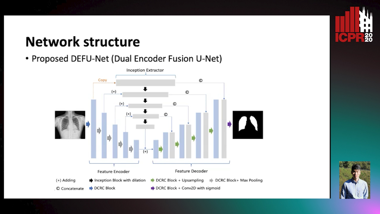
Auto-TLDR; Inception Convolutional Neural Network with Dilation for Chest X-Ray Segmentation

Slides Similar

A number of methods based on the deep learning have been applied to medical image segmentation and have achieved state-of-the-art performance. The most famous technique is U-Net which has been used to many medical datasets including the Chest X-ray. Due to the importance of chest x- ray data in studying COVID-19, there is a demand for state-of- art models capable of precisely segmenting chest x-rays. In this paper, we propose a dual encoder fusion U-Net framework for Chest X-rays based on Inception Convolutional Neural Network with dilation, Densely Connected Recurrent Convolutional Neural Network, which is named DEFU-Net. The densely connected recurrent path extends the network deeper for facilitating context feature extraction. In order to increase the width of network and enrich representation of features, the inception blocks with dilation have been used. The inception blocks can capture globally and locally spatial information with various receptive fields to avoid information loss caused by max-pooling. Meanwhile, the features fusion of two path by summation preserve the context and the spatial information for decoding part. We applied this model in Chest X-ray dataset from two different manufacturers (Montgomery and Shenzhen hospital). The DEFU-Net achieves the better performance than basic U-Net, residual U-Net, BCDU- Net, R2U-Net and attention R2U-Net. This model approaches state-of-the-art in this mixed dataset. The open source code for this proposed framework is public available.

Merged 1D-2D Deep Convolutional Neural Networks for Nerve Detection in Ultrasound Images

Mohammad Alkhatib, Adel Hafiane, Pierre Vieyres

Responsive image

Auto-TLDR; A Deep Neural Network for Deep Neural Networks to Detect Median Nerve in Ultrasound-Guided Regional Anesthesia

Slides Poster Similar

Ultrasound-Guided Regional Anesthesia (UGRA) becomes a standard procedure in surgical operations and contributes to pain management. It offers the advantages of the targeted nerve detection and provides the visualization of regions of interest such as anatomical structures. However, nerve detection is one of the most challenging tasks that anesthetists can encounter in the UGRA procedure. A computer-aided system that can detect automatically the nerve region would facilitate the anesthetist's daily routine and allow them to concentrate more on the anesthetic delivery. In this paper, we propose a new method based on merging deep learning models from different data to detect the median nerve. The merged architecture consists of two branches, one being one dimensional (1D) convolutional neural networks (CNN) branch and another 2D CNN branch. The merged architecture aims to learn the high-level features from 1D handcrafted noise-robust features and 2D ultrasound images. The obtained results show the validity and high accuracy of the proposed approach and its robustness.

Epileptic Seizure Prediction: A Semi-Dilated Convolutional Neural Network Architecture

Ramy Hussein, Rabab K. Ward, Soojin Lee, Martin Mckeown

Responsive image

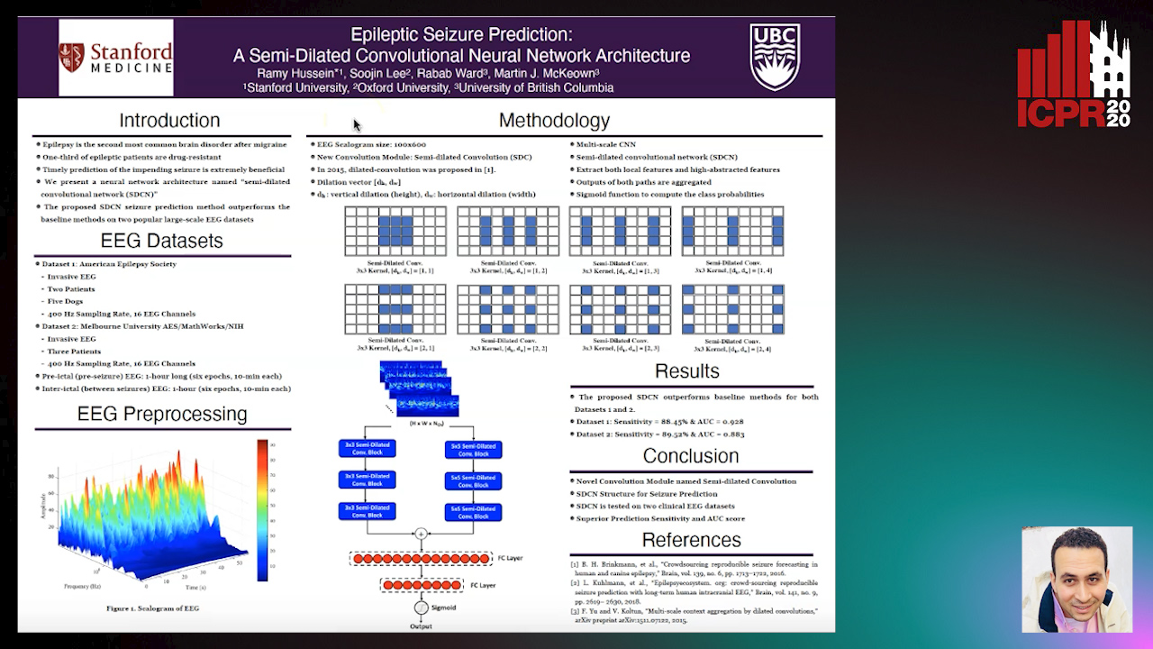
Auto-TLDR; Semi-Dilated Convolutional Network for Seizure Prediction using EEG Scalograms

Poster Similar

Despite many recent advances in machine learning and time-series classification, accurate prediction of seizures remains elusive. In this work, we develop a convolutional network module that uses Electroencephalogram (EEG) scalograms to distinguish between the pre-seizure and normal brain activities. Since the EEG scalogram takes rectangular image format with many more temporal bins than spectral bins, the presented module uses "semi-dilated convolutions" to also create a proportional non-square receptive field. The proposed semi-dilated convolutions support exponential expansion of the receptive field over the long dimension (image width, i.e. time) while maintaining high resolution over the short dimension (image height, i.e., frequency). The proposed architecture comprises a set of co-operative semi-dilated convolutional blocks, each block has a stack of parallel semi-dilated convolutional modules with different dilation rates. Results show that our proposed seizure prediction solution outperforms the state-of-the-art methods, achieving a seizure prediction sensitivity of 88.45% and 89.52% for the American Epilepsy Society and Melbourne University EEG datasets, respectively.

Trainable Spectrally Initializable Matrix Transformations in Convolutional Neural Networks

Michele Alberti, Angela Botros, Schuetz Narayan, Rolf Ingold, Marcus Liwicki, Mathias Seuret

Responsive image

Auto-TLDR; Trainable and Spectrally Initializable Matrix Transformations for Neural Networks

Slides Poster Similar

In this work, we introduce a new architectural component to Neural Networks (NN), i.e., trainable and spectrally initializable matrix transformations on feature maps. While previous literature has already demonstrated the possibility of adding static spectral transformations as feature processors, our focus is on more general trainable transforms. We study the transforms in various architectural configurations on four datasets of different nature: from medical (ColorectalHist, HAM10000) and natural (Flowers) images to historical documents (CB55). With rigorous experiments that control for the number of parameters and randomness, we show that networks utilizing the introduced matrix transformations outperform vanilla neural networks. The observed accuracy increases appreciably across all datasets. In addition, we show that the benefit of spectral initialization leads to significantly faster convergence, as opposed to randomly initialized matrix transformations. The transformations are implemented as auto-differentiable PyTorch modules that can be incorporated into any neural network architecture. The entire code base is open-source.

Deep Learning on Active Sonar Data Using Bayesian Optimization for Hyperparameter Tuning

Henrik Berg, Karl Thomas Hjelmervik

Responsive image

Auto-TLDR; Bayesian Optimization for Sonar Operations in Littoral Environments

Slides Poster Similar

Sonar operations in littoral environments may be challenging due to an increased probability of false alarms. Machine learning can be used to train classifiers that are able to filter out most of the false alarms automatically, however, this is a time consuming process, with many hyperparameters that need to be tuned in order to yield useful results. In this paper, Bayesian optimization is used to search for good values for some of the hyperparameters, like topology and training parameters, resulting in performance superior to earlier trial-and-error based training. Additionally, we analyze some of the parameters involved in the Bayesian optimization, as well as the resulting hyperparameter values.

AdaFilter: Adaptive Filter Design with Local Image Basis Decomposition for Optimizing Image Recognition Preprocessing

Aiga Suzuki, Keiichi Ito, Takahide Ibe, Nobuyuki Otsu

Responsive image

Auto-TLDR; Optimal Preprocessing Filtering for Pattern Recognition Using Higher-Order Local Auto-Correlation

Slides Poster Similar

Image preprocessing is an important process during pattern recognition which increases the recognition performance. Linear convolution filtering is a primary preprocessing method used to enhance particular local patterns of the image which are essential for recognizing the images. However, because of the vast search space of the preprocessing filter, almost no earlier studies have tackled the problem of identifying an optimal preprocessing filter that yields effective features for input images. This paper proposes a novel design method for the optimal preprocessing filter corresponding to a given task. Our method calculates local image bases of the training dataset and represents the optimal filter as a linear combination of these local image bases with the optimized coefficients to maximize the expected generalization performance. Thereby, the optimization problem of the preprocessing filter is converted to a lower-dimensional optimization problem. Our proposed method combined with a higher-order local auto-correlation (HLAC) feature extraction exhibited the best performance both in the anomaly detection task with the conventional pattern recognition algorithm and in the classification task using the deep convolutional neural network compared with typical preprocessing filters.

3D Facial Matching by Spiral Convolutional Metric Learning and a Biometric Fusion-Net of Demographic Properties

Soha Sadat Mahdi, Nele Nauwelaers, Philip Joris, Giorgos Bouritsas, Imperial London, Sergiy Bokhnyak, Susan Walsh, Mark Shriver, Michael Bronstein, Peter Claes

Responsive image

Auto-TLDR; Multi-biometric Fusion for Biometric Verification using 3D Facial Mesures

Slides Similar

Face recognition is a widely accepted biometric verification tool, as the face contains a lot of information about the identity of a person. In this study, a 2-step neural-based pipeline is presented for matching 3D facial shape to multiple DNA-related properties (sex, age, BMI and genomic background). The first step consists of a triplet loss-based metric learner that compresses facial shape into a lower dimensional embedding while preserving information about the property of interest. Most studies in the field of metric learning have only focused on Euclidean data. In this work, geometric deep learning is employed to learn directly from 3D facial meshes. To this end, spiral convolutions are used along with a novel mesh-sampling scheme that retains uniformly sampled 3D points at different levels of resolution. The second step is a multi-biometric fusion by a fully connected neural network. The network takes an ensemble of embeddings and property labels as input and returns genuine and imposter scores. Since embeddings are accepted as an input, there is no need to train classifiers for the different properties and available data can be used more efficiently. Results obtained by a 10-fold cross-validation for biometric verification show that combining multiple properties leads to stronger biometric systems. Furthermore, the proposed neural-based pipeline outperforms a linear baseline, which consists of principal component analysis, followed by classification with linear support vector machines and a Naïve Bayes-based score-fuser.

Fall Detection by Human Pose Estimation and Kinematic Theory

Vincenzo Dentamaro, Donato Impedovo, Giuseppe Pirlo

Responsive image

Auto-TLDR; A Decision Support System for Automatic Fall Detection on Le2i and URFD Datasets

Slides Poster Similar

In a society with increasing age, the understanding of human falls it is of paramount importance. This paper presents a Decision Support System whose pipeline is designed to extract and compute physical domain’s features achieving the state of the art accuracy on the Le2i and UR fall detection datasets. The paper uses the Kinematic Theory of Rapid Human Movement and its sigma-lognormal model together with classic physical features to achieve 98% and 99% of accuracy in automatic fall detection on respectively Le2i and URFD datasets. The effort made in the design of this work is toward recognition of falls by using physical models whose laws are clear and understandable.

Early Wildfire Smoke Detection in Videos

Taanya Gupta, Hengyue Liu, Bir Bhanu

Responsive image

Auto-TLDR; Semi-supervised Spatio-Temporal Video Object Segmentation for Automatic Detection of Smoke in Videos during Forest Fire

Poster Similar

Recent advances in unmanned aerial vehicles and camera technology have proven useful for the detection of smoke that emerges above the trees during a forest fire. Automatic detection of smoke in videos is of great interest to Fire department. To date, in most parts of the world, the fire is not detected in its early stage and generally it turns catastrophic. This paper introduces a novel technique that integrates spatial and temporal features in a deep learning framework using semi-supervised spatio-temporal video object segmentation and dense optical flow. However, detecting this smoke in the presence of haze and without the labeled data is difficult. Considering the visibility of haze in the sky, a dark channel pre-processing method is used that reduces the amount of haze in video frames and consequently improves the detection results. Online training is performed on a video at the time of testing that reduces the need for ground-truth data. Tests using the publicly available video datasets show that the proposed algorithms outperform previous work and they are robust across different wildfire-threatened locations.

A Benchmark Dataset for Segmenting Liver, Vasculature and Lesions from Large-Scale Computed Tomography Data

Bo Wang, Zhengqing Xu, Wei Xu, Qingsen Yan, Liang Zhang, Zheng You

Responsive image

Auto-TLDR; The Biggest Treatment-Oriented Liver Cancer Dataset for Segmentation

Slides Poster Similar

How to build a high-performance liver-related computer assisted diagnosis system is an open question of great interest. However, the performance of the state-of-art algorithm is always limited by the amount of data and quality of the label. To address this problem, we propose the biggest treatment-oriented liver cancer dataset for liver surgery and treatment planning. This dataset provides 216 cases (totally about 268K frames) scanned images in contrast-enhanced computed tomography (CT). We labeled all the CT images with the liver, liver vasculature and liver tumor segmentation ground truth for train and tune segmentation algorithms in advance. Based on that, we evaluate several recent and state-of-the-art segmentation algorithms, including 7 deep learning methods, on CT sequences. All results are compared to reference segmentations five error metrics that highlight different aspects of segmentation accuracy. In general, compared with previous datasets, our dataset is really a challenging dataset. To our knowledge, the proposed dataset and benchmark allow for the first time systematic exploration of such issues, and will be made available to allow for further research in this field.

Mean Decision Rules Method with Smart Sampling for Fast Large-Scale Binary SVM Classification

Alexandra Makarova, Mikhail Kurbakov, Valentina Sulimova

Responsive image

Auto-TLDR; Improving Mean Decision Rule for Large-Scale Binary SVM Problems

Slides Poster Similar

This paper relies on the Mean Decision Rule (MDR) method for solving large-scale binary SVM problems. It consists in taking small random samples of the full dataset and separate training for each of them with consecutive averaging the respective individual decision rules to obtain a final one. This paper proposes two new approaches to improve it. The first proposed approach is a new sampling technique that exploits SVM and MDR properties to fast form so called smart samples by selecting only the objects, that are candidates to be the support ones. The proposed technique essentially increases MDR convergence and allows to reach the highest quality in less time. In the case of kernel-based MDR (KMDR) the proposed sampling technique allows additionally to reduce the number of support objects in the final decision rule and, as a result, to decrease the recognition time. The second proposed approach is a new data strategy to accelerate random access to large datasets stored in the traditional libsvm format. The proposed strategy allows to quickly extract random subsets of objects from a file and load them into RAM, and is it also suitable for any sampling-based methods, including stochastic gradient methods. Joint using of the proposed approaches with (K)MDR allows to obtain the best (or near the best) decision of large-scale binary SVM problems faster, compared to the existing SVM solvers.

Transfer Learning through Weighted Loss Function and Group Normalization for Vessel Segmentation from Retinal Images

Abdullah Sarhan, Jon Rokne, Reda Alhajj, Andrew Crichton

Responsive image

Auto-TLDR; Deep Learning for Segmentation of Blood Vessels in Retinal Images

Slides Poster Similar

The vascular structure of blood vessels is important in diagnosing retinal conditions such as glaucoma and diabetic retinopathy. Accurate segmentation of these vessels can help in detecting retinal objects such as the optic disc and optic cup and hence determine if there are damages to these areas. Moreover, the structure of the vessels can help in diagnosing glaucoma. The rapid development of digital imaging and computer-vision techniques has increased the potential for developing approaches for segmenting retinal vessels. In this paper, we propose an approach for segmenting retinal vessels that uses deep learning along with transfer learning. We adapted the U-Net structure to use a customized InceptionV3 as the encoder and used multiple skip connections to form the decoder. Moreover, we used a weighted loss function to handle the issue of class imbalance in retinal images. Furthermore, we contributed a new dataset to this field. We tested our approach on six publicly available datasets and a newly created dataset. We achieved an average accuracy of 95.60\% and a Dice coefficient of 80.98\%. The results obtained from comprehensive experiments demonstrate the robustness of our approach to the segmentation of blood vessels in retinal images obtained from different sources. Our approach results in greater segmentation accuracy than other approaches.

Fused 3-Stage Image Segmentation for Pleural Effusion Cell Clusters

Sike Ma, Meng Zhao, Hao Wang, Fan Shi, Xuguo Sun, Shengyong Chen, Hong-Ning Dai

Responsive image

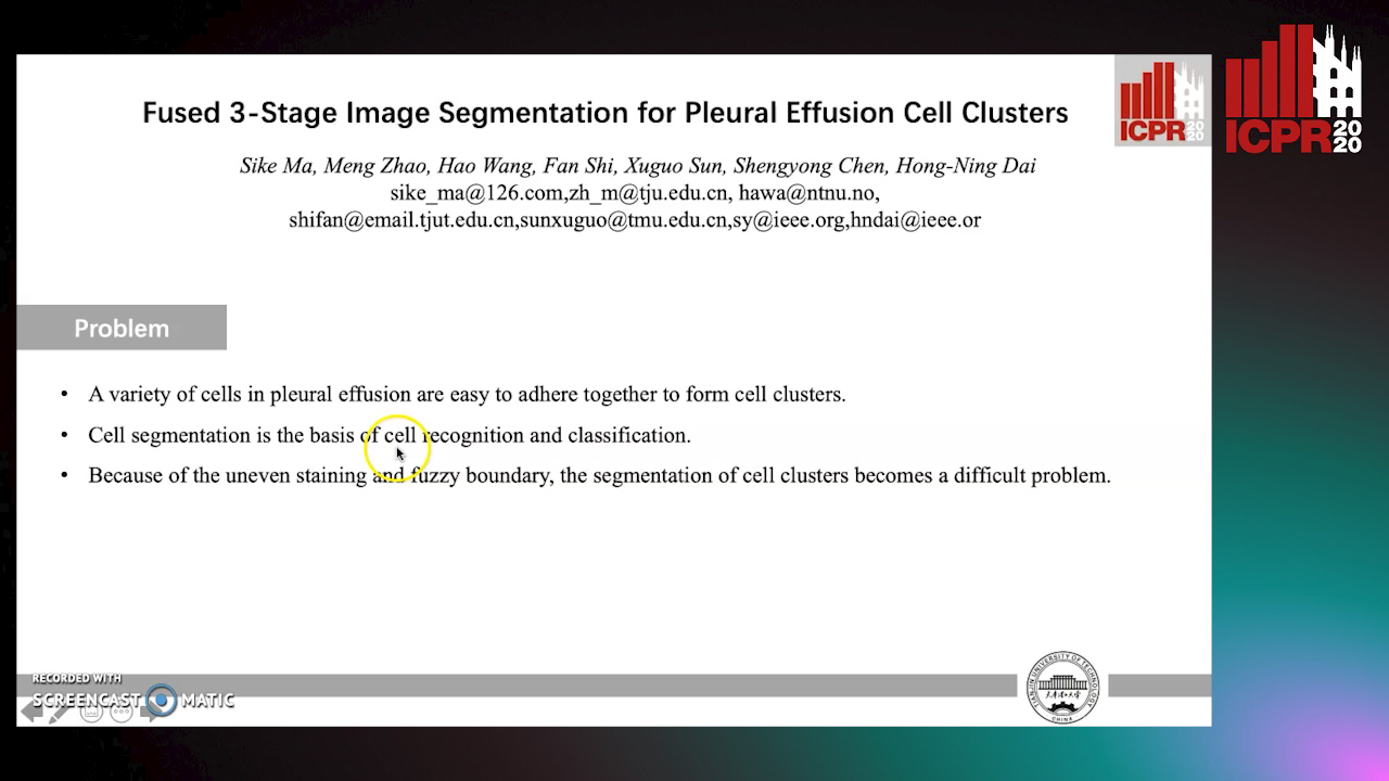
Auto-TLDR; Coarse Segmentation of Stained and Stained Unstained Cell Clusters in pleural effusion using 3-stage segmentation method

Slides Poster Similar

The appearance of tumor cell clusters in pleural effusion is usually a vital sign of cancer metastasis. Segmentation, as an indispensable basis, is of crucial importance for diagnosing, chemical treatment, and prognosis in patients. However, accurate segmentation of unstained cell clusters containing more detailed features than the fluorescent staining images remains to be a challenging problem due to the complex background and the unclear boundary. Therefore, in this paper, we propose a fused 3-stage image segmentation algorithm, namely Coarse segmentation-Mapping-Fine segmentation (CMF) to achieve unstained cell clusters from whole slide images. Firstly, we establish a tumor cell cluster dataset consisting of 107 sets of images, with each set containing one unstained image, one stained image, and one ground-truth image. Then, according to the features of the unstained and stained cell clusters, we propose a three-stage segmentation method: 1) Coarse segmentation on stained images to extract suspicious cell regions-Region of Interest (ROI); 2) Mapping this ROI to the corresponding unstained image to get the ROI of the unstained image (UI-ROI); 3) Fine Segmentation using improved automatic fuzzy clustering framework (AFCF) on the UI-ROI to get precise cell cluster boundaries. Experimental results on 107 sets of images demonstrate that the proposed algorithm can achieve better performance on unstained cell clusters with an F1 score of 90.40%.

Attribute-Based Quality Assessment for Demographic Estimation in Face Videos

Fabiola Becerra-Riera, Annette Morales-González, Heydi Mendez-Vazquez, Jean-Luc Dugelay

Responsive image

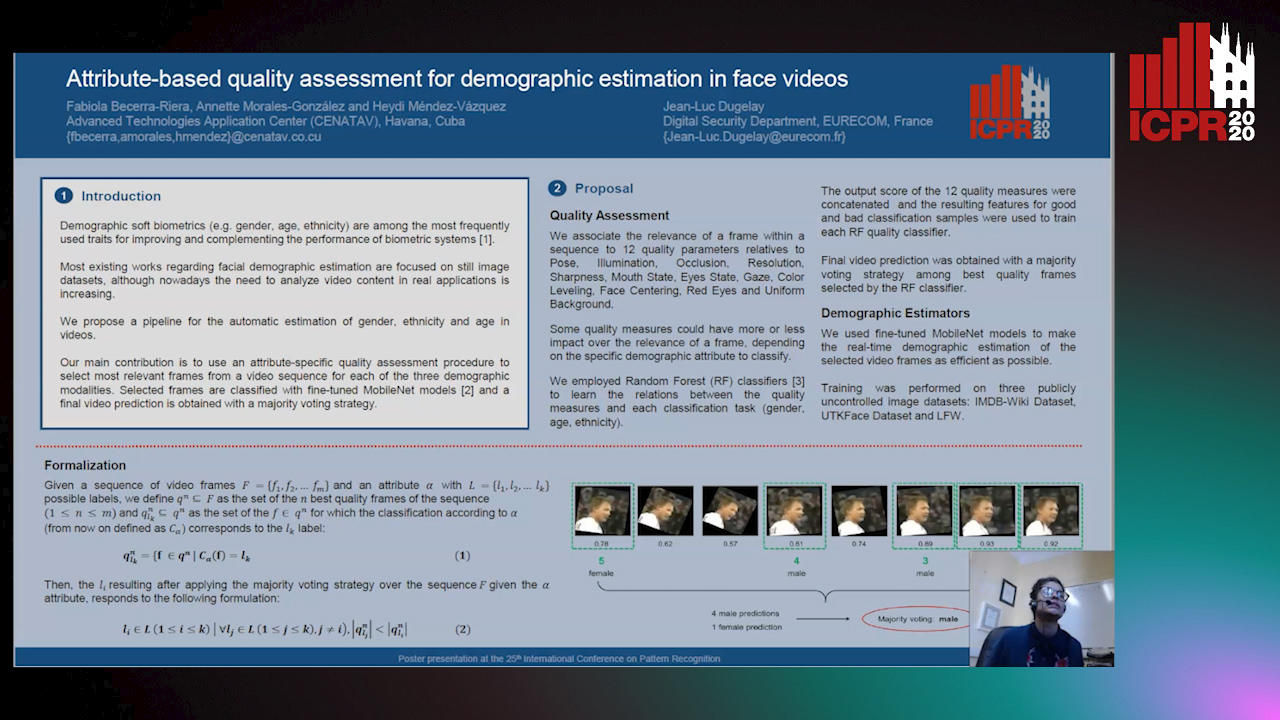
Auto-TLDR; Facial Demographic Estimation in Video Scenarios Using Quality Assessment

Slides Similar

Most existing works regarding facial demographic estimation are focused on still image datasets, although nowadays the need to analyze video content in real applications is increasing. We propose to tackle gender, age and ethnicity estimation in the context of video scenarios. Our main contribution is to use an attribute-specific quality assessment procedure to select best quality frames from a video sequence for each of the three demographic modalities. Best quality frames are classified with fine-tuned MobileNet models and a final video prediction is obtained with a majority voting strategy among the best selected frames. Our validation on three different datasets and our comparison with state-of-the-art models, show the effectiveness of the proposed demographic classifiers and the quality pipeline, which allows to reduce both: the number of frames to be classified and the processing time in practical applications; and improves the soft biometrics prediction accuracy.

Multi-Scale and Attention Based ResNet for Heartbeat Classification

Haojie Zhang, Gongping Yang, Yuwen Huang, Feng Yuan, Yilong Yin

Responsive image

Auto-TLDR; A Multi-Scale and Attention based ResNet for ECG heartbeat classification in intra-patient and inter-patient paradigms

Slides Poster Similar

This paper presents a novel deep learning framework for the electrocardiogram (ECG) heartbeat classification. Although there have been some studies with excellent overall accuracy, these studies have not been very accurate in the diagnosis of arrhythmia classes especially such as supraventricular ectopic beat (SVEB) and ventricular ectopic beat (VEB). In our work, we propose a Multi-Scale and Attention based ResNet for heartbeat classification in intra-patient and inter-patient paradigms respectively. Firstly, we extract shallow features from a convolutional layer. Secondly, the shallow features are sent into three branches with different convolution kernels in order to combine receptive fields of different sizes. Finally, fully connected layers are used to classify the heartbeat. Besides, we design a new attention mechanism based on the characteristics of heartbeat data. At last, extensive experiments on benchmark dataset demonstrate the effectiveness of our proposed model.

Using Machine Learning to Refer Patients with Chronic Kidney Disease to Secondary Care

Lee Au-Yeung, Xianghua Xie, Timothy Marcus Scale, James Anthony Chess

Responsive image

Auto-TLDR; A Machine Learning Approach for Chronic Kidney Disease Prediction using Blood Test Data

Slides Poster Similar

There has been growing interest recently in using machine learning techniques as an aid in clinical medicine. Machine learning offers a range of classification algorithms which can be applied to medical data to aid in making clinical predictions. Recent studies have demonstrated the high predictive accuracy of various classification algorithms applied to clinical data. Several studies have already been conducted in diagnosing or predicting chronic kidney disease at various stages using different sets of variables. In this study we are investigating the use machine learning techniques with blood test data. Such a system could aid renal teams in making recommendations to primary care general practitioners to refer patients to secondary care where patients may benefit from earlier specialist assessment and medical intervention. We are able to achieve an overall accuracy of 88.48\% using logistic regression, 87.12\% using ANN and 85.29\% using SVM. ANNs performed with the highest sensitivity at 89.74\% compared to 86.67\% for logistic regression and 85.51\% for SVM.

Deep Learning-Based Type Identification of Volumetric MRI Sequences

Jean Pablo De Mello, Thiago Paixão, Rodrigo Berriel, Mauricio Reyes, Alberto F. De Souza, Claudine Badue, Thiago Oliveira-Santos

Responsive image

Auto-TLDR; Deep Learning for Brain MRI Sequences Identification Using Convolutional Neural Network

Slides Poster Similar

The analysis of Magnetic Resonance Imaging (MRI) sequences enables clinical professionals to monitor the progression of a brain tumor. As the interest for automatizing brain volume MRI analysis increases, it becomes convenient to have each sequence well identified. However, the unstandardized naming of MRI sequences make their identification difficult for automated systems, as well as make it difficult for researches to generate or use datasets for machine learning research. In face of that, we propose a system for identifying types of brain MRI sequences based on deep learning. By training a Convolutional Neural Network (CNN) based on 18-layer ResNet architecture, our system is able to classify a volumetric brain MRI as a T1, T1c, T2 or FLAIR sequence, or whether it does not belong to any of these classes. The network was trained with both pre-processed (BraTS dataset) and non-pre-processed (TCGA-GBM dataset) images with diverse acquisition protocols, requiring only a few layers of the volume for training. Our system is able to classify among sequence types with an accuracy of 96.27%.

A Novel Adaptive Minority Oversampling Technique for Improved Classification in Data Imbalanced Scenarios

Ayush Tripathi, Rupayan Chakraborty, Sunil Kumar Kopparapu

Responsive image

Auto-TLDR; Synthetic Minority OverSampling Technique for Imbalanced Data

Slides Poster Similar

Imbalance in the proportion of training samples belonging to different classes often poses performance degradation of conventional classifiers. This is primarily due to the tendency of the classifier to be biased towards the majority classes in the imbalanced dataset. In this paper, we propose a novel three step technique to address imbalanced data. As a first step we significantly oversample the minority class distribution by employing the traditional Synthetic Minority OverSampling Technique (SMOTE) algorithm using the neighborhood of the minority class samples and in the next step we partition the generated samples using a Gaussian-Mixture Model based clustering algorithm. In the final step synthetic data samples are chosen based on the weight associated with the cluster, the weight itself being determined by the distribution of the majority class samples. Extensive experiments on several standard datasets from diverse domains show the usefulness of the proposed technique in comparison with the original SMOTE and its state-of-the-art variants algorithms.

Supporting Skin Lesion Diagnosis with Content-Based Image Retrieval

Stefano Allegretti, Federico Bolelli, Federico Pollastri, Sabrina Longhitano, Giovanni Pellacani, Costantino Grana

Responsive image

Auto-TLDR; Skin Images Retrieval Using Convolutional Neural Networks for Skin Lesion Classification and Segmentation

Slides Poster Similar

Given the relevance of skin cancer, many attempts have been dedicated to the creation of automated devices that could assist both expert and beginner dermatologists towards fast and early diagnosis of skin lesions. In recent years, tasks such as skin lesion classification and segmentation have been extensively addressed with deep learning algorithms, which in some cases reach a diagnostic accuracy comparable to that of expert physicians. However, the general lack of interpretability and reliability severely hinders the ability of those approaches to actually support dermatologists in the diagnosis process. In this paper a novel skin images retrieval system is presented, which exploits features extracted by Convolutional Neural Networks to gather similar images from a publicly available dataset, in order to assist the diagnosis process of both expert and novice practitioners. In the proposed framework, Resnet-50 is initially trained for the classification of dermoscopic images; then, the feature extraction part is isolated, and an embedding network is build on top of it. The embedding learns an alternative representation, which allows to check image similarity by means of a distance measure. Experimental results reveal that the proposed method is able to select meaningful images, which can effectively boost the classification accuracy of human dermatologists.

Depth Videos for the Classification of Micro-Expressions

Ankith Jain Rakesh Kumar, Bir Bhanu, Christopher Casey, Sierra Cheung, Aaron Seitz

Responsive image

Auto-TLDR; RGB-D Dataset for the Classification of Facial Micro-expressions

Slides Poster Similar

Facial micro-expressions are spontaneous, subtle, involuntary muscle movements occurring briefly on the face. The spotting and recognition of these expressions are difficult due to the subtle behavior, and the time duration of these expressions is about half a second, which makes it difficult for humans to identify them. These micro-expressions have many applications in our daily life, such as in the field of online learning, game playing, lie detection, and therapy sessions. Traditionally, researchers use RGB images/videos to spot and classify these micro-expressions, which pose challenging problems, such as illumination, privacy concerns and pose variation. The use of depth videos solves these issues to some extent, as the depth videos are not susceptible to the variation in illumination. This paper describes the collection of a first RGB-D dataset for the classification of facial micro-expressions into 6 universal expressions: Anger, Happy, Sad, Fear, Disgust, and Surprise. This paper shows the comparison between the RGB and Depth videos for the classification of facial micro-expressions. Further, a comparison of results shows that depth videos alone can be used to classify facial micro-expressions correctly in a decision tree structure by using the traditional and deep learning approaches with good classification accuracy. The dataset will be released to the public in the near future.

Inferring Functional Properties from Fluid Dynamics Features

Andrea Schillaci, Maurizio Quadrio, Carlotta Pipolo, Marcello Restelli, Giacomo Boracchi

Responsive image

Auto-TLDR; Exploiting Convective Properties of Computational Fluid Dynamics for Medical Diagnosis

Slides Poster Similar

In a wide range of applied problems involving fluid flows, Computational Fluid Dynamics (CFD) provides detailed quantitative information on the flow field, at various levels of fidelity and computational cost. However, CFD alone cannot predict high-level functional properties of the system that are not easily obtained from the equations of fluid motion. In this work, we present a data-driven framework to extract additional information, such as medical diagnostic output, from CFD solutions. The task is made difficult by the huge data dimensionality of CFD, together with the limited amount of training data implied by its high computational cost. By pursuing a traditional ML pipeline of pre-processing, feature extraction, and model training, we demonstrate that informative features can be extracted from CFD data. Two experiments, pertaining to different application domains, support the claim that the convective properties implicit into a CFD solution can be leveraged to retrieve functional information for which an analytical definition is missing. Despite the preliminary nature of our study and the relative simplicity of both the geometrical and CFD models, for the first time we demonstrate that the combination of ML and CFD can diagnose a complex system in terms of high-level functional information.