Deep Learning in the Ultrasound Evaluation of Neonatal Respiratory Status

Michela Gravina, Diego Gragnaniello, Giovanni Poggi, Luisa Verdoliva, Carlo Sansone, Iuri Corsini, Carlo Dani, Fabio Meneghin, Gianluca Lista, Salvatore Aversa, Migliaro Migliaro, Raimondi Francesco

Responsive image

Auto-TLDR; Lung Ultrasound Imaging with Deep Learning Networks and Training Strategies: An Analysis and Adaptation

Slides Poster

Lung ultrasound imaging is reaching growing interest from the scientific community. On one side, thanks to its harmlessness and high descriptive power, this kind of diagnostic imaging became largely adopted in sensitive applications, like the diagnosis and follow-up of preterm newborns in neonatal intensive care units. At the same time, novel image analysis and pattern recognition approaches can fully exploit the rich information contained in this data, making them attractive for the research community. In this work, we present a thorough analysis of recent deep learning networks and training strategies conducted on a vast and challenging multicenter dataset comprising 87 patients with different diseases and gestational ages. These approaches are firstly discussed in the context of lung respiratory status assessing through ultrasound imaging and then evaluated against a reference marker. The conducted analysis shed some light on this problem, by relating the criticisms that can mislead the training procedure and proposing some adaptations to the specific problem. The achieved results sensibly outperform that obtained by previous work, based on textural features, and narrow the gap with the visual score predicted by the human experts.

Similar papers

Dealing with Scarce Labelled Data: Semi-Supervised Deep Learning with Mix Match for Covid-19 Detection Using Chest X-Ray Images

Saúl Calderón Ramirez, Raghvendra Giri, Shengxiang Yang, Armaghan Moemeni, Mario Umaña, David Elizondo, Jordina Torrents-Barrena, Miguel A. Molina-Cabello

Responsive image

Auto-TLDR; Semi-supervised Deep Learning for Covid-19 Detection using Chest X-rays

Slides Poster Similar

Coronavirus (Covid-19) is spreading fast, infecting people through contact in various forms including droplets from sneezing and coughing. Therefore, the detection of infected subjects in an early, quick and cheap manner is urgent. Currently available tests are scarce and limited to people in danger of serious illness. The application of deep learning to chest X-ray images for Covid-19 detection is an attractive approach. However, this technology usually relies on the availability of large labelled datasets, a requirement hard to meet in the context of a virus outbreak. To overcome this challenge, a semi-supervised deep learning model using both labelled and unlabelled data is proposed. We developed and tested a semi-supervised deep learning framework based on the Mix Match architecture to classify chest X-rays into Covid-19, pneumonia and healthy cases. The presented approach was calibrated using two publicly available datasets. The results show an accuracy increase of around $15\%$ under low labelled / unlabelled data ratio. This indicates that our semi-supervised framework can help improve performance levels towards Covid-19 detection when the amount of high-quality labelled data is scarce. Also, we introduce a semi-supervised deep learning boost coefficient which is meant to ease the scalability of our approach and performance comparison.

A Systematic Investigation on Deep Architectures for Automatic Skin Lesions Classification

Pierluigi Carcagni, Marco Leo, Andrea Cuna, Giuseppe Celeste, Cosimo Distante

Responsive image

Auto-TLDR; RegNet: Deep Investigation of Convolutional Neural Networks for Automatic Classification of Skin Lesions

Slides Poster Similar

Computer vision-based techniques are more and more employed in healthcare and medical fields nowadays in order, principally, to be as a support to the experienced medical staff to help them to make a quick and correct diagnosis. One of the hot topics in this arena concerns the automatic classification of skin lesions. Several promising works exist about it, mainly leveraging Convolutional Neural Networks (CNN), but proposed pipeline mainly rely on complex data preprocessing and there is no systematic investigation about how available deep models can actually reach the accuracy needed for real applications. In order to overcome these drawbacks, in this work, an end-to-end pipeline is introduced and some of the most recent Convolutional Neural Networks (CNNs) architectures are included in it and compared on the largest common benchmark dataset recently introduced. To this aim, for the first time in this application context, a new network design paradigm, namely RegNet, has been exploited to get the best models among a population of configurations. The paper introduces a threefold level of contribution and novelty with respect the previous literature: the deep investigation of several CNN architectures driving to a consistent improvement of the lesions recognition accuracy, the exploitation of a new network design paradigm able to study the behavior of populations of models and a deep discussion about pro and cons of each analyzed method paving the path towards new research lines.

Confidence Calibration for Deep Renal Biopsy Immunofluorescence Image Classification

Federico Pollastri, Juan Maroñas, Federico Bolelli, Giulia Ligabue, Roberto Paredes, Riccardo Magistroni, Costantino Grana

Responsive image

Auto-TLDR; A Probabilistic Convolutional Neural Network for Immunofluorescence Classification in Renal Biopsy

Slides Poster Similar

With this work we tackle immunofluorescence classification in renal biopsy, employing state-of-the-art Convolutional Neural Networks. In this setting, the aim of the probabilistic model is to assist an expert practitioner towards identifying the location pattern of antibody deposits within a glomerulus. Since modern neural networks often provide overconfident outputs, we stress the importance of having a reliable prediction, demonstrating that Temperature Scaling, a recently introduced re-calibration technique, can be successfully applied to immunofluorescence classification in renal biopsy. Experimental results demonstrate that the designed model yields good accuracy on the specific task, and that Temperature Scaling is able to provide reliable probabilities, which are highly valuable for such a task given the low inter-rater agreement.

Prediction of Obstructive Coronary Artery Disease from Myocardial Perfusion Scintigraphy using Deep Neural Networks

Ida Arvidsson, Niels Christian Overgaard, Miguel Ochoa Figueroa, Jeronimo Rose, Anette Davidsson, Kalle Åström, Anders Heyden

Responsive image

Auto-TLDR; A Deep Learning Algorithm for Multi-label Classification of Myocardial Perfusion Scintigraphy for Stable Ischemic Heart Disease

Slides Poster Similar

For diagnosis and risk assessment in patients with stable ischemic heart disease, myocardial perfusion scintigraphy is one of the most common cardiological examinations performed today. There are however many motivations for why an artificial intelligence algorithm would provide useful input to this task. For example to reduce the subjectiveness and save time for the nuclear medicine physicians working with this time consuming task. In this work we have developed a deep learning algorithm for multi-label classification based on a modified convolutional neural network to estimate probability of obstructive coronary artery disease in the left anterior artery, left circumflex artery and right coronary artery. The prediction is based on data from myocardial perfusion scintigraphy studies conducted in a dedicated Cadmium-Zinc-Telluride cardio camera (D-SPECT Spectrum Dynamics). Data from 588 patients was available, with stress images in both upright and supine position, as well as a number of auxiliary parameters such as angina symptoms and BMI. The data was used to train and evaluate the algorithm using 5-fold cross-validation. We achieve state-of-the-art results for this task with an area under the receiver operating characteristics curve of 0.89 as average on per-vessel level and 0.94 on per-patient level.

A Systematic Investigation on End-To-End Deep Recognition of Grocery Products in the Wild

Marco Leo, Pierluigi Carcagni, Cosimo Distante

Responsive image

Auto-TLDR; Automatic Recognition of Products on grocery shelf images using Convolutional Neural Networks

Slides Poster Similar

Automatic recognition of products on grocery shelf images is a new and attractive topic in computer vision and machine learning since, it can be exploited in different application areas. This paper introduces a complete end-to-end pipeline (without preliminary radiometric and spatial transformations usually involved while dealing with the considered issue) and it provides a systematic investigation of recent machine learning models based on convolutional neural networks for addressing the product recognition task by exploiting the proposed pipeline on a recent challenging grocery product dataset. The investigated models were never been used in this context: they derive from the successful and more generic object recognition task and have been properly tuned to address this specific issue. Besides, also ensembles of nets built by most advanced theoretical fundaments have been taken into account. Gathered classification results were very encouraging since the recognition accuracy has been improved up to 15\% with respect to the leading approaches in the state of art on the same dataset. A discussion about the pros and cons of the investigated solutions are discussed by paving the path towards new research lines.

Weight Estimation from an RGB-D Camera in Top-View Configuration

Marco Mameli, Marina Paolanti, Nicola Conci, Filippo Tessaro, Emanuele Frontoni, Primo Zingaretti

Responsive image

Auto-TLDR; Top-View Weight Estimation using Deep Neural Networks

Slides Poster Similar

The development of so-called soft-biometrics aims at providing information related to the physical and behavioural characteristics of a person. This paper focuses on bodyweight estimation based on the observation from a top-view RGB-D camera. In fact, the capability to estimate the weight of a person can be of help in many different applications, from health-related scenarios to business intelligence and retail analytics. To deal with this issue, a TVWE (Top-View Weight Estimation) framework is proposed with the aim of predicting the weight. The approach relies on the adoption of Deep Neural Networks (DNNs) that have been trained on depth data. Each network has also been modified in its top section to replace classification with prediction inference. The performance of five state-of-art DNNs has been compared, namely VGG16, ResNet, Inception, DenseNet and Efficient-Net. In addition, a convolutional auto-encoder has also been included for completeness. Considering the limited literature in this domain, the TVWE framework has been evaluated on a new publicly available dataset: “VRAI Weight estimation Dataset”, which also collects, for each subject, labels related to weight, gender, and height. The experimental results have demonstrated that the proposed methods are suitable for this task, bringing different and significant insights for the application of the solution in different domains.

A Comparison of Neural Network Approaches for Melanoma Classification

Maria Frasca, Michele Nappi, Michele Risi, Genoveffa Tortora, Alessia Auriemma Citarella

Responsive image

Auto-TLDR; Classification of Melanoma Using Deep Neural Network Methodologies

Slides Poster Similar

Melanoma is the deadliest form of skin cancer and it is diagnosed mainly visually, starting from initial clinical screening and followed by dermoscopic analysis, biopsy and histopathological examination. A dermatologist’s recognition of melanoma may be subject to errors and may take some time to diagnose it. In this regard, deep learning can be useful in the study and classification of skin cancer. In particular, by classifying images with Deep Neural Network methodologies, it is possible to obtain comparable or even superior results compared to those of dermatologists. In this paper, we propose a methodology for the classification of melanoma by adopting different deep learning techniques applied to a common dataset, composed of images from the ISIC dataset and consisting of different types of skin diseases, including melanoma on which we applied a specific pre-processing phase. In particular, a comparison of the results is performed in order to select the best effective neural network to be applied to the problem of recognition and classification of melanoma. Moreover, we also evaluate the impact of the pre- processing phase on the final classification. Different metrics such as accuracy, sensitivity, and specificity have been selected to assess the goodness of the adopted neural networks and compare them also with the manual classification of dermatologists.

Automatic Semantic Segmentation of Structural Elements related to the Spinal Cord in the Lumbar Region by Using Convolutional Neural Networks

Jhon Jairo Sáenz Gamboa, Maria De La Iglesia-Vaya, Jon Ander Gómez

Responsive image

Auto-TLDR; Semantic Segmentation of Lumbar Spine Using Convolutional Neural Networks

Slides Poster Similar

This work addresses the problem of automatically segmenting the MR images corresponding to the lumbar spine. The purpose is to detect and delimit the different structural elements like vertebrae, intervertebral discs, nerves, blood vessels, etc. This task is known as semantic segmentation. The approach proposed in this work is based on convolutional neural networks whose output is a mask where each pixel from the input image is classified into one of the possible classes. Classes were defined by radiologists and correspond to structural elements and tissues. The proposed network architectures are variants of the U-Net. Several complementary blocks were used to define the variants: spatial attention models, deep supervision and multi-kernels at input, this last block type is based on the idea of inception. Those architectures which got the best results are described in this paper, and their results are discussed. Two of the proposed architectures outperform the standard U-Net used as baseline.

Unsupervised Detection of Pulmonary Opacities for Computer-Aided Diagnosis of COVID-19 on CT Images

Rui Xu, Xiao Cao, Yufeng Wang, Yen-Wei Chen, Xinchen Ye, Lin Lin, Wenchao Zhu, Chao Chen, Fangyi Xu, Yong Zhou, Hongjie Hu, Shoji Kido, Noriyuki Tomiyama

Responsive image

Auto-TLDR; A computer-aided diagnosis of COVID-19 from CT images using unsupervised pulmonary opacity detection

Slides Poster Similar

COVID-19 emerged towards the end of 2019 which was identified as a global pandemic by the world heath organization (WHO). With the rapid spread of COVID-19, the number of infected and suspected patients has increased dramatically. Chest computed tomography (CT) has been recognized as an efficient tool for the diagnosis of COVID-19. However, the huge CT data make it difficult for radiologist to fully exploit them on the diagnosis. In this paper, we propose a computer-aided diagnosis system that can automatically analyze CT images to distinguish the COVID-19 against to community-acquired pneumonia (CAP). The proposed system is based on an unsupervised pulmonary opacity detection method that locates opacity regions by a detector unsupervisedly trained from CT images with normal lung tissues. Radiomics based features are extracted insides the opacity regions, and fed into classifiers for classification. We evaluate the proposed CAD system by using 200 CT images collected from different patients in several hospitals. The accuracy, precision, recall, f1-score and AUC achieved are 95.5%, 100%, 91%, 95.1% and 95.9% respectively, exhibiting the promising capacity on the differential diagnosis of COVID-19 from CT images.

Fine-Tuning Convolutional Neural Networks: A Comprehensive Guide and Benchmark Analysis for Glaucoma Screening

Amed Mvoulana, Rostom Kachouri, Mohamed Akil

Responsive image

Auto-TLDR; Fine-tuning Convolutional Neural Networks for Glaucoma Screening

Slides Poster Similar

This work aimed at giving a comprehensive and in-detailed guide on the route to fine-tuning Convolutional Neural Networks (CNNs) for glaucoma screening. Transfer learning consists in a promising alternative to train CNNs from stratch, to avoid the huge data and resources requirements. After a thorough study of five state-of-the-art CNNs architectures, a complete and well-explained strategy for fine-tuning these networks is proposed, using hyperparameter grid-searching and two-phase training approach. Excellent performance is reached on model evaluation, with a 0.9772 AUROC validation rate, giving arise to reliable glaucoma diagosis-help systems. Also, a benchmark analysis is conducted across all fine-tuned models, studying them according to performance indices such as model complexity and size, AUROC density and inference time. This in-depth analysis allows a rigorous comparison between model characteristics, and is useful for giving practioners important trademarks for prospective applications and deployments.

Automatic Tuberculosis Detection Using Chest X-Ray Analysis with Position Enhanced Structural Information

Hermann Jepdjio Nkouanga, Szilard Vajda

Responsive image

Auto-TLDR; Automatic Chest X-ray Screening for Tuberculosis in Rural Population using Localized Region on Interest

Slides Poster Similar

For Tuberculosis (TB) detection beside the more expensive diagnosis solutions such as culture or sputum smear analysis one could consider the automatic analysis of the chest X-ray (CXR). This could mimic the lung region reading by the radiologist and it could provide a cheap solution to analyze and diagnose pulmonary abnormalities such as TB which often co- occurs with HIV. This software based pulmonary screening can be a reliable and affordable solution for rural population in different parts of the world such as India, Africa, etc. Our fully automatic system is processing the incoming CXR image by applying image processing techniques to detect the region on interest (ROI) followed by a computationally cheap feature extraction involving edge detection using Laplacian of Gaussian which we enrich by counting the local distribution of the intensities. The choice to ”zoom in” the ROI and look for abnormalities locally is motivated by the fact that some pulmonary abnormalities are localized in specific regions of the lungs. Later on the classifiers can decide about the normal or abnormal nature of each lung X-ray. Our goal is to find a simple feature, instead of a combination of several ones, -proposed and promoted in recent years’ literature, which can properly describe the different pathological alterations in the lungs. Our experiments report results on two publicly available data collections1, namely the Shenzhen and the Montgomery collection. For performance evaluation, measures such as area under the curve (AUC), and accuracy (ACC) were considered, achieving AUC = 0.81 (ACC = 83.33%) and AUC = 0.96 (ACC = 96.35%) for the Montgomery and Schenzen collections, respectively. Several comparisons are also provided to other state- of-the-art systems reported recently in the field.

Bridging the Gap between Natural and Medical Images through Deep Colorization

Lia Morra, Luca Piano, Fabrizio Lamberti, Tatiana Tommasi

Responsive image

Auto-TLDR; Transfer Learning for Diagnosis on X-ray Images Using Color Adaptation

Slides Poster Similar

Deep learning has thrived by training on large-scale datasets. However, in many applications, as for medical image diagnosis, getting massive amount of data is still prohibitive due to privacy, lack of acquisition homogeneity and annotation cost. In this scenario transfer learning from natural image collections is a standard practice that attempts to tackle shape, texture and color discrepancy all at once through pretrained model fine-tuning. In this work we propose to disentangle those challenges and design a dedicated network module that focuses on color adaptation. We combine learning from scratch of the color module with transfer learning of different classification backbones obtaining an end-to-end, easy-to-train architecture for diagnostic image recognition on X-ray images. Extensive experiments show how our approach is particularly efficient in case of data scarcity and provides a new path for further transferring the learned color information across multiple medical datasets.

Merged 1D-2D Deep Convolutional Neural Networks for Nerve Detection in Ultrasound Images

Mohammad Alkhatib, Adel Hafiane, Pierre Vieyres

Responsive image

Auto-TLDR; A Deep Neural Network for Deep Neural Networks to Detect Median Nerve in Ultrasound-Guided Regional Anesthesia

Slides Poster Similar

Ultrasound-Guided Regional Anesthesia (UGRA) becomes a standard procedure in surgical operations and contributes to pain management. It offers the advantages of the targeted nerve detection and provides the visualization of regions of interest such as anatomical structures. However, nerve detection is one of the most challenging tasks that anesthetists can encounter in the UGRA procedure. A computer-aided system that can detect automatically the nerve region would facilitate the anesthetist's daily routine and allow them to concentrate more on the anesthetic delivery. In this paper, we propose a new method based on merging deep learning models from different data to detect the median nerve. The merged architecture consists of two branches, one being one dimensional (1D) convolutional neural networks (CNN) branch and another 2D CNN branch. The merged architecture aims to learn the high-level features from 1D handcrafted noise-robust features and 2D ultrasound images. The obtained results show the validity and high accuracy of the proposed approach and its robustness.

A Lumen Segmentation Method in Ureteroscopy Images Based on a Deep Residual U-Net Architecture

Jorge Lazo, Marzullo Aldo, Sara Moccia, Michele Catellani, Benoit Rosa, Elena De Momi, Michel De Mathelin, Francesco Calimeri

Responsive image

Auto-TLDR; A Deep Neural Network for Ureteroscopy with Residual Units

Slides Poster Similar

Ureteroscopy is becoming the first surgical treatment option for the majority of urinary affections. This procedure is carried out using an endoscope which provides the surgeon with the visual and spatial information necessary to navigate inside the urinary tract. Having in mind the development of surgical assistance systems, that could enhance the performance of surgeon, the task of lumen segmentation is a fundamental part since this is the visual reference which marks the path that the endoscope should follow. This is something that has not been analyzed in ureteroscopy data before. However, this task presents several challenges given the image quality and the conditions itself of ureteroscopy procedures. In this paper, we study the implementation of a Deep Neural Network which exploits the advantage of residual units in an architecture based on U-Net. For the training of these networks, we analyze the use of two different color spaces: gray-scale and RGB data images. We found that training on gray-scale images gives the best results obtaining mean values of Dice Score, Precision, and Recall of 0.73, 0.58, and 0.92 respectively. The results obtained show that the use of residual U-Net could be a suitable model for further development for a computer-aided system for navigation and guidance through the urinary system.

Inception Based Deep Learning Architecture for Tuberculosis Screening of Chest X-Rays

Dipayan Das, K.C. Santosh, Umapada Pal

Responsive image

Auto-TLDR; End to End CNN-based Chest X-ray Screening for Tuberculosis positive patients in the severely resource constrained regions of the world

Slides Poster Similar

The motivation for this work is the primary need of screening Tuberculosis (TB) positive patients in the severely resource constrained regions of the world. Chest X-ray (CXR) is considered to be a promising indicator for the onset of TB, but the lack of skilled radiologists in such regions degrades the situation. Therefore, several computer aided diagnosis (CAD) systems have been proposed to solve the decision making problem, which includes hand engineered feature extraction methods to deep learning or Convolutional Neural Network (CNN) based methods. Feature extraction, being a time and resource intensive process, often delays the process of mass screening. Hence an end to end CNN architecture is proposed in this work to solve the problem. Two benchmark CXR datasets have been used in this work, collected from Shenzhen (China) and Montgomery County (USA), on which the proposed methodology achieved a maximum abnormality detection accuracy (ACC) of 91.7\% (0.96 AUC) and 87.47\% (0.92 AUC) respectively. To the greatest of our knowledge, the obtained results are marginally superior to the state of the art results that have solely used deep learning methodologies on the aforementioned datasets.

Video Face Manipulation Detection through Ensemble of CNNs

Nicolo Bonettini, Edoardo Daniele Cannas, Sara Mandelli, Luca Bondi, Paolo Bestagini, Stefano Tubaro

Responsive image

Auto-TLDR; Face Manipulation Detection in Video Sequences Using Convolutional Neural Networks

Slides Similar

In the last few years, several techniques for facial manipulation in videos have been successfully developed and made available to the masses (i.e., FaceSwap, deepfake, etc.). These methods enable anyone to easily edit faces in video sequences with incredibly realistic results and a very little effort. Despite the usefulness of these tools in many fields, if used maliciously, they can have a significantly bad impact on society (e.g., fake news spreading, cyber bullying through fake revenge porn). The ability of objectively detecting whether a face has been manipulated in a video sequence is then a task of utmost importance. In this paper, we tackle the problem of face manipulation detection in video sequences targeting modern facial manipulation techniques. In particular, we study the ensembling of different trained Convolutional Neural Network (CNN) models. In the proposed solution, different models are obtained starting from a base network (i.e., EfficientNetB4) making use of two different concepts: (i) attention layers; (ii) siamese training. We show that combining these networks leads to promising face manipulation detection results on two publicly available datasets with more than 119000 videos.

The Color Out of Space: Learning Self-Supervised Representations for Earth Observation Imagery

Stefano Vincenzi, Angelo Porrello, Pietro Buzzega, Marco Cipriano, Pietro Fronte, Roberto Cuccu, Carla Ippoliti, Annamaria Conte, Simone Calderara

Responsive image

Auto-TLDR; Satellite Image Representation Learning for Remote Sensing

Slides Poster Similar

The recent growth in the number of satellite images fosters the development of effective deep-learning techniques for Remote Sensing (RS). However, their full potential is untapped due to the lack of large annotated datasets. Such a problem is usually countered by fine-tuning a feature extractor that is previously trained on the ImageNet dataset. Unfortunately, the domain of natural images differs from the RS one, which hinders the final performance. In this work, we propose to learn meaningful representations from satellite imagery, leveraging its high-dimensionality spectral bands to reconstruct the visible colors. We conduct experiments on land cover classification (BigEarthNet) and West Nile Virus detection, showing that colorization is a solid pretext task for training a feature extractor. Furthermore, we qualitatively observe that guesses based on natural images and colorization rely on different parts of the input. This paves the way to an ensemble model that eventually outperforms both the above-mentioned techniques.

Deep Transfer Learning for Alzheimer’s Disease Detection

Nicole Cilia, Claudio De Stefano, Francesco Fontanella, Claudio Marrocco, Mario Molinara, Alessandra Scotto Di Freca

Responsive image

Auto-TLDR; Automatic Detection of Handwriting Alterations for Alzheimer's Disease Diagnosis using Dynamic Features

Slides Poster Similar

Early detection of Alzheimer’s Disease (AD) is essential in order to initiate therapies that can reduce the effects of such a disease, improving both life quality and life expectancy of patients. Among all the activities carried out in our daily life, handwriting seems one of the first to be influenced by the arise of neurodegenerative diseases. For this reason, the analysis of handwriting and the study of its alterations has become of great interest in this research field in order to make a diagnosis as early as possible. In recent years, many studies have tried to use classification algorithms applied to handwritings to implement decision support systems for AD diagnosis. A key issue for the use of these techniques is the detection of effective features, that allow the system to distinguish the natural handwriting alterations due to age, from those caused by neurodegenerative disorders. In this context, many interesting results have been published in the literature in which the features have been typically selected by hand, generally considering the dynamics of the handwriting process in order to detect motor disorders closely related to AD. Features directly derived from handwriting generation models can be also very helpful for AD diagnosis. It should be remarked, however, that the above features do not consider changes in the shape of handwritten traces, which may occur as a consequence of neurodegenerative diseases, as well as the correlation among shape alterations and changes in the dynamics of the handwriting process. Moving from these considerations, the aim of this study is to verify if the combined use of both shape and dynamic features allows a decision support system to improve performance for AD diagnosis. To this purpose, starting from a database of on-line handwriting samples, we generated for each of them a synthetic off-line colour image, where the colour of each elementary trait encodes, in the three RGB channels, the dynamic information associated to that trait. Finally, we exploited the capability of Deep Neural Networks (DNN) to automatically extract features from raw images. The experimental comparison of the results obtained by using standard features and features extracted according the above procedure, confirmed the effectiveness of our approach.

BG-Net: Boundary-Guided Network for Lung Segmentation on Clinical CT Images

Rui Xu, Yi Wang, Tiantian Liu, Xinchen Ye, Lin Lin, Yen-Wei Chen, Shoji Kido, Noriyuki Tomiyama

Responsive image

Auto-TLDR; Boundary-Guided Network for Lung Segmentation on CT Images

Slides Poster Similar

Lung segmentation on CT images is a crucial step for a computer-aided diagnosis system of lung diseases. The existing deep learning based lung segmentation methods are less efficient to segment lungs on clinical CT images, especially that the segmentation on lung boundaries is not accurate enough due to complex pulmonary opacities in practical clinics. In this paper, we propose a boundary-guided network (BG-Net) to address this problem. It contains two auxiliary branches that separately segment lungs and extract the lung boundaries, and an aggregation branch that efficiently exploits lung boundary cues to guide the network for more accurate lung segmentation on clinical CT images. We evaluate the proposed method on a private dataset collected from the Osaka university hospital and four public datasets including StructSeg, HUG, VESSEL12, and a Novel Coronavirus 2019 (COVID-19) dataset. Experimental results show that the proposed method can segment lungs more accurately and outperform several other deep learning based methods.

From Early Biological Models to CNNs: Do They Look Where Humans Look?

Marinella Iole Cadoni, Andrea Lagorio, Enrico Grosso, Jia Huei Tan, Chee Seng Chan

Responsive image

Auto-TLDR; Comparing Neural Networks to Human Fixations for Semantic Learning

Slides Poster Similar

Early hierarchical computational visual models as well as recent deep neural networks have been inspired by the functioning of the primate visual cortex system. Although much effort has been made to dissect neural networks to visualize the features they learn at the individual units, the scope of the visualizations has been limited to a categorization of the features in terms of their semantic level. Considering the ability humans have to select high semantic level regions of a scene, the question whether neural networks can match this ability, and if similarity with humans attention is correlated with neural networks performance naturally arise. To address this question we propose a pipeline to select and compare sets of feature points that maximally activate individual networks units to human fixations. We extract features from a variety of neural networks, from early hierarchical models such as HMAX up to recent deep convolutional neural netwoks such as Densnet, to compare them to human fixations. Experiments over the ETD database show that human fixations correlate with CNNs features from deep layers significantly better than with random sets of points, while they do not with features extracted from the first layers of CNNs, nor with the HMAX features, which seem to have low semantic level compared with the features that respond to the automatically learned filters of CNNs. It also turns out that there is a correlation between CNN’s human similarity and classification performance.

DR2S: Deep Regression with Region Selection for Camera Quality Evaluation

Marcelin Tworski, Stéphane Lathuiliere, Salim Belkarfa, Attilio Fiandrotti, Marco Cagnazzo

Responsive image

Auto-TLDR; Texture Quality Estimation Using Deep Learning

Slides Poster Similar

In this work, we tackle the problem of estimating a camera capability to preserve fine texture details at a given lighting condition. Importantly, our texture preservation measurement should coincide with human perception. Consequently, we formulate our problem as a regression one and we introduce a deep convolutional network to estimate texture quality score. At training time, we use ground-truth quality scores provided by expert human annotators in order to obtain a subjective quality measure. In addition, we propose a region selection method to identify the image regions that are better suited at measuring perceptual quality. Finally, our experimental evaluation shows that our learning-based approach outperforms existing methods and that our region selection algorithm consistently improves the quality estimation.

Supporting Skin Lesion Diagnosis with Content-Based Image Retrieval

Stefano Allegretti, Federico Bolelli, Federico Pollastri, Sabrina Longhitano, Giovanni Pellacani, Costantino Grana

Responsive image

Auto-TLDR; Skin Images Retrieval Using Convolutional Neural Networks for Skin Lesion Classification and Segmentation

Slides Poster Similar

Given the relevance of skin cancer, many attempts have been dedicated to the creation of automated devices that could assist both expert and beginner dermatologists towards fast and early diagnosis of skin lesions. In recent years, tasks such as skin lesion classification and segmentation have been extensively addressed with deep learning algorithms, which in some cases reach a diagnostic accuracy comparable to that of expert physicians. However, the general lack of interpretability and reliability severely hinders the ability of those approaches to actually support dermatologists in the diagnosis process. In this paper a novel skin images retrieval system is presented, which exploits features extracted by Convolutional Neural Networks to gather similar images from a publicly available dataset, in order to assist the diagnosis process of both expert and novice practitioners. In the proposed framework, Resnet-50 is initially trained for the classification of dermoscopic images; then, the feature extraction part is isolated, and an embedding network is build on top of it. The embedding learns an alternative representation, which allows to check image similarity by means of a distance measure. Experimental results reveal that the proposed method is able to select meaningful images, which can effectively boost the classification accuracy of human dermatologists.

A Benchmark Dataset for Segmenting Liver, Vasculature and Lesions from Large-Scale Computed Tomography Data

Bo Wang, Zhengqing Xu, Wei Xu, Qingsen Yan, Liang Zhang, Zheng You

Responsive image

Auto-TLDR; The Biggest Treatment-Oriented Liver Cancer Dataset for Segmentation

Slides Poster Similar

How to build a high-performance liver-related computer assisted diagnosis system is an open question of great interest. However, the performance of the state-of-art algorithm is always limited by the amount of data and quality of the label. To address this problem, we propose the biggest treatment-oriented liver cancer dataset for liver surgery and treatment planning. This dataset provides 216 cases (totally about 268K frames) scanned images in contrast-enhanced computed tomography (CT). We labeled all the CT images with the liver, liver vasculature and liver tumor segmentation ground truth for train and tune segmentation algorithms in advance. Based on that, we evaluate several recent and state-of-the-art segmentation algorithms, including 7 deep learning methods, on CT sequences. All results are compared to reference segmentations five error metrics that highlight different aspects of segmentation accuracy. In general, compared with previous datasets, our dataset is really a challenging dataset. To our knowledge, the proposed dataset and benchmark allow for the first time systematic exploration of such issues, and will be made available to allow for further research in this field.

Deep Learning-Based Type Identification of Volumetric MRI Sequences

Jean Pablo De Mello, Thiago Paixão, Rodrigo Berriel, Mauricio Reyes, Alberto F. De Souza, Claudine Badue, Thiago Oliveira-Santos

Responsive image

Auto-TLDR; Deep Learning for Brain MRI Sequences Identification Using Convolutional Neural Network

Slides Poster Similar

The analysis of Magnetic Resonance Imaging (MRI) sequences enables clinical professionals to monitor the progression of a brain tumor. As the interest for automatizing brain volume MRI analysis increases, it becomes convenient to have each sequence well identified. However, the unstandardized naming of MRI sequences make their identification difficult for automated systems, as well as make it difficult for researches to generate or use datasets for machine learning research. In face of that, we propose a system for identifying types of brain MRI sequences based on deep learning. By training a Convolutional Neural Network (CNN) based on 18-layer ResNet architecture, our system is able to classify a volumetric brain MRI as a T1, T1c, T2 or FLAIR sequence, or whether it does not belong to any of these classes. The network was trained with both pre-processed (BraTS dataset) and non-pre-processed (TCGA-GBM dataset) images with diverse acquisition protocols, requiring only a few layers of the volume for training. Our system is able to classify among sequence types with an accuracy of 96.27%.

Loop-closure detection by LiDAR scan re-identification

Jukka Peltomäki, Xingyang Ni, Jussi Puura, Joni-Kristian Kamarainen, Heikki Juhani Huttunen

Responsive image

Auto-TLDR; Loop-Closing Detection from LiDAR Scans Using Convolutional Neural Networks

Slides Poster Similar

In this work, loop-closure detection from LiDAR scans is defined as an image re-identification problem. Re-identification is performed by computing Euclidean distances of a query scan to a gallery set of previous scans. The distances are computed in a feature embedding space where the scans are mapped by a convolutional neural network (CNN). The network is trained using the triplet loss training strategy. In our experiments we compare different backbone networks, variants of the triplet loss and generic and LiDAR specific data augmentation techniques. With a realistic indoor dataset the best architecture obtains the mean average precision (mAP) above 90%.

A Close Look at Deep Learning with Small Data

Lorenzo Brigato, Luca Iocchi

Responsive image

Auto-TLDR; Low-Complex Neural Networks for Small Data Conditions

Slides Poster Similar

In this work, we perform a wide variety of experiments with different Deep Learning architectures in small data conditions. We show that model complexity is a critical factor when only a few samples per class are available. Differently from the literature, we improve the state of the art using low complexity models. We show that standard convolutional neural networks with relatively few parameters are effective in this scenario. In many of our experiments, low complexity models outperform state-of-the-art architectures. Moreover, we propose a novel network that uses an unsupervised loss to regularize its training. Such architecture either improves the results either performs comparably well to low capacity networks. Surprisingly, experiments show that the dynamic data augmentation pipeline is not beneficial in this particular domain. Statically augmenting the dataset might be a promising research direction while dropout maintains its role as a good regularizer.

Deep Recurrent-Convolutional Model for AutomatedSegmentation of Craniomaxillofacial CT Scans

Francesca Murabito, Simone Palazzo, Federica Salanitri Proietto, Francesco Rundo, Ulas Bagci, Daniela Giordano, Rosalia Leonardi, Concetto Spampinato

Responsive image

Auto-TLDR; Automated Segmentation of Anatomical Structures in Craniomaxillofacial CT Scans using Fully Convolutional Deep Networks

Slides Poster Similar

In this paper we define a deep learning architecture for automated segmentation of anatomical structures in Craniomaxillofacial (CMF) CT scans that leverages the recent success of encoder-decoder models for semantic segmentation of natural images. In particular, we propose a fully convolutional deep network that combines the advantages of recent fully convolutional models, such as Tiramisu, with squeeze-and-excitation blocks for feature recalibration, integrated with convolutional LSTMs to model spatio-temporal correlations between consecutive slices. The proposed segmentation network shows superior performance and generalization capabilities (to different structures and imaging modalities) than state of the art methods on automated segmentation of CMF structures (e.g., mandibles and airways) in several standard benchmarks (e.g., MICCAI datasets) and on new datasets proposed herein, effectively facing shape variability.

Relevance Detection in Cataract Surgery Videos by Spatio-Temporal Action Localization

Negin Ghamsarian, Mario Taschwer, Doris Putzgruber, Stephanie. Sarny, Klaus Schoeffmann

Responsive image

Auto-TLDR; relevance-based retrieval in cataract surgery videos

Slides Similar

In cataract surgery, the operation is performed with the help of a microscope. Since the microscope enables watching real-time surgery by up to two people only, a major part of surgical training is conducted using the recorded videos. To optimize the training procedure with the video content, the surgeons require an automatic relevance detection approach. In addition to relevance-based retrieval, these results can be further used for skill assessment and irregularity detection in cataract surgery videos. In this paper, a three-module framework is proposed to detect and classify the relevant phase segments in cataract videos. Taking advantage of an idle frame recognition network, the video is divided into idle and action segments. To boost the performance in relevance detection Mask R-CNN is utilized to detect the cornea in each frame where the relevant surgical actions are conducted. The spatio-temporal localized segments containing higher-resolution information about the pupil texture and actions, and complementary temporal information from the same phase are fed into the relevance detection module. This module consists of four parallel recurrent CNNs being responsible to detect four relevant phases that have been defined with medical experts. The results will then be integrated to classify the action phases as irrelevant or one of four relevant phases. Experimental results reveal that the proposed approach outperforms static CNNs and different configurations of feature-based and end-to-end recurrent networks.

A Novel Computer-Aided Diagnostic System for Early Assessment of Hepatocellular Carcinoma

Ahmed Alksas, Mohamed Shehata, Gehad Saleh, Ahmed Shaffie, Ahmed Soliman, Mohammed Ghazal, Hadil Abukhalifeh, Abdel Razek Ahmed, Ayman El-Baz

Responsive image

Auto-TLDR; Classification of Liver Tumor Lesions from CE-MRI Using Structured Structural Features and Functional Features

Slides Poster Similar

Early assessment of liver cancer patients with hepatocellular carcinoma (HCC) is of immense importance to provide the proper treatment plan. In this paper, we have developed a two-stage classification computer-aided diagnostic (CAD) system that has the ability to detect and grade the liver observations from multiphase contrast enhanced magnetic resonance imaging (CE-MRI). The proposed approach consists of three main steps. First, a pre-processing is applied to the CE-MRI scans to delineate the tumor lesions that will be used as an ROI across the four different phases of the CE-MRI, (namely, the pre-contrast, late-arterial, portal-venous, and delayed-contrast). Second, a group of three features are modeled to provide a quantitative discrimination between the tumor lesions; namely: i) the tumor appearance that is modeled using a set of texture features, (namely; the first-order histogram, second-order gray-level co-occurrence matrix, and second-order gray-level run-length matrix), to capture any discrimination that may appear in the lesion texture, ii) the spherical harmonics (SH) based shape features that have the ability to describe the shape complexity of the liver tumors, and iii) the functional features that are based on the calculation of the wash-in/wash-out through that evaluate the intensity changes across the post-contrast phases. Finally, the aforementioned individual features were then integrated together to obtain the combined features to be fed to a machine learning classifier towards getting the final diagnostic decision. The proposed CAD system has been tested using hepatic observations that was obtained from 85 participating patients, 34 patients with benign tumors, 34 patients with intermediate tumors and 34 with malignant tumors. Using a random forests based classifier with a leave-one-subject-out (LOSO) cross-validation, the developed CAD system achieved an 87.1% accuracy in distinguishing the malignant, intermediate and benign tumors. The classification performance is then evaluated using k-fold (5/10-fold) cross-validation approach to examine the robustness of the system. The LR-1 lesions were classified from LR-2 benign lesions with 91.2% accuracy, while 85.3% accuracy was achieved differentiating between LR-4 and LR-5 malignant tumors. The obtained results hold a promise of the proposed framework to be reliably used as a noninvasive diagnostic tool for the early detection and grading of liver cancer tumors.

Dual Stream Network with Selective Optimization for Skin Disease Recognition in Consumer Grade Images

Krishnam Gupta, Jaiprasad Rampure, Monu Krishnan, Ajit Narayanan, Nikhil Narayan

Responsive image

Auto-TLDR; A Deep Network Architecture for Skin Disease Localisation and Classification on Consumer Grade Images

Slides Poster Similar

Skin disease localisation and classification on consumer-grade images is more challenging compared to that on dermoscopic imaging. Consumer grade images refer to the images taken using commonly available imaging devices such as a mobile camera or a hand held digital camera. Such images, in addition to having the skin condition of interest in a very small area of the image, has other noisy non-clinical details introduced due to the lighting conditions and the distance of the hand held device from the anatomy at the time of acquisition. We propose a novel deep network architecture \& a new optimization strategy for classification with implicit localisation of skin diseases from clinical/consumer grade images. A weakly supervised segmentation algorithm is first employed to extract Region of Interests (RoI) from the image, the RoI and the original image form the two input streams of the proposed architecture. Each stream of the architecture learns high level and low level features from the original image and the RoI, respectively. The two streams are independently optimised until the loss stops decreasing after which both the streams are optimised collectively with the help of a third combiner sub-network. Such a strategy resulted in a 5% increase of accuracy over the current state-of-the-art methods on SD-198 dataset, which is publicly available. The proposed algorithm is also validated on a new dataset containing over 12,000 images across 75 different skin conditions. We intend to release this dataset as SD-75 to aid in the advancement of research on skin condition classification on consumer grade images.

Influence of Event Duration on Automatic Wheeze Classification

Bruno M Rocha, Diogo Pessoa, Alda Marques, Paulo Carvalho, Rui Pedro Paiva

Responsive image

Auto-TLDR; Experimental Design of the Non-wheeze Class for Wheeze Classification

Slides Poster Similar

Patients with respiratory conditions typically exhibit adventitious respiratory sounds, such as wheezes. Wheeze events have variable duration. In this work we studied the influence of event duration on wheeze classification, namely how the creation of the non-wheeze class affected the classifiers' performance. First, we evaluated several classifiers on an open access respiratory sound database, with the best one reaching sensitivity and specificity values of 98% and 95%, respectively. Then, by changing one parameter in the design of the non-wheeze class, i.e., event duration, the best classifier only reached sensitivity and specificity values of 53% and 75%, respectively. These results demonstrate the importance of experimental design on the assessment of wheeze classification algorithms' performance.

NephCNN: A Deep-Learning Framework for Vessel Segmentation in Nephrectomy Laparoscopic Videos

Alessandro Casella, Sara Moccia, Chiara Carlini, Emanuele Frontoni, Elena De Momi, Leonardo Mattos

Responsive image

Auto-TLDR; Adversarial Fully Convolutional Neural Networks for kidney vessel segmentation from nephrectomy laparoscopic videos

Slides Poster Similar

Objective: In the last years, Robot-assisted partial nephrectomy (RAPN) is establishing as elected treatment for renal cell carcinoma (RCC). Reduced field of view, field occlusions by surgical tools, and reduced maneuverability may potentially cause accidents, such as unwanted vessel resection with consequent bleeding. Surgical Data Science (SDS) can provide effective context-aware tools for supporting surgeons. However, currently no tools have been exploited for automatic vessels segmentation from nephrectomy laparoscopic videos. Herein, we propose a new approach based on adversarial Fully Convolutional Neural Networks (FCNNs) to kidney vessel segmentation from nephrectomy laparoscopic vision. Methods: The proposed approach enhances existing segmentation framework by (i) encoding 3D kernels for spatio-temporal features extraction to enforce pixel connectivity in time, and (ii) perform training in adversarial fashion, which constrains vessels shape. Results: We performed a preliminary study using 8 different RAPN videos (1871 frames), the first in the field, achieving a median Dice Similarity Coefficient of 71.76%. Conclusions: Results showed that the proposed approach could be a valuable solution with a view to assist surgeon during RAPN.

Planar 3D Transfer Learning for End to End Unimodal MRI Unbalanced Data Segmentation

Martin Kolarik, Radim Burget, Carlos M. Travieso-Gonzalez, Jan Kocica

Responsive image

Auto-TLDR; Planar 3D Res-U-Net Network for Unbalanced 3D Image Segmentation using Fluid Attenuation Inversion Recover

Slides Similar

We present a novel approach of 2D to 3D transfer learning based on mapping pre-trained 2D convolutional neural network weights into planar 3D kernels. The method is validated by proposed planar 3D res-u-net network with encoder transferred from the 2D VGG-16 which is applied for a single-stage unbalanced 3D image data segmentation. In particular, we evaluate the method on the MICCAI 2016 MS lesion segmentation challenge dataset utilizing solely Fluid Attenuation Inversion Recover (FLAIR) sequence without brain extraction for training and inference to simulate real medical praxis. The planar 3D res-u-net network performed the best both in sensitivity and Dice score amongst end to end methods processing raw MRI scans and achieved comparable Dice score to a state-of-the-art unimodal not end to end approach. Complete source code was released under the open-source license and this paper is in compliance with the Machine learning Reproducibility Checklist. By implementing practical transfer learning for 3D data representation we were able to successfully segment heavily unbalanced data without selective sampling and achieved more reliable results using less training data in single modality. From medical perspective, the unimodal approach gives an advantage in real praxis as it does not require co-registration nor additional scanning time during examination. Although modern medical imaging methods capture high resolution 3D anatomy scans suitable for computer aided detection system processing, deployment of automatic systems for interpretation of radiology imaging is still rather theoretical in many medical areas. Our work aims to bridge the gap offering solution for partial research questions.

End-To-End Multi-Task Learning for Lung Nodule Segmentation and Diagnosis

Wei Chen, Qiuli Wang, Dan Yang, Xiaohong Zhang, Chen Liu, Yucong Li

Responsive image

Auto-TLDR; A novel multi-task framework for lung nodule diagnosis based on deep learning and medical features

Slides Similar

Computer-Aided Diagnosis (CAD) systems for lung nodule diagnosis based on deep learning have attracted much attention in recent years. However, most existing methods ignore the relationships between the segmentation and classification tasks, which leads to unstable performances. To address this problem, we propose a novel multi-task framework, which can provide lung nodule segmentation mask, malignancy prediction, and medical features for interpretable diagnosis at the same time. Our framework mainly contains two sub-network: (1) Multi-Channel Segmentation Sub-network (MSN) for lung nodule segmentation, and (2) Joint Classification Sub-network (JCN) for interpretable lung nodule diagnosis. In the proposed framework, we use U-Net down-sampling processes for extracting low-level deep learning features, which are shared by two sub-networks. The JCN forces the down-sampling processes to learn better lowlevel deep features, which lead to a better construct of segmentation masks. Meanwhile, two additional channels constructed by OTSU and super-pixel (SLIC) methods, are utilized as the guideline of the feature extraction. The proposed framework takes advantages of deep learning methods and classical methods, which can significantly improve the performances of all tasks. We evaluate the proposed framework on public dataset LIDCIDRI. Our framework achieves a promising Dice score of 86.43% in segmentation, 87.07% in malignancy level prediction, and convincing results in interpretable medical feature predictions.

Segmentation of Intracranial Aneurysm Remnant in MRA Using Dual-Attention Atrous Net

Subhashis Banerjee, Ashis Kumar Dhara, Johan Wikström, Robin Strand

Responsive image

Auto-TLDR; Dual-Attention Atrous Net for Segmentation of Intracranial Aneurysm Remnant from MRA Images

Slides Poster Similar

Due to the advancement of non-invasive medical imaging modalities like Magnetic Resonance Angiography (MRA), an increasing number of Intracranial Aneurysm (IA) cases are being reported in recent years. The IAs are typically treated by so-called endovascular coiling, where blood flow in the IA is prevented by embolization with a platinum coil. Accurate quantification of the IA Remnant (IAR), i.e. the volume with blood flow present post treatment is the utmost important factor in choosing the right treatment planning. This is typically done by manually segmenting the aneurysm remnant from the MRA volume. Since manual segmentation of volumetric images is a labour-intensive and error-prone process, development of an automatic volumetric segmentation method is required. Segmentation of small structures such as IA, that may largely vary in size, shape, and location is considered extremely difficult. Similar intensity distribution of IAs and surrounding blood vessels makes it more challenging and susceptible to false positive. In this paper we propose a novel 3D CNN architecture called Dual-Attention Atrous Net (DAtt-ANet), which can efficiently segment IAR volumes from MRA images by reconciling features at different scales using the proposed Parallel Atrous Unit (PAU) along with the use of self-attention mechanism for extracting fine-grained features and intra-class correlation. The proposed DAtt-ANet model is trained and evaluated on a clinical MRA image dataset (prospective research project, approved by the local ethical committee) of IAR consisting of 46 subjects, annotated by an expert radiologist from our group. We compared the proposed DAtt-ANet with five state-of-the-art CNN models based on their segmentation performance. The proposed DAtt-ANet outperformed all other methods and was able to achieve a five-fold cross-validation DICE score of $0.73\pm0.06$.

Robust Localization of Retinal Lesions Via Weakly-Supervised Learning

Ruohan Zhao, Qin Li, Jane You

Responsive image

Auto-TLDR; Weakly Learning of Lesions in Fundus Images Using Multi-level Feature Maps and Classification Score

Slides Poster Similar

Retinal fundus images reveal the condition of retina, blood vessels and optic nerve. Retinal imaging is becoming widely adopted in clinical work because any subtle changes to the structures at the back of the eyes can affect the eyes and indicate the overall health. Machine learning, in particular deep learning by convolutional neural network (CNN), has been increasingly adopted for computer-aided detection (CAD) of retinal lesions. However, a significant barrier to the high performance of CNN based CAD approach is caused by the lack of sufficient labeled ground-truth image samples for training. Unlike the fully-supervised learning which relies on pixel-level annotation of pathology in fundus images, this paper presents a new approach to discriminate the location of various lesions based on image-level labels via weakly learning. More specifically, our proposed method leverages multi-level feature maps and classification score to cope with both bright and red lesions in fundus images. To enhance capability of learning less discriminative parts of objects (e.g. small blobs of microaneurysms opposed to bulk of exudates), the classifier is regularized by refining images with corresponding labels. The experimental results of the performance evaluation and benchmarking at both image-level and pixel-level on the public DIARETDB1 dataset demonstrate the feasibility and excellent potentials of our method in practice.

A Deep Learning Approach for the Segmentation of Myocardial Diseases

Khawala Brahim, Abdull Qayyum, Alain Lalande, Arnaud Boucher, Anis Sakly, Fabrice Meriaudeau

Responsive image

Auto-TLDR; Segmentation of Myocardium Infarction Using Late GADEMRI and SegU-Net

Slides Poster Similar

Cardiac left ventricular (LV) segmentation is of paramount essential step for both diagnosis and treatment of cardiac pathologies such as ischemia, myocardial infarction, arrhythmia and myocarditis. However, this segmentation is challenging due to high variability across patients and the potential lack of contrast between structures. In this work, we propose and evaluate a (2.5D) SegU-Net model based on the fusion of two deep learning techniques (U-Net and Seg-Net) for automated LGEMRI (Late gadolinium enhanced magnetic resonance imaging) myocardial disease (infarct core and no reflow region) quantification in a new multifield expert annotated dataset. Given that the scar tissue represents a small part of the whole MRI slices, we focused on myocardium area. Segmentation results show that this preprocessing step facilitate the learning procedure. In order to solve the class imbalance problem, we propose to apply the Jaccard loss and the Focal Loss as optimization loss function and to integrate a class weights strategy into the objective function. Late combination has been used to merge the output of the best trained models on a different set of hyperparameters. The final network segmentation performances will be useful for future comparison of new method to the current related work for this task. A total number of 2237 of slices (320 cases) were used for training/validation and 210 slices (35 cases) were used for testing. Experiments over our proposed dataset, using several evaluation metrics such Jaccard distance (IOU), Accuracy and Dice similarity coefficient (DSC), demonstrate efficiency performance in quantifying different zones of myocardium infarction across various patients. As compared to the second intra-observer study, our testing results showed that the SegUNet prediction model leads to these average dice coefficients over all segmented tissue classes, respectively : 'Background': 0.99999, 'Myocardium': 0.99434, 'Infarctus': 0.95587, 'Noreflow': 0.78187.

Temporal Binary Representation for Event-Based Action Recognition

Simone Undri Innocenti, Federico Becattini, Federico Pernici, Alberto Del Bimbo

Responsive image

Auto-TLDR; Temporal Binary Representation for Gesture Recognition

Slides Poster Similar

In this paper we present an event aggregation strategy to convert the output of an event camera into frames processable by traditional Computer Vision algorithms. The proposed method first generates sequences of intermediate binary representations, which are then losslessly transformed into a compact format by simply applying a binary-to-decimal conversion. This strategy allows us to encode temporal information directly into pixel values, which are then interpreted by deep learning models. We apply our strategy, called Temporal Binary Representation, to the task of Gesture Recognition, obtaining state of the art results on the popular DVS128 Gesture Dataset. To underline the effectiveness of the proposed method compared to existing ones, we also collect an extension of the dataset under more challenging conditions on which to perform experiments.

Trainable Spectrally Initializable Matrix Transformations in Convolutional Neural Networks

Michele Alberti, Angela Botros, Schuetz Narayan, Rolf Ingold, Marcus Liwicki, Mathias Seuret

Responsive image

Auto-TLDR; Trainable and Spectrally Initializable Matrix Transformations for Neural Networks

Slides Poster Similar

In this work, we introduce a new architectural component to Neural Networks (NN), i.e., trainable and spectrally initializable matrix transformations on feature maps. While previous literature has already demonstrated the possibility of adding static spectral transformations as feature processors, our focus is on more general trainable transforms. We study the transforms in various architectural configurations on four datasets of different nature: from medical (ColorectalHist, HAM10000) and natural (Flowers) images to historical documents (CB55). With rigorous experiments that control for the number of parameters and randomness, we show that networks utilizing the introduced matrix transformations outperform vanilla neural networks. The observed accuracy increases appreciably across all datasets. In addition, we show that the benefit of spectral initialization leads to significantly faster convergence, as opposed to randomly initialized matrix transformations. The transformations are implemented as auto-differentiable PyTorch modules that can be incorporated into any neural network architecture. The entire code base is open-source.

Neural Machine Registration for Motion Correction in Breast DCE-MRI

Federica Aprea, Stefano Marrone, Carlo Sansone

Responsive image

Auto-TLDR; A Neural Registration Network for Dynamic Contrast Enhanced-Magnetic Resonance Imaging

Slides Poster Similar

Cancer is one of the leading causes of death in the western world, with medical imaging playing a key role for early diagnosis. Focusing on breast cancer, one of the emerging imaging methodologies is Dynamic Contrast Enhanced-Magnetic Resonance Imaging (DCE-MRI). The flip side of using DCE-MRI is in its long acquisition times, often causing the patient to move, resulting in motion artefacts, namely distortions in the acquired image that can affect DCE-MRI analysis. A possible solution consists in the use of Motion Correction Techniques (MCTs), i.e. procedures intended to re-align the post-contrast image to the corresponding pre-contrast (reference) one. This task is particularly critic in DCE-MRI, due to brightness variations introduced in post-contrast images by the contrast-agent flowing. To face this problem, in this work we introduce a new MCT for breast DCE-MRI leveraging Physiologically Based PharmacoKinetic (PBPK) modelling and Artificial Neural Networks (ANN) to determine the most suitable physiologically-compliant transformation. To this aim, we propose a Neural Registration Network relying on a very task-specific loss function explicitly designed to take into account the contrast agent flowing while enforcing a correct re-alignment. We compared the obtained results against some conventional motion correction techniques, evaluating the performance on a patient-by-patient basis. Results clearly show the effectiveness of the proposed approach, resulting as the best performing even when compares against other techniques designed to take into account for brightness variations.

Which are the factors affecting the performance of audio surveillance systems?

Antonio Greco, Antonio Roberto, Alessia Saggese, Mario Vento

Responsive image

Auto-TLDR; Sound Event Recognition Using Convolutional Neural Networks and Visual Representations on MIVIA Audio Events

Slides Similar

Sound event recognition systems are rapidly becoming part of our life, since they can be profitably used in several vertical markets, ranging from audio security applications to scene classification and multi-modal analysis in social robotics. In the last years, a not negligible part of the scientific community started to apply Convolutional Neural Networks (CNNs) to image-based representations of the audio stream, due to their successful adoption in almost all the computer vision tasks. In this paper, we carry out a detailed benchmark of various widely used CNN architectures and visual representations on a popular dataset, namely the MIVIA Audio Events database. Our analysis is aimed at understanding how these factors affect the sound event recognition performance with a particular focus on the false positive rate, very relevant in audio surveillance solutions. In fact, although most of the proposed solutions achieve a high recognition rate, the capability of distinguishing the events-of-interest from the background is often not yet sufficient for real systems, and prevent its usage in real applications. Our comprehensive experimental analysis investigates this aspect and allows to identify useful design guidelines for increasing the specificity of sound event recognition systems.

MaxDropout: Deep Neural Network Regularization Based on Maximum Output Values

Claudio Filipi Gonçalves Santos, Danilo Colombo, Mateus Roder, Joao Paulo Papa

Responsive image

Auto-TLDR; MaxDropout: A Regularizer for Deep Neural Networks

Slides Poster Similar

Different techniques have emerged in the deep learning scenario, such as Convolutional Neural Networks, Deep Belief Networks, and Long Short-Term Memory Networks, to cite a few. In lockstep, regularization methods, which aim to prevent overfitting by penalizing the weight connections, or turning off some units, have been widely studied either. In this paper, we present a novel approach called MaxDropout, a regularizer for deep neural network models that works in a supervised fashion by removing (shutting off) the prominent neurons (i.e., most active) in each hidden layer. The model forces fewer activated units to learn more representative information, thus providing sparsity. Regarding the experiments, we show that it is possible to improve existing neural networks and provide better results in neural networks when Dropout is replaced by MaxDropout. The proposed method was evaluated in image classification, achieving comparable results to existing regularizers, such as Cutout and RandomErasing, also improving the accuracy of neural networks that uses Dropout by replacing the existing layer by MaxDropout.

Conditional Multi-Task Learning for Plant Disease Identification

Sue Han Lee, Herve Goëau, Pierre Bonnet, Alexis Joly

Responsive image

Auto-TLDR; A conditional multi-task learning approach for plant disease identification

Slides Poster Similar

Several recent studies have proposed an automatic plant disease identification system based on deep learning. Although successful, these approaches are generally based on learned classification models with target classes of joint host species-disease pairs that may not allow optimal use of the available information. This is due to the fact that they require distinguishing between similar host species or diseases. In fact, these approaches have limited scalability because the size of a network gradually increases as new classes are added, even if information on host species or diseases is already available. This constraint is all the more important as it can be difficult to collect/establish a specific list of all diseases for each host plant species in an actual application. In this paper, we address the problems by proposing a new conditional multi-task learning (CMTL) approach which allows the distribution of host species and disease characteristics learned simultaneously with a conditional link between them. This conditioning is formed in such a way that the knowledge to infer the prediction of one concept (the diseases) depends on the other concept (the host species), which corresponds to the way plant pathologists used to infer the diseases of the host species. We show that our approach can improve the performance of plant disease identification compared to the usual species-disease pair modeling in the previous studies. Meanwhile, we also compose a new dataset on plant disease identification that could serve as an important benchmark in this field.

FOANet: A Focus of Attention Network with Application to Myocardium Segmentation

Zhou Zhao, Elodie Puybareau, Nicolas Boutry, Thierry Geraud

Responsive image

Auto-TLDR; FOANet: A Hybrid Loss Function for Myocardium Segmentation of Cardiac Magnetic Resonance Images

Slides Poster Similar

In myocardium segmentation of cardiac magnetic resonance images, ambiguities often appear near the boundaries of the target domains due to tissue similarities. To address this issue, we propose a new architecture, called FOANet, which can be decomposed in three main steps: a localization step, a Gaussian-based contrast enhancement step, and a segmentation step. This architecture is supplied with a hybrid loss function that guides the FOANet to study the transformation relationship between the input image and the corresponding label in a threelevel hierarchy (pixel-, patch- and map-level), which is helpful to improve segmentation and recovery of the boundaries. We demonstrate the efficiency of our approach on two public datasets in terms of regional and boundary segmentations.

The Effect of Image Enhancement Algorithmson Convolutional Neural Networks

José A. Rodríguez-Rodríguez, Miguel A. Molina-Cabello, Rafaela Benítez-Rochel, Ezequiel López-Rubio

Responsive image

Auto-TLDR; Optimization of Convolutional Neural Networks for Image Classification

Slides Poster Similar

Convolutional Neural Networks (CNNs) are widely used due to their high performance in many tasks related to computer vision. In particular, image classification is one of the fields where CNNs are employed with success. However, images can be heavily affected by several inconveniences such as noise or illumination. Therefore, image enhancement algorithms have been developed to improve the quality of the images. In this work, the impact that brightness and image contrast enhancement techniques have on the performance achieved by CNNs in classification tasks is analyzed. More specifically, several well known CNNs architectures such as Alexnet or Googlenet, and image contrast enhancement techniques such as Gamma Correction or Logarithm Transformation are studied. Different experiments have been carried out, and the obtained qualitative and quantitative results are reported.

BAT Optimized CNN Model Identifies Water Stress in Chickpea Plant Shoot Images

Shiva Azimi, Taranjit Kaur, Tapan Gandhi

Responsive image

Auto-TLDR; BAT Optimized ResNet-18 for Stress Classification of chickpea shoot images under water deficiency

Slides Poster Similar

Stress due to water deficiency in plants can significantly lower the agricultural yield. It can affect many visible plant traits such as size and surface area, the number of leaves and their color, etc. In recent years, computer vision-based plant phenomics has emerged as a promising tool for plant research and management. Such techniques have the advantage of being non-destructive, non-evasive, fast, and offer high levels of automation. Pulses like chickpeas play an important role in ensuring food security in poor countries owing to their high protein and nutrition content. In the present work, we have built a dataset comprising of two varieties of chickpea plant shoot images under different moisture stress conditions. Specifically, we propose a BAT optimized ResNet-18 model for classifying stress induced by water deficiency using chickpea shoot images. BAT algorithm identifies the optimal value of the mini-batch size to be used for training rather than employing the traditional manual approach of trial and error. Experimentation on two crop varieties (JG and Pusa) reveals that BAT optimized approach achieves an accuracy of 96% and 91% for JG and Pusa varieties that is better than the traditional method by 4%. The experimental results are also compared with state of the art CNN models like Alexnet, GoogleNet, and ResNet-50. The comparison results demonstrate that the proposed BAT optimized ResNet-18 model achieves higher performance than the comparison counterparts.

Improving Model Accuracy for Imbalanced Image Classification Tasks by Adding a Final Batch Normalization Layer: An Empirical Study

Veysel Kocaman, Ofer M. Shir, Thomas Baeck

Responsive image

Auto-TLDR; Exploiting Batch Normalization before the Output Layer in Deep Learning for Minority Class Detection in Imbalanced Data Sets

Slides Poster Similar

Some real-world domains, such as Agriculture and Healthcare, comprise early-stage disease indications whose recording constitutes a rare event, and yet, whose precise detection at that stage is critical. In this type of highly imbalanced classification problems, which encompass complex features, deep learning (DL) is much needed because of its strong detection capabilities. At the same time, DL is observed in practice to favor majority over minority classes and consequently suffer from inaccurate detection of the targeted early-stage indications. To simulate such scenarios, we artificially generate skewness (99% vs. 1%) for certain plant types out of the PlantVillage dataset as a basis for classification of scarce visual cues through transfer learning. By randomly and unevenly picking healthy and unhealthy samples from certain plant types to form a training set, we consider a base experiment as fine-tuning ResNet34 and VGG19 architectures and then testing the model performance on a balanced dataset of healthy and unhealthy images. We empirically observe that the initial F1 test score jumps from 0.29 to 0.95 for the minority class upon adding a final Batch Normalization (BN) layer just before the output layer in VGG19. We demonstrate that utilizing an additional BN layer before the output layer in modern CNN architectures has a considerable impact in terms of minimizing the training time and testing error for minority classes in highly imbalanced data sets. Moreover, when the final BN is employed, trying to minimize validation and training losses may not be an optimal way for getting a high F1 test score for minority classes in anomaly detection problems. That is, the network might perform better even if it is not ‘confident’ enough while making a prediction; leading to another discussion about why softmax output is not a good uncertainty measure for DL models.

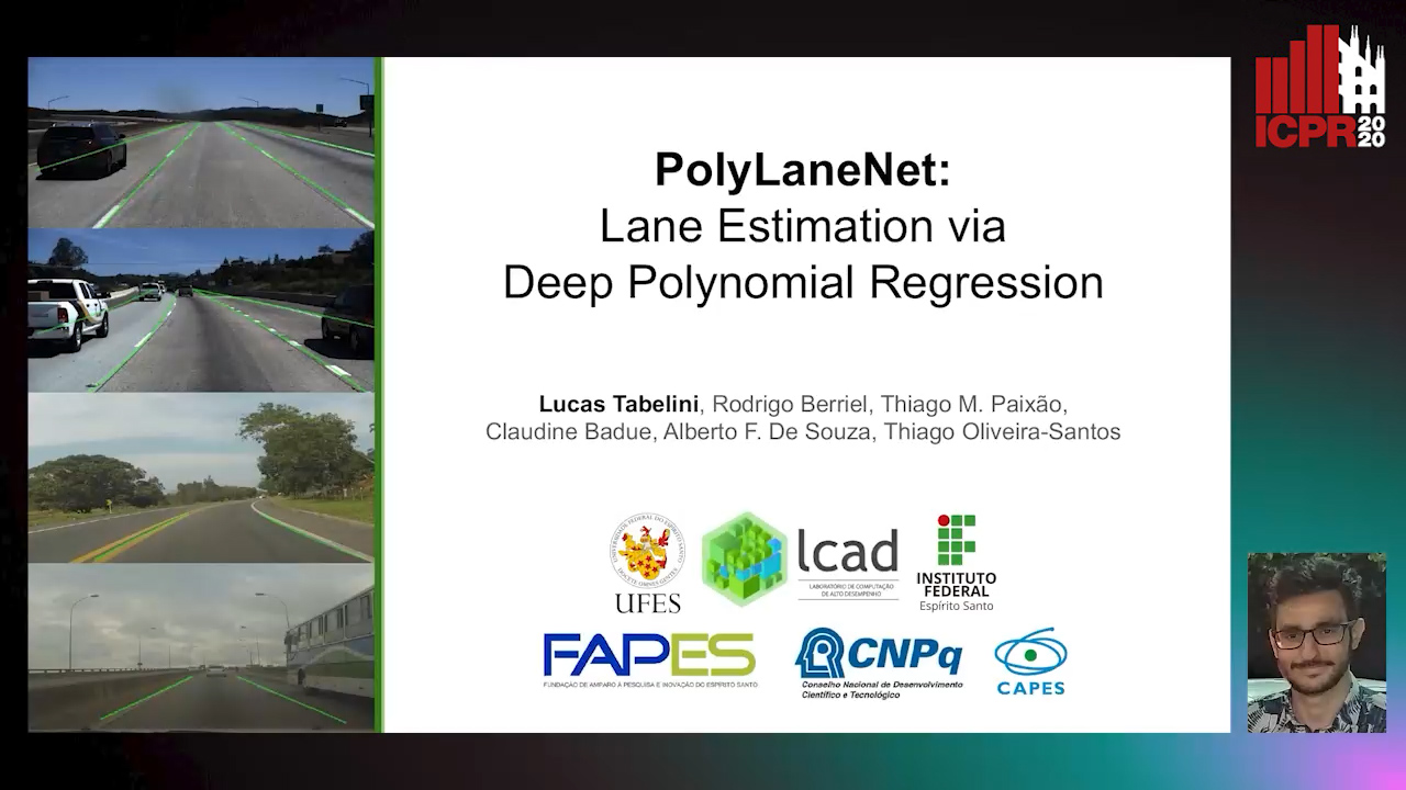
PolyLaneNet: Lane Estimation Via Deep Polynomial Regression

Talles Torres, Rodrigo Berriel, Thiago Paixão, Claudine Badue, Alberto F. De Souza, Thiago Oliveira-Santos

Responsive image

Auto-TLDR; Real-Time Lane Detection with Deep Polynomial Regression

Slides Poster Similar

One of the main factors that contributed to the large advances in autonomous driving is the advent of deep learning. For safer self-driving vehicles, one of the problems that has yet to be solved completely is lane detection. Since methods for this task have to work in real time (+30 FPS), they not only have to be effective (i.e., have high accuracy) but they also have to be efficient (i.e., fast). In this work, we present a novel method for lane detection that uses as input an image from a forward-looking camera mounted in the vehicle and outputs polynomials representing each lane marking in the image, via deep polynomial regression. The proposed method is shown to be competitive with existing state-of-the-art methods in the TuSimple dataset, while maintaining its efficiency (115 FPS). Additionally, extensive qualitative results on two additional public datasets are presented, alongside with limitations in the evaluation metrics used by recent works for lane detection. Finally, we provide source code and trained models that allow others to replicate all the results shown in this paper, which is surprisingly rare in state-of-the-art lane detection methods.