A Novel Computer-Aided Diagnostic System for Early Assessment of Hepatocellular Carcinoma

Ahmed Alksas, Mohamed Shehata, Gehad Saleh, Ahmed Shaffie, Ahmed Soliman, Mohammed Ghazal, Hadil Abukhalifeh, Abdel Razek Ahmed, Ayman El-Baz

Responsive image

Auto-TLDR; Classification of Liver Tumor Lesions from CE-MRI Using Structured Structural Features and Functional Features

Slides Poster

Early assessment of liver cancer patients with hepatocellular carcinoma (HCC) is of immense importance to provide the proper treatment plan. In this paper, we have developed a two-stage classification computer-aided diagnostic (CAD) system that has the ability to detect and grade the liver observations from multiphase contrast enhanced magnetic resonance imaging (CE-MRI). The proposed approach consists of three main steps. First, a pre-processing is applied to the CE-MRI scans to delineate the tumor lesions that will be used as an ROI across the four different phases of the CE-MRI, (namely, the pre-contrast, late-arterial, portal-venous, and delayed-contrast). Second, a group of three features are modeled to provide a quantitative discrimination between the tumor lesions; namely: i) the tumor appearance that is modeled using a set of texture features, (namely; the first-order histogram, second-order gray-level co-occurrence matrix, and second-order gray-level run-length matrix), to capture any discrimination that may appear in the lesion texture, ii) the spherical harmonics (SH) based shape features that have the ability to describe the shape complexity of the liver tumors, and iii) the functional features that are based on the calculation of the wash-in/wash-out through that evaluate the intensity changes across the post-contrast phases. Finally, the aforementioned individual features were then integrated together to obtain the combined features to be fed to a machine learning classifier towards getting the final diagnostic decision. The proposed CAD system has been tested using hepatic observations that was obtained from 85 participating patients, 34 patients with benign tumors, 34 patients with intermediate tumors and 34 with malignant tumors. Using a random forests based classifier with a leave-one-subject-out (LOSO) cross-validation, the developed CAD system achieved an 87.1% accuracy in distinguishing the malignant, intermediate and benign tumors. The classification performance is then evaluated using k-fold (5/10-fold) cross-validation approach to examine the robustness of the system. The LR-1 lesions were classified from LR-2 benign lesions with 91.2% accuracy, while 85.3% accuracy was achieved differentiating between LR-4 and LR-5 malignant tumors. The obtained results hold a promise of the proposed framework to be reliably used as a noninvasive diagnostic tool for the early detection and grading of liver cancer tumors.

Similar papers

A Benchmark Dataset for Segmenting Liver, Vasculature and Lesions from Large-Scale Computed Tomography Data

Bo Wang, Zhengqing Xu, Wei Xu, Qingsen Yan, Liang Zhang, Zheng You

Responsive image

Auto-TLDR; The Biggest Treatment-Oriented Liver Cancer Dataset for Segmentation

Slides Poster Similar

How to build a high-performance liver-related computer assisted diagnosis system is an open question of great interest. However, the performance of the state-of-art algorithm is always limited by the amount of data and quality of the label. To address this problem, we propose the biggest treatment-oriented liver cancer dataset for liver surgery and treatment planning. This dataset provides 216 cases (totally about 268K frames) scanned images in contrast-enhanced computed tomography (CT). We labeled all the CT images with the liver, liver vasculature and liver tumor segmentation ground truth for train and tune segmentation algorithms in advance. Based on that, we evaluate several recent and state-of-the-art segmentation algorithms, including 7 deep learning methods, on CT sequences. All results are compared to reference segmentations five error metrics that highlight different aspects of segmentation accuracy. In general, compared with previous datasets, our dataset is really a challenging dataset. To our knowledge, the proposed dataset and benchmark allow for the first time systematic exploration of such issues, and will be made available to allow for further research in this field.

A Comparison of Neural Network Approaches for Melanoma Classification

Maria Frasca, Michele Nappi, Michele Risi, Genoveffa Tortora, Alessia Auriemma Citarella

Responsive image

Auto-TLDR; Classification of Melanoma Using Deep Neural Network Methodologies

Slides Poster Similar

Melanoma is the deadliest form of skin cancer and it is diagnosed mainly visually, starting from initial clinical screening and followed by dermoscopic analysis, biopsy and histopathological examination. A dermatologist’s recognition of melanoma may be subject to errors and may take some time to diagnose it. In this regard, deep learning can be useful in the study and classification of skin cancer. In particular, by classifying images with Deep Neural Network methodologies, it is possible to obtain comparable or even superior results compared to those of dermatologists. In this paper, we propose a methodology for the classification of melanoma by adopting different deep learning techniques applied to a common dataset, composed of images from the ISIC dataset and consisting of different types of skin diseases, including melanoma on which we applied a specific pre-processing phase. In particular, a comparison of the results is performed in order to select the best effective neural network to be applied to the problem of recognition and classification of melanoma. Moreover, we also evaluate the impact of the pre- processing phase on the final classification. Different metrics such as accuracy, sensitivity, and specificity have been selected to assess the goodness of the adopted neural networks and compare them also with the manual classification of dermatologists.

Segmentation of Axillary and Supraclavicular Tumoral Lymph Nodes in PET/CT: A Hybrid CNN/Component-Tree Approach

Diana Lucia Farfan Cabrera, Nicolas Gogin, David Morland, Benoît Naegel, Dimitri Papathanassiou, Nicolas Passat

Responsive image

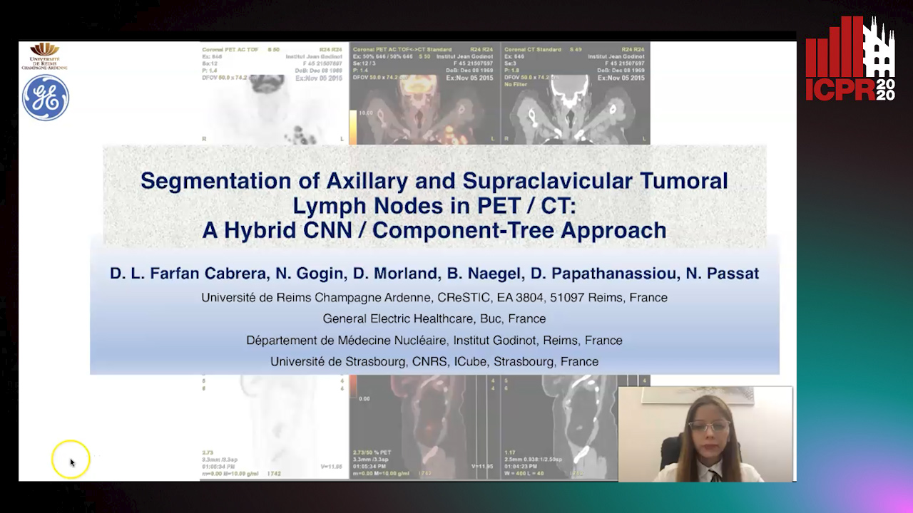
Auto-TLDR; Coupling Convolutional Neural Networks and Component-Trees for Lymph node Segmentation from PET/CT Images

Slides Similar

The analysis of axillary and supraclavicular lymph nodes is a primary prognostic factor for the staging of breast cancer. However, due to the size of lymph nodes and the low resolution of PET data, their segmentation is challenging. We investigate the relevance of considering axillary and supraclavicular lymph node segmentation from PET/CT images by coupling Convolutional Neural Networks (CNNs) and Component-Trees (C-Trees). Building upon the U-Net architecture, we propose a framework that couples a multi-modal U-Net fed with PET and CT, coupled with a hierarchical model obtained from the PET that provides additional high-level region-based features as input channels. Our working hypotheses are twofold. First, we take advantage of both anatomical information from CT for detecting the nodes, and from functional information from PET for detecting the pathological ones. Second, we consider region-based attributes extracted from C-Tree analysis of 3D PET/CT images to improve the CNN segmentation. We carried out experiments on a dataset of 240 pathological lymph nodes from 52 patients scans, and compared our outputs with human expert-defined ground-truth, leading to promising results.

Automatic Classification of Human Granulosa Cells in Assisted Reproductive Technology Using Vibrational Spectroscopy Imaging

Marina Paolanti, Emanuele Frontoni, Giorgia Gioacchini, Giorgini Elisabetta, Notarstefano Valentina, Zacà Carlotta, Carnevali Oliana, Andrea Borini, Marco Mameli

Responsive image

Auto-TLDR; Predicting Oocyte Quality in Assisted Reproductive Technology Using Machine Learning Techniques

Slides Poster Similar

In the field of reproductive technology, the biochemical composition of female gametes has been successfully investigated with the use of vibrational spectroscopy. Currently, in assistive reproductive technology (ART), there are no shared criteria for the choice of oocyte, and automatic classification methods for the best quality oocytes have not yet been applied. In this paper, considering the lack of criteria in Assisted Reproductive Technology (ART), we use Machine Learning (ML) techniques to predict oocyte quality for a successful pregnancy. To improve the chances of successful implantation and minimize any complications during the pregnancy, Fourier transform infrared microspectroscopy (FTIRM) analysis has been applied on granulosa cells (GCs) collected along with the oocytes during oocyte aspiration, as it is routinely done in ART, and specific spectral biomarkers were selected by multivariate statistical analysis. A proprietary biological reference dataset (BRD) was successfully collected to predict the best oocyte for a successful pregnancy. Personal health information are stored, maintained and backed up using a cloud computing service. Using a user-friendly interface, the user will evaluate whether or not the selected oocyte will have a positive result. This interface includes a dashboard for retrospective analysis, reporting, real-time processing, and statistical analysis. The experimental results are promising and confirm the efficiency of the method in terms of classification metrics: precision, recall, and F1-score (F1) measures.

Leveraging Unlabeled Data for Glioma Molecular Subtype and Survival Prediction

Nicholas Nuechterlein, Beibin Li, Mehmet Saygin Seyfioglu, Sachin Mehta, Patrick Cimino, Linda Shapiro

Responsive image

Auto-TLDR; Multimodal Brain Tumor Segmentation Using Unlabeled MR Data and Genomic Data for Cancer Prediction

Slides Poster Similar

In this paper, we address two long-standing challenges in neuro-oncology: (1) how to leverage large amounts of unlabeled magnetic resonance (MR) imaging data for radiogenomic tasks and (2) how to unite glioma MR imaging with genomic data. We examine multi-parametric MR data from 542 patients in the combined training, validation, and testing sets of the 2018 Multimodal Brain Tumor Segmentation Challenge and somatic copy number alteration (SCNA) data from 1090 patients in The Cancer Genome Archive's (TCGA) lower-grade glioma and glioblastoma projects. We propose a novel application of multi-task learning (MTL) that leverages unlabeled MR data by jointly learning tumor segmentation masks with glioma molecular subtype markers and allows for SCNA input when available. There are 235 patients in the intersection of these MR and SCNA datasets, which we divide into an unlabeled training set, a labeled training set, and a validation set. Our MTL model significantly outperforms comparable classification models trained only on labeled MR data for both IDH1/2 mutation and 1p/19q co-deletion glioma subtype marker prediction tasks. We also observe that models trained on genomic and imaging data improve survival prediction results achieved by models trained on either alone. We will release our source code for future research.

Unsupervised Detection of Pulmonary Opacities for Computer-Aided Diagnosis of COVID-19 on CT Images

Rui Xu, Xiao Cao, Yufeng Wang, Yen-Wei Chen, Xinchen Ye, Lin Lin, Wenchao Zhu, Chao Chen, Fangyi Xu, Yong Zhou, Hongjie Hu, Shoji Kido, Noriyuki Tomiyama

Responsive image

Auto-TLDR; A computer-aided diagnosis of COVID-19 from CT images using unsupervised pulmonary opacity detection

Slides Poster Similar

COVID-19 emerged towards the end of 2019 which was identified as a global pandemic by the world heath organization (WHO). With the rapid spread of COVID-19, the number of infected and suspected patients has increased dramatically. Chest computed tomography (CT) has been recognized as an efficient tool for the diagnosis of COVID-19. However, the huge CT data make it difficult for radiologist to fully exploit them on the diagnosis. In this paper, we propose a computer-aided diagnosis system that can automatically analyze CT images to distinguish the COVID-19 against to community-acquired pneumonia (CAP). The proposed system is based on an unsupervised pulmonary opacity detection method that locates opacity regions by a detector unsupervisedly trained from CT images with normal lung tissues. Radiomics based features are extracted insides the opacity regions, and fed into classifiers for classification. We evaluate the proposed CAD system by using 200 CT images collected from different patients in several hospitals. The accuracy, precision, recall, f1-score and AUC achieved are 95.5%, 100%, 91%, 95.1% and 95.9% respectively, exhibiting the promising capacity on the differential diagnosis of COVID-19 from CT images.

Neural Machine Registration for Motion Correction in Breast DCE-MRI

Federica Aprea, Stefano Marrone, Carlo Sansone

Responsive image

Auto-TLDR; A Neural Registration Network for Dynamic Contrast Enhanced-Magnetic Resonance Imaging

Slides Poster Similar

Cancer is one of the leading causes of death in the western world, with medical imaging playing a key role for early diagnosis. Focusing on breast cancer, one of the emerging imaging methodologies is Dynamic Contrast Enhanced-Magnetic Resonance Imaging (DCE-MRI). The flip side of using DCE-MRI is in its long acquisition times, often causing the patient to move, resulting in motion artefacts, namely distortions in the acquired image that can affect DCE-MRI analysis. A possible solution consists in the use of Motion Correction Techniques (MCTs), i.e. procedures intended to re-align the post-contrast image to the corresponding pre-contrast (reference) one. This task is particularly critic in DCE-MRI, due to brightness variations introduced in post-contrast images by the contrast-agent flowing. To face this problem, in this work we introduce a new MCT for breast DCE-MRI leveraging Physiologically Based PharmacoKinetic (PBPK) modelling and Artificial Neural Networks (ANN) to determine the most suitable physiologically-compliant transformation. To this aim, we propose a Neural Registration Network relying on a very task-specific loss function explicitly designed to take into account the contrast agent flowing while enforcing a correct re-alignment. We compared the obtained results against some conventional motion correction techniques, evaluating the performance on a patient-by-patient basis. Results clearly show the effectiveness of the proposed approach, resulting as the best performing even when compares against other techniques designed to take into account for brightness variations.

Automatic Tuberculosis Detection Using Chest X-Ray Analysis with Position Enhanced Structural Information

Hermann Jepdjio Nkouanga, Szilard Vajda

Responsive image

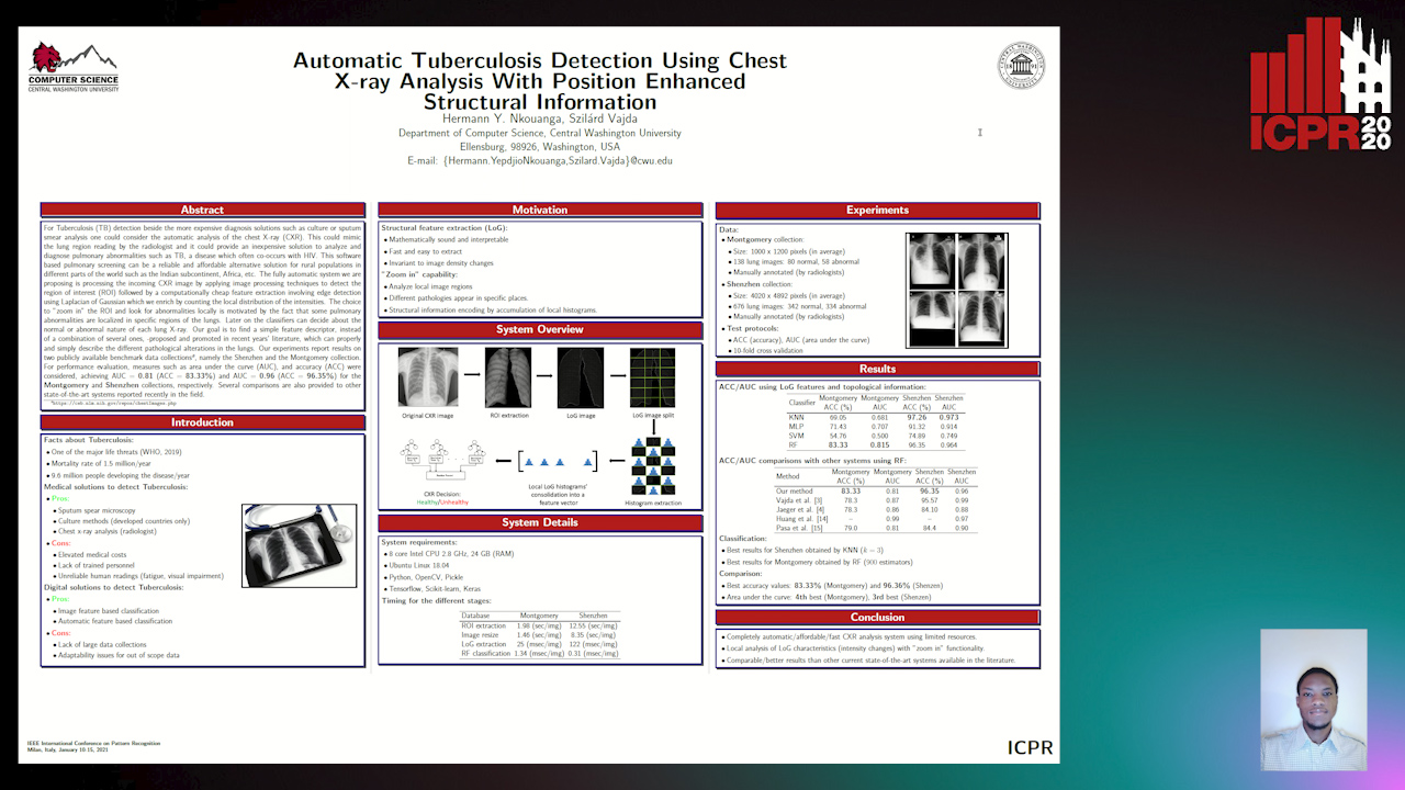
Auto-TLDR; Automatic Chest X-ray Screening for Tuberculosis in Rural Population using Localized Region on Interest

Slides Poster Similar

For Tuberculosis (TB) detection beside the more expensive diagnosis solutions such as culture or sputum smear analysis one could consider the automatic analysis of the chest X-ray (CXR). This could mimic the lung region reading by the radiologist and it could provide a cheap solution to analyze and diagnose pulmonary abnormalities such as TB which often co- occurs with HIV. This software based pulmonary screening can be a reliable and affordable solution for rural population in different parts of the world such as India, Africa, etc. Our fully automatic system is processing the incoming CXR image by applying image processing techniques to detect the region on interest (ROI) followed by a computationally cheap feature extraction involving edge detection using Laplacian of Gaussian which we enrich by counting the local distribution of the intensities. The choice to ”zoom in” the ROI and look for abnormalities locally is motivated by the fact that some pulmonary abnormalities are localized in specific regions of the lungs. Later on the classifiers can decide about the normal or abnormal nature of each lung X-ray. Our goal is to find a simple feature, instead of a combination of several ones, -proposed and promoted in recent years’ literature, which can properly describe the different pathological alterations in the lungs. Our experiments report results on two publicly available data collections1, namely the Shenzhen and the Montgomery collection. For performance evaluation, measures such as area under the curve (AUC), and accuracy (ACC) were considered, achieving AUC = 0.81 (ACC = 83.33%) and AUC = 0.96 (ACC = 96.35%) for the Montgomery and Schenzen collections, respectively. Several comparisons are also provided to other state- of-the-art systems reported recently in the field.

Automatic Semantic Segmentation of Structural Elements related to the Spinal Cord in the Lumbar Region by Using Convolutional Neural Networks

Jhon Jairo Sáenz Gamboa, Maria De La Iglesia-Vaya, Jon Ander Gómez

Responsive image

Auto-TLDR; Semantic Segmentation of Lumbar Spine Using Convolutional Neural Networks

Slides Poster Similar

This work addresses the problem of automatically segmenting the MR images corresponding to the lumbar spine. The purpose is to detect and delimit the different structural elements like vertebrae, intervertebral discs, nerves, blood vessels, etc. This task is known as semantic segmentation. The approach proposed in this work is based on convolutional neural networks whose output is a mask where each pixel from the input image is classified into one of the possible classes. Classes were defined by radiologists and correspond to structural elements and tissues. The proposed network architectures are variants of the U-Net. Several complementary blocks were used to define the variants: spatial attention models, deep supervision and multi-kernels at input, this last block type is based on the idea of inception. Those architectures which got the best results are described in this paper, and their results are discussed. Two of the proposed architectures outperform the standard U-Net used as baseline.

Classify Breast Histopathology Images with Ductal Instance-Oriented Pipeline

Beibin Li, Ezgi Mercan, Sachin Mehta, Stevan Knezevich, Corey Arnold, Donald Weaver, Joann Elmore, Linda Shapiro

Responsive image

Auto-TLDR; DIOP: Ductal Instance-Oriented Pipeline for Diagnostic Classification

Slides Poster Similar

In this study, we propose the Ductal Instance-Oriented Pipeline (DIOP) that contains a duct-level instance segmentation model, a tissue-level semantic segmentation model, and three-levels of features for diagnostic classification. Based on recent advancements in instance segmentation and the Mask R-CNN model, our duct-level segmenter tries to identify each ductal individual inside a microscopic image; then, it extracts tissue-level information from the identified ductal instances. Leveraging three levels of information obtained from these ductal instances and also the histopathology image, the proposed DIOP outperforms previous approaches (both feature-based and CNN-based) in all diagnostic tasks; for the four-way classification task, the DIOP achieves comparable performance to general pathologists in this unique dataset. The proposed DIOP only takes a few seconds to run in the inference time, which could be used interactively on most modern computers. More clinical explorations are needed to study the robustness and generalizability of this system in the future.

Vesselness Filters: A Survey with Benchmarks Applied to Liver Imaging

Jonas Lamy, Odyssée Merveille, Bertrand Kerautret, Nicolas Passat, Antoine Vacavant

Responsive image

Auto-TLDR; Comparison of Vessel Enhancement Filters for Liver Vascular Network Segmentation

Slides Poster Similar

The accurate knowledge of vascular network geometry is crucial for many clinical applications such as cardiovascular disease diagnosis and surgery planning. Vessel enhancement algorithms are often a key step to improve the robustness of vessel segmentation. A wide variety of enhancement filters exists in the literature, but they are often difficult to compare as the applications and datasets differ from a paper to another and the code is rarely available. In this article, we compare seven vessel enhancement filters covering the last twenty years literature in a unique common framework. We focus our study on the liver vascular network which is under-represented in the literature. The evaluation is made from three points of view: in the whole liver, in the vessel neighborhood and near the bifurcations. The study is performed on two publicly available datasets: the Ircad dataset (CT images) and the VascuSynth dataset adapted for MRI simulation. We discuss the strengths and weaknesses of each method in the hepatic context. In addition, the benchmark framework including a C++ implementation of each compared method is provided. An online demonstration ensures the reproducibility of the results without requiring any additional software.

A Systematic Investigation on Deep Architectures for Automatic Skin Lesions Classification

Pierluigi Carcagni, Marco Leo, Andrea Cuna, Giuseppe Celeste, Cosimo Distante

Responsive image

Auto-TLDR; RegNet: Deep Investigation of Convolutional Neural Networks for Automatic Classification of Skin Lesions

Slides Poster Similar

Computer vision-based techniques are more and more employed in healthcare and medical fields nowadays in order, principally, to be as a support to the experienced medical staff to help them to make a quick and correct diagnosis. One of the hot topics in this arena concerns the automatic classification of skin lesions. Several promising works exist about it, mainly leveraging Convolutional Neural Networks (CNN), but proposed pipeline mainly rely on complex data preprocessing and there is no systematic investigation about how available deep models can actually reach the accuracy needed for real applications. In order to overcome these drawbacks, in this work, an end-to-end pipeline is introduced and some of the most recent Convolutional Neural Networks (CNNs) architectures are included in it and compared on the largest common benchmark dataset recently introduced. To this aim, for the first time in this application context, a new network design paradigm, namely RegNet, has been exploited to get the best models among a population of configurations. The paper introduces a threefold level of contribution and novelty with respect the previous literature: the deep investigation of several CNN architectures driving to a consistent improvement of the lesions recognition accuracy, the exploitation of a new network design paradigm able to study the behavior of populations of models and a deep discussion about pro and cons of each analyzed method paving the path towards new research lines.

Tensor Factorization of Brain Structural Graph for Unsupervised Classification in Multiple Sclerosis

Berardino Barile, Marzullo Aldo, Claudio Stamile, Françoise Durand-Dubief, Dominique Sappey-Marinier

Responsive image

Auto-TLDR; A Fully Automated Tensor-based Algorithm for Multiple Sclerosis Classification based on Structural Connectivity Graph of the White Matter Network

Slides Poster Similar

Analysis of longitudinal changes in brain diseases is essential for a better characterization of pathological processes and evaluation of the prognosis. This is particularly important in Multiple Sclerosis (MS) which is the first traumatic disease in young adults, with unknown etiology and characterized by complex inflammatory and degenerative processes leading to different clinical courses. In this work, we propose a fully automated tensor-based algorithm for the classification of MS clinical forms based on the structural connectivity graph of the white matter (WM) network. Using non-negative tensor factorization (NTF), we first focused on the detection of pathological patterns of the brain WM network affected by significant longitudinal variations. Second, we performed unsupervised classification of different MS phenotypes based on these longitudinal patterns, and finally, we used the latent factors obtained by the factorization algorithm to identify the most affected brain regions.

Supporting Skin Lesion Diagnosis with Content-Based Image Retrieval

Stefano Allegretti, Federico Bolelli, Federico Pollastri, Sabrina Longhitano, Giovanni Pellacani, Costantino Grana

Responsive image

Auto-TLDR; Skin Images Retrieval Using Convolutional Neural Networks for Skin Lesion Classification and Segmentation

Slides Poster Similar

Given the relevance of skin cancer, many attempts have been dedicated to the creation of automated devices that could assist both expert and beginner dermatologists towards fast and early diagnosis of skin lesions. In recent years, tasks such as skin lesion classification and segmentation have been extensively addressed with deep learning algorithms, which in some cases reach a diagnostic accuracy comparable to that of expert physicians. However, the general lack of interpretability and reliability severely hinders the ability of those approaches to actually support dermatologists in the diagnosis process. In this paper a novel skin images retrieval system is presented, which exploits features extracted by Convolutional Neural Networks to gather similar images from a publicly available dataset, in order to assist the diagnosis process of both expert and novice practitioners. In the proposed framework, Resnet-50 is initially trained for the classification of dermoscopic images; then, the feature extraction part is isolated, and an embedding network is build on top of it. The embedding learns an alternative representation, which allows to check image similarity by means of a distance measure. Experimental results reveal that the proposed method is able to select meaningful images, which can effectively boost the classification accuracy of human dermatologists.

Prediction of Obstructive Coronary Artery Disease from Myocardial Perfusion Scintigraphy using Deep Neural Networks

Ida Arvidsson, Niels Christian Overgaard, Miguel Ochoa Figueroa, Jeronimo Rose, Anette Davidsson, Kalle Åström, Anders Heyden

Responsive image

Auto-TLDR; A Deep Learning Algorithm for Multi-label Classification of Myocardial Perfusion Scintigraphy for Stable Ischemic Heart Disease

Slides Poster Similar

For diagnosis and risk assessment in patients with stable ischemic heart disease, myocardial perfusion scintigraphy is one of the most common cardiological examinations performed today. There are however many motivations for why an artificial intelligence algorithm would provide useful input to this task. For example to reduce the subjectiveness and save time for the nuclear medicine physicians working with this time consuming task. In this work we have developed a deep learning algorithm for multi-label classification based on a modified convolutional neural network to estimate probability of obstructive coronary artery disease in the left anterior artery, left circumflex artery and right coronary artery. The prediction is based on data from myocardial perfusion scintigraphy studies conducted in a dedicated Cadmium-Zinc-Telluride cardio camera (D-SPECT Spectrum Dynamics). Data from 588 patients was available, with stress images in both upright and supine position, as well as a number of auxiliary parameters such as angina symptoms and BMI. The data was used to train and evaluate the algorithm using 5-fold cross-validation. We achieve state-of-the-art results for this task with an area under the receiver operating characteristics curve of 0.89 as average on per-vessel level and 0.94 on per-patient level.

Planar 3D Transfer Learning for End to End Unimodal MRI Unbalanced Data Segmentation

Martin Kolarik, Radim Burget, Carlos M. Travieso-Gonzalez, Jan Kocica

Responsive image

Auto-TLDR; Planar 3D Res-U-Net Network for Unbalanced 3D Image Segmentation using Fluid Attenuation Inversion Recover

Slides Similar

We present a novel approach of 2D to 3D transfer learning based on mapping pre-trained 2D convolutional neural network weights into planar 3D kernels. The method is validated by proposed planar 3D res-u-net network with encoder transferred from the 2D VGG-16 which is applied for a single-stage unbalanced 3D image data segmentation. In particular, we evaluate the method on the MICCAI 2016 MS lesion segmentation challenge dataset utilizing solely Fluid Attenuation Inversion Recover (FLAIR) sequence without brain extraction for training and inference to simulate real medical praxis. The planar 3D res-u-net network performed the best both in sensitivity and Dice score amongst end to end methods processing raw MRI scans and achieved comparable Dice score to a state-of-the-art unimodal not end to end approach. Complete source code was released under the open-source license and this paper is in compliance with the Machine learning Reproducibility Checklist. By implementing practical transfer learning for 3D data representation we were able to successfully segment heavily unbalanced data without selective sampling and achieved more reliable results using less training data in single modality. From medical perspective, the unimodal approach gives an advantage in real praxis as it does not require co-registration nor additional scanning time during examination. Although modern medical imaging methods capture high resolution 3D anatomy scans suitable for computer aided detection system processing, deployment of automatic systems for interpretation of radiology imaging is still rather theoretical in many medical areas. Our work aims to bridge the gap offering solution for partial research questions.

Deep Learning-Based Type Identification of Volumetric MRI Sequences

Jean Pablo De Mello, Thiago Paixão, Rodrigo Berriel, Mauricio Reyes, Alberto F. De Souza, Claudine Badue, Thiago Oliveira-Santos

Responsive image

Auto-TLDR; Deep Learning for Brain MRI Sequences Identification Using Convolutional Neural Network

Slides Poster Similar

The analysis of Magnetic Resonance Imaging (MRI) sequences enables clinical professionals to monitor the progression of a brain tumor. As the interest for automatizing brain volume MRI analysis increases, it becomes convenient to have each sequence well identified. However, the unstandardized naming of MRI sequences make their identification difficult for automated systems, as well as make it difficult for researches to generate or use datasets for machine learning research. In face of that, we propose a system for identifying types of brain MRI sequences based on deep learning. By training a Convolutional Neural Network (CNN) based on 18-layer ResNet architecture, our system is able to classify a volumetric brain MRI as a T1, T1c, T2 or FLAIR sequence, or whether it does not belong to any of these classes. The network was trained with both pre-processed (BraTS dataset) and non-pre-processed (TCGA-GBM dataset) images with diverse acquisition protocols, requiring only a few layers of the volume for training. Our system is able to classify among sequence types with an accuracy of 96.27%.

Using Machine Learning to Refer Patients with Chronic Kidney Disease to Secondary Care

Lee Au-Yeung, Xianghua Xie, Timothy Marcus Scale, James Anthony Chess

Responsive image

Auto-TLDR; A Machine Learning Approach for Chronic Kidney Disease Prediction using Blood Test Data

Slides Poster Similar

There has been growing interest recently in using machine learning techniques as an aid in clinical medicine. Machine learning offers a range of classification algorithms which can be applied to medical data to aid in making clinical predictions. Recent studies have demonstrated the high predictive accuracy of various classification algorithms applied to clinical data. Several studies have already been conducted in diagnosing or predicting chronic kidney disease at various stages using different sets of variables. In this study we are investigating the use machine learning techniques with blood test data. Such a system could aid renal teams in making recommendations to primary care general practitioners to refer patients to secondary care where patients may benefit from earlier specialist assessment and medical intervention. We are able to achieve an overall accuracy of 88.48\% using logistic regression, 87.12\% using ANN and 85.29\% using SVM. ANNs performed with the highest sensitivity at 89.74\% compared to 86.67\% for logistic regression and 85.51\% for SVM.

SAGE: Sequential Attribute Generator for Analyzing Glioblastomas Using Limited Dataset

Padmaja Jonnalagedda, Brent Weinberg, Jason Allen, Taejin Min, Shiv Bhanu, Bir Bhanu

Responsive image

Auto-TLDR; SAGE: Generative Adversarial Networks for Imaging Biomarker Detection and Prediction

Slides Poster Similar

While deep learning approaches have shown remarkable performance in many imaging tasks, most of these methods rely on availability of large quantities of data. Medical image data, however, is scarce and fragmented. Generative Adversarial Networks (GANs) have recently been very effective in handling such datasets by generating more data. If the datasets are very small, however, GANs cannot learn the data distribution properly, resulting in less diverse or low-quality results. One such limited dataset is that for the concurrent gain of 19/20 chromosomes (19/20 co-gain), a mutation with positive prognostic value in Glioblastomas (GBM). In this paper, we detect imaging biomarkers for the mutation to streamline the extensive and invasive prognosis pipeline. Since this mutation is relatively rare, i.e. small dataset, we propose a novel generative framework – the Sequential Attribute GEnerator (SAGE), that generates detailed tumor imaging features while learning from a limited dataset. Experiments show that not only does SAGE generate high quality tumors when compared to standard Deep Convolutional GAN (DC-GAN) and Wasserstein GAN with Gradient Penalty (WGAN-GP), it also captures the imaging biomarkers accurately.

3D Medical Multi-Modal Segmentation Network Guided by Multi-Source Correlation Constraint

Tongxue Zhou, Stéphane Canu, Pierre Vera, Su Ruan

Responsive image

Auto-TLDR; Multi-modality Segmentation with Correlation Constrained Network

Slides Poster Similar

In the field of multimodal segmentation, the correlation between different modalities can be considered for improving the segmentation results. In this paper, we propose a multi-modality segmentation network with a correlation constraint. Our network includes N model-independent encoding paths with N image sources, a correlation constrain block, a feature fusion block, and a decoding path. The model-independent encoding path can capture modality-specific features from the N modalities. Since there exists a strong correlation between different modalities, we first propose a linear correlation block to learn the correlation between modalities, then a loss function is used to guide the network to learn the correlated features based on the correlation representation block. This block forces the network to learn the latent correlated features which are more relevant for segmentation. Considering that not all the features extracted from the encoders are useful for segmentation, we propose to use dual attention based fusion block to recalibrate the features along the modality and spatial paths, which can suppress less informative features and emphasize the useful ones. The fused feature representation is finally projected by the decoder to obtain the segmentation result. Our experiment results tested on BraTS-2018 dataset for brain tumor segmentation demonstrate the effectiveness of our proposed method.

A Multi-Task Contextual Atrous Residual Network for Brain Tumor Detection & Segmentation

Ngan Le, Kashu Yamazaki, Quach Kha Gia, Thanh-Dat Truong, Marios Savvides

Responsive image

Auto-TLDR; Contextual Brain Tumor Segmentation Using 3D atrous Residual Networks and Cascaded Structures

Poster Similar

In recent years, deep neural networks have achieved state-of-the-art performance in a variety of recognition and segmentation tasks in medical imaging including brain tumor segmentation. We investigate that segmenting brain tumor is facing to the imbalanced data problem where the number of pixels belonging to background class (non tumor pixel) is much larger than the number of pixels belonging to foreground class (tumor pixel). To address this problem, we propose a multi-task network which is formed as a cascaded structure and designed to share the feature maps. Our model consists of two targets, i.e., (i) effectively differentiating brain tumor regions and (ii) estimating brain tumor masks. The first task is performed by our proposed contextual brain tumor detection network, which plays the role of an attention gate and focuses on the region around brain tumor only while ignore the background (non tumor area). Instead of processing every pixel, our contextual brain tumor detection network only processes contextual regions around ground-truth instances and this strategy helps to produce meaningful regions proposals. The second task is built upon a 3D atrous residual network and under an encode-decode network in order to effectively segment both large and small objects (brain tumor). Our 3D atrous residual network is designed with a skip connection to enables the gradient from the deep layers to be directly propagated to shallow layers, thus, features of different depths are preserved and used for refining each other. In order to incorporate larger contextual information in volume MRI data, our network is designed by 3D atrous convolution with various kernel sizes, which enlarges the receptive field of filters. Our proposed network has been evaluated on various datasets including BRATS2015, BRATS2017 and BRATS2018 datasets with both validation set and testing set. Our performance has been benchmarked by both region-based metrics and surface-based metrics. We also have conducted comparisons against state-of-the-art approaches.

DARN: Deep Attentive Refinement Network for Liver Tumor Segmentation from 3D CT Volume

Yao Zhang, Jiang Tian, Cheng Zhong, Yang Zhang, Zhongchao Shi, Zhiqiang He

Responsive image

Auto-TLDR; Deep Attentive Refinement Network for Liver Tumor Segmentation from 3D Computed Tomography Using Multi-Level Features

Slides Poster Similar

Automatic liver tumor segmentation from 3D Computed Tomography (CT) is a necessary prerequisite in the interventions of hepatic abnormalities and surgery planning. However, accurate liver tumor segmentation remains challenging due to the large variability of tumor sizes and inhomogeneous texture. Recent advances based on Fully Convolutional Network (FCN) in liver tumor segmentation draw on success of learning discriminative multi-level features. In this paper, we propose a Deep Attentive Refinement Network (DARN) for improved liver tumor segmentation from CT volumes by fully exploiting both low and high level features embedded in different layers of FCN. Different from existing works, we exploit attention mechanism to leverage the relation of different levels of features encoded in different layers of FCN. Specifically, we introduce a Semantic Attention Refinement (SemRef) module to selectively emphasize global semantic information in low level features with the guidance of high level ones, and a Spatial Attention Refinement (SpaRef) module to adaptively enhance spatial details in high level features with the guidance of low level ones. We evaluate our network on the public MICCAI 2017 Liver Tumor Segmentation Challenge dataset (LiTS dataset) and it achieves state-of-the-art performance. The proposed refinement modules are an effective strategy to exploit multi-level features and has great potential to generalize to other medical image segmentation tasks.

A Deep Learning Approach for the Segmentation of Myocardial Diseases

Khawala Brahim, Abdull Qayyum, Alain Lalande, Arnaud Boucher, Anis Sakly, Fabrice Meriaudeau

Responsive image

Auto-TLDR; Segmentation of Myocardium Infarction Using Late GADEMRI and SegU-Net

Slides Poster Similar

Cardiac left ventricular (LV) segmentation is of paramount essential step for both diagnosis and treatment of cardiac pathologies such as ischemia, myocardial infarction, arrhythmia and myocarditis. However, this segmentation is challenging due to high variability across patients and the potential lack of contrast between structures. In this work, we propose and evaluate a (2.5D) SegU-Net model based on the fusion of two deep learning techniques (U-Net and Seg-Net) for automated LGEMRI (Late gadolinium enhanced magnetic resonance imaging) myocardial disease (infarct core and no reflow region) quantification in a new multifield expert annotated dataset. Given that the scar tissue represents a small part of the whole MRI slices, we focused on myocardium area. Segmentation results show that this preprocessing step facilitate the learning procedure. In order to solve the class imbalance problem, we propose to apply the Jaccard loss and the Focal Loss as optimization loss function and to integrate a class weights strategy into the objective function. Late combination has been used to merge the output of the best trained models on a different set of hyperparameters. The final network segmentation performances will be useful for future comparison of new method to the current related work for this task. A total number of 2237 of slices (320 cases) were used for training/validation and 210 slices (35 cases) were used for testing. Experiments over our proposed dataset, using several evaluation metrics such Jaccard distance (IOU), Accuracy and Dice similarity coefficient (DSC), demonstrate efficiency performance in quantifying different zones of myocardium infarction across various patients. As compared to the second intra-observer study, our testing results showed that the SegUNet prediction model leads to these average dice coefficients over all segmented tissue classes, respectively : 'Background': 0.99999, 'Myocardium': 0.99434, 'Infarctus': 0.95587, 'Noreflow': 0.78187.

Encoding Brain Networks through Geodesic Clustering of Functional Connectivity for Multiple Sclerosis Classification

Muhammad Abubakar Yamin, Valsasina Paola, Michael Dayan, Sebastiano Vascon, Tessadori Jacopo, Filippi Massimo, Vittorio Murino, A Rocca Maria, Diego Sona

Responsive image

Auto-TLDR; Geodesic Clustering of Connectivity Matrices for Multiple Sclerosis Classification

Slides Poster Similar

An important task in brain connectivity research is the classification of patients from healthy subjects. In this work, we present a two-step mathematical framework allowing to discriminate between two groups of people with an application to multiple sclerosis. The proposed approach exploits the properties of the connectivity matrices determined using the covariances between signals of a fixed set of brain areas. These positive semi-definite matrices lay on a Riemannian manifold, allowing to use a geodesic distance defined on this space. In order to generate a vector representation useful for classification purpose, but still preserving the network structures, we encoded the data exploiting the network attractors determined by a geodesic clustering of connectivity matrices. Then clustering centroids were used as a dictionary allowing to encode subject’s connectivity matrices as a vector of geodesic distances. A Linear Support Vector Machine was then used to perform classification between subjects. To demonstrate the advantage of using geodesic metrics in this framework, we conducted the same analysis using Euclidean metric. Experimental results validate the fact that employing geodesic metric in this framework leads to a higher classification performance, whereas with Euclidean metric performance was suboptimal.

Inferring Functional Properties from Fluid Dynamics Features

Andrea Schillaci, Maurizio Quadrio, Carlotta Pipolo, Marcello Restelli, Giacomo Boracchi

Responsive image

Auto-TLDR; Exploiting Convective Properties of Computational Fluid Dynamics for Medical Diagnosis

Slides Poster Similar

In a wide range of applied problems involving fluid flows, Computational Fluid Dynamics (CFD) provides detailed quantitative information on the flow field, at various levels of fidelity and computational cost. However, CFD alone cannot predict high-level functional properties of the system that are not easily obtained from the equations of fluid motion. In this work, we present a data-driven framework to extract additional information, such as medical diagnostic output, from CFD solutions. The task is made difficult by the huge data dimensionality of CFD, together with the limited amount of training data implied by its high computational cost. By pursuing a traditional ML pipeline of pre-processing, feature extraction, and model training, we demonstrate that informative features can be extracted from CFD data. Two experiments, pertaining to different application domains, support the claim that the convective properties implicit into a CFD solution can be leveraged to retrieve functional information for which an analytical definition is missing. Despite the preliminary nature of our study and the relative simplicity of both the geometrical and CFD models, for the first time we demonstrate that the combination of ML and CFD can diagnose a complex system in terms of high-level functional information.

Dealing with Scarce Labelled Data: Semi-Supervised Deep Learning with Mix Match for Covid-19 Detection Using Chest X-Ray Images

Saúl Calderón Ramirez, Raghvendra Giri, Shengxiang Yang, Armaghan Moemeni, Mario Umaña, David Elizondo, Jordina Torrents-Barrena, Miguel A. Molina-Cabello

Responsive image

Auto-TLDR; Semi-supervised Deep Learning for Covid-19 Detection using Chest X-rays

Slides Poster Similar

Coronavirus (Covid-19) is spreading fast, infecting people through contact in various forms including droplets from sneezing and coughing. Therefore, the detection of infected subjects in an early, quick and cheap manner is urgent. Currently available tests are scarce and limited to people in danger of serious illness. The application of deep learning to chest X-ray images for Covid-19 detection is an attractive approach. However, this technology usually relies on the availability of large labelled datasets, a requirement hard to meet in the context of a virus outbreak. To overcome this challenge, a semi-supervised deep learning model using both labelled and unlabelled data is proposed. We developed and tested a semi-supervised deep learning framework based on the Mix Match architecture to classify chest X-rays into Covid-19, pneumonia and healthy cases. The presented approach was calibrated using two publicly available datasets. The results show an accuracy increase of around $15\%$ under low labelled / unlabelled data ratio. This indicates that our semi-supervised framework can help improve performance levels towards Covid-19 detection when the amount of high-quality labelled data is scarce. Also, we introduce a semi-supervised deep learning boost coefficient which is meant to ease the scalability of our approach and performance comparison.

A Deep Learning-Based Method for Predicting Volumes of Nasopharyngeal Carcinoma for Adaptive Radiation Therapy Treatment

Bilel Daoud, Ken'Ichi Morooka, Shoko Miyauchi, Ryo Kurazume, Wafa Mnejja, Leila Farhat, Jamel Daoud

Responsive image

Auto-TLDR; TEP-Net: Tumor Evolution Prediction of Nasopharyngeal Carcinoma and Organ-at-risks Using CT Images

Slides Poster Similar

This paper presents a new system for predicting the spatial change of Nasopharyngeal carcinoma(NPC) and organ-at-risks (OARs) volumes over the course of the radiation therapy (RT) treatment for facilitating the workflow of adaptive radiotherapy. The proposed system, called " Tumor Evolution Prediction (TEP-Net)", predicts the spatial distributions of NPC and 5 OARs, separately, in response to RT in the coming week, week n. Here, TEP-Net has (n-1)-inputs that are week 1 to week n-1 of CT axial, coronal or sagittal images acquired once the patient complete the planned RT treatment of the corresponding week. As a result, three predicted results of each target region are obtained from the three-view CT images. To determine the final prediction of NPC and 5 OARs, two integration methods, weighted fully connected layers and weighted voting methods, are introduced. From the experiments using weekly CT images of 140 NPC patients, our proposed system achieves the best performance for predicting NPC and OARs compared with conventional methods.

MTGAN: Mask and Texture-Driven Generative Adversarial Network for Lung Nodule Segmentation

Wei Chen, Qiuli Wang, Kun Wang, Dan Yang, Xiaohong Zhang, Chen Liu, Yucong Li

Responsive image

Auto-TLDR; Mask and Texture-driven Generative Adversarial Network for Lung Nodule Segmentation

Slides Poster Similar

Accurate segmentation for lung nodules in lung computed tomography (CT) scans plays a key role in the early diagnosis of lung cancer. Many existing methods, especially UNet, have made significant progress in lung nodule segmentation. However, due to the complex shapes of lung nodules and the similarity of visual characteristics between nodules and lung tissues, an accurate segmentation with few false positives of lung nodules is still a challenging problem. Considering the fact that both boundary and texture information of lung nodules are important for obtaining an accurate segmentation result, we propose a novel Mask and Texture-driven Generative Adversarial Network (MTGAN) with a joint multi-scale L1 loss for lung nodule segmentation, which takes full advantages of U-Net and adversarial training. The proposed MTGAN leverages adversarial learning strategy guided by the boundary and texture information of lung nodules to generate more accurate segmentation results with lesser false positives. We validate our model with the LIDC–IDRI dataset, and experimental results show that our method achieves excellent segmentation results for a variety of lung nodules, especially for juxtapleural nodules and low-dense nodules. Without any bells and whistles, the proposed MTGAN achieves significant segmentation performance with the Dice similarity coefficient (DSC) of 85.24% on the LIDC–IDRI dataset.

Inception Based Deep Learning Architecture for Tuberculosis Screening of Chest X-Rays

Dipayan Das, K.C. Santosh, Umapada Pal

Responsive image

Auto-TLDR; End to End CNN-based Chest X-ray Screening for Tuberculosis positive patients in the severely resource constrained regions of the world

Slides Poster Similar

The motivation for this work is the primary need of screening Tuberculosis (TB) positive patients in the severely resource constrained regions of the world. Chest X-ray (CXR) is considered to be a promising indicator for the onset of TB, but the lack of skilled radiologists in such regions degrades the situation. Therefore, several computer aided diagnosis (CAD) systems have been proposed to solve the decision making problem, which includes hand engineered feature extraction methods to deep learning or Convolutional Neural Network (CNN) based methods. Feature extraction, being a time and resource intensive process, often delays the process of mass screening. Hence an end to end CNN architecture is proposed in this work to solve the problem. Two benchmark CXR datasets have been used in this work, collected from Shenzhen (China) and Montgomery County (USA), on which the proposed methodology achieved a maximum abnormality detection accuracy (ACC) of 91.7\% (0.96 AUC) and 87.47\% (0.92 AUC) respectively. To the greatest of our knowledge, the obtained results are marginally superior to the state of the art results that have solely used deep learning methodologies on the aforementioned datasets.

A Lumen Segmentation Method in Ureteroscopy Images Based on a Deep Residual U-Net Architecture

Jorge Lazo, Marzullo Aldo, Sara Moccia, Michele Catellani, Benoit Rosa, Elena De Momi, Michel De Mathelin, Francesco Calimeri

Responsive image

Auto-TLDR; A Deep Neural Network for Ureteroscopy with Residual Units

Slides Poster Similar

Ureteroscopy is becoming the first surgical treatment option for the majority of urinary affections. This procedure is carried out using an endoscope which provides the surgeon with the visual and spatial information necessary to navigate inside the urinary tract. Having in mind the development of surgical assistance systems, that could enhance the performance of surgeon, the task of lumen segmentation is a fundamental part since this is the visual reference which marks the path that the endoscope should follow. This is something that has not been analyzed in ureteroscopy data before. However, this task presents several challenges given the image quality and the conditions itself of ureteroscopy procedures. In this paper, we study the implementation of a Deep Neural Network which exploits the advantage of residual units in an architecture based on U-Net. For the training of these networks, we analyze the use of two different color spaces: gray-scale and RGB data images. We found that training on gray-scale images gives the best results obtaining mean values of Dice Score, Precision, and Recall of 0.73, 0.58, and 0.92 respectively. The results obtained show that the use of residual U-Net could be a suitable model for further development for a computer-aided system for navigation and guidance through the urinary system.

A New Geodesic-Based Feature for Characterization of 3D Shapes: Application to Soft Tissue Organ Temporal Deformations

Karim Makki, Amine Bohi, Augustin Ogier, Marc-Emmanuel Bellemare

Responsive image

Auto-TLDR; Spatio-Temporal Feature Descriptors for 3D Shape Characterization from Point Clouds

Slides Poster Similar

Spatio-temporal feature descriptors are of great importance for characterizing the local changes of 3D deformable shapes. In this study, we propose a method for characterizing 3D shapes from point clouds and we show a direct application on a study of organ temporal deformations. As an example, we characterize the behavior of the bladder during forced respiratory motion with a reduced number of 3D surface points: first, a set of equidistant points representing the vertices of quadrilateral mesh for the organ surface are tracked throughout a long dynamic MRI sequence using a large deformation diffeomorphic metric mapping (LDDMM) framework. Second, a novel 3D shape descriptor invariant to translation, scale and rotation is proposed for characterizing the temporal organ deformations by employing an Eulerian Partial Differential Equations (PDEs) methodology. We demonstrate the robustness of our feature on both synthetic 3D shapes and realistic dynamic Magnetic Resonance Imaging (MRI) data sequences portraying the bladder deformation during a forced breathing exercise. Promising results are obtained, showing that the proposed feature may be useful for several computer vision applications such as medical imaging, aerodynamics and robotics.

Breast Anatomy Enriched Tumor Saliency Estimation

Fei Xu, Yingtao Zhang, Heng-Da Cheng, Jianrui Ding, Boyu Zhang, Chunping Ning, Ying Wang

Responsive image

Auto-TLDR; Tumor Saliency Estimation for Breast Ultrasound using enriched breast anatomy knowledge

Slides Poster Similar

Breast cancer investigation is of great significance and developing tumor detection methodologies is a critical need. However, it is a challenging task for breast cancer detection using breast ultrasound (BUS) images due to the complicated breast structure and poor quality of the images. In this paper, we propose a novel tumor saliency estimation (TSE) model guided by enriched breast anatomy knowledge to localize the tumor. First, the breast anatomy layers are generated by a deep neural network. Then we refine the layers by integrating a non-semantic breast anatomy model to solve the problems of incomplete mammary layers. Meanwhile, a new background map generation method weighted by the semantic probability and spatial distance is proposed to improve the performance. The experiment demonstrates that the proposed method with the new background map outperforms four state-of-the-art TSE models with increasing 10% of F_meansure on the public BUS dataset.

Trainable Spectrally Initializable Matrix Transformations in Convolutional Neural Networks

Michele Alberti, Angela Botros, Schuetz Narayan, Rolf Ingold, Marcus Liwicki, Mathias Seuret

Responsive image

Auto-TLDR; Trainable and Spectrally Initializable Matrix Transformations for Neural Networks

Slides Poster Similar

In this work, we introduce a new architectural component to Neural Networks (NN), i.e., trainable and spectrally initializable matrix transformations on feature maps. While previous literature has already demonstrated the possibility of adding static spectral transformations as feature processors, our focus is on more general trainable transforms. We study the transforms in various architectural configurations on four datasets of different nature: from medical (ColorectalHist, HAM10000) and natural (Flowers) images to historical documents (CB55). With rigorous experiments that control for the number of parameters and randomness, we show that networks utilizing the introduced matrix transformations outperform vanilla neural networks. The observed accuracy increases appreciably across all datasets. In addition, we show that the benefit of spectral initialization leads to significantly faster convergence, as opposed to randomly initialized matrix transformations. The transformations are implemented as auto-differentiable PyTorch modules that can be incorporated into any neural network architecture. The entire code base is open-source.

Deep Multi-Stage Model for Automated Landmarking of Craniomaxillofacial CT Scans

Simone Palazzo, Giovanni Bellitto, Luca Prezzavento, Francesco Rundo, Ulas Bagci, Daniela Giordano, Rosalia Leonardi, Concetto Spampinato

Responsive image

Auto-TLDR; Automated Landmarking of Craniomaxillofacial CT Images Using Deep Multi-Stage Architecture

Slides Similar

In this paper we define a deep multi-stage architecture for automated landmarking of craniomaxillofacial (CMF) CT images. Our model is composed of three subnetworks that first localize, on reduced-resolution images, areas where land-marks may be found and then refine the search, at full-resolution scale, through a hierarchical structure aiming at increasing the granularity of the investigated region. The multi-stage pipeline is designed to deal with full resolution data and does not require any additional pre-processing step to reduce search space, as opposed to existing methods that can be only adopted for searching landmarks located in well-defined anatomical structures (e.g.,mandibles). The automated landmarking system is tested on identifying landmarks located in several CMF regions, achieving an average error of 0.8 mm, significantly lower than expert readings. The proposed model also outperforms baselines and is on par with existing models that employ additional upstream segmentation, on state-of-the-art benchmarks.

Deep Transfer Learning for Alzheimer’s Disease Detection

Nicole Cilia, Claudio De Stefano, Francesco Fontanella, Claudio Marrocco, Mario Molinara, Alessandra Scotto Di Freca

Responsive image

Auto-TLDR; Automatic Detection of Handwriting Alterations for Alzheimer's Disease Diagnosis using Dynamic Features

Slides Poster Similar

Early detection of Alzheimer’s Disease (AD) is essential in order to initiate therapies that can reduce the effects of such a disease, improving both life quality and life expectancy of patients. Among all the activities carried out in our daily life, handwriting seems one of the first to be influenced by the arise of neurodegenerative diseases. For this reason, the analysis of handwriting and the study of its alterations has become of great interest in this research field in order to make a diagnosis as early as possible. In recent years, many studies have tried to use classification algorithms applied to handwritings to implement decision support systems for AD diagnosis. A key issue for the use of these techniques is the detection of effective features, that allow the system to distinguish the natural handwriting alterations due to age, from those caused by neurodegenerative disorders. In this context, many interesting results have been published in the literature in which the features have been typically selected by hand, generally considering the dynamics of the handwriting process in order to detect motor disorders closely related to AD. Features directly derived from handwriting generation models can be also very helpful for AD diagnosis. It should be remarked, however, that the above features do not consider changes in the shape of handwritten traces, which may occur as a consequence of neurodegenerative diseases, as well as the correlation among shape alterations and changes in the dynamics of the handwriting process. Moving from these considerations, the aim of this study is to verify if the combined use of both shape and dynamic features allows a decision support system to improve performance for AD diagnosis. To this purpose, starting from a database of on-line handwriting samples, we generated for each of them a synthetic off-line colour image, where the colour of each elementary trait encodes, in the three RGB channels, the dynamic information associated to that trait. Finally, we exploited the capability of Deep Neural Networks (DNN) to automatically extract features from raw images. The experimental comparison of the results obtained by using standard features and features extracted according the above procedure, confirmed the effectiveness of our approach.

End-To-End Multi-Task Learning for Lung Nodule Segmentation and Diagnosis

Wei Chen, Qiuli Wang, Dan Yang, Xiaohong Zhang, Chen Liu, Yucong Li

Responsive image

Auto-TLDR; A novel multi-task framework for lung nodule diagnosis based on deep learning and medical features

Slides Similar

Computer-Aided Diagnosis (CAD) systems for lung nodule diagnosis based on deep learning have attracted much attention in recent years. However, most existing methods ignore the relationships between the segmentation and classification tasks, which leads to unstable performances. To address this problem, we propose a novel multi-task framework, which can provide lung nodule segmentation mask, malignancy prediction, and medical features for interpretable diagnosis at the same time. Our framework mainly contains two sub-network: (1) Multi-Channel Segmentation Sub-network (MSN) for lung nodule segmentation, and (2) Joint Classification Sub-network (JCN) for interpretable lung nodule diagnosis. In the proposed framework, we use U-Net down-sampling processes for extracting low-level deep learning features, which are shared by two sub-networks. The JCN forces the down-sampling processes to learn better lowlevel deep features, which lead to a better construct of segmentation masks. Meanwhile, two additional channels constructed by OTSU and super-pixel (SLIC) methods, are utilized as the guideline of the feature extraction. The proposed framework takes advantages of deep learning methods and classical methods, which can significantly improve the performances of all tasks. We evaluate the proposed framework on public dataset LIDCIDRI. Our framework achieves a promising Dice score of 86.43% in segmentation, 87.07% in malignancy level prediction, and convincing results in interpretable medical feature predictions.

A Heuristic-Based Decision Tree for Connected Components Labeling of 3D Volumes

Maximilian Söchting, Stefano Allegretti, Federico Bolelli, Costantino Grana

Responsive image

Auto-TLDR; Entropy Partitioning Decision Tree for Connected Components Labeling

Slides Poster Similar

Connected Components Labeling represents a fundamental step for many Computer Vision and Image Processing pipelines. Since the first appearance of the task in the sixties, many algorithmic solutions to optimize the computational load needed to label an image have been proposed. Among them, block-based scan approaches and decision trees revealed to be some of the most valuable strategies. However, due to the cost of the manual construction of optimal decision trees and the computational limitations of automatic strategies employed in the past, the application of blocks and decision trees has been restricted to small masks, and thus to 2D algorithms. With this paper we present a novel heuristic algorithm based on decision tree learning methodology, called Entropy Partitioning Decision Tree (EPDT). It allows to compute near-optimal decision trees for large scan masks. Experimental results demonstrate that algorithms based on the generated decision trees outperform state-of-the-art competitors.

Investigating and Exploiting Image Resolution for Transfer Learning-Based Skin Lesion Classification

Amirreza Mahbod, Gerald Schaefer, Chunliang Wang, Rupert Ecker, Georg Dorffner, Isabella Ellinger

Responsive image

Auto-TLDR; Fine-tuned Neural Networks for Skin Lesion Classification Using Dermoscopic Images

Slides Poster Similar

Skin cancer is among the most common cancer types. Dermoscopic image analysis improves the diagnostic accuracy for detection of malignant melanoma and other pigmented skin lesions when compared to unaided visual inspection. Hence, computer-based methods to support medical experts in the diagnostic procedure are of great interest. Fine-tuning pre-trained convolutional neural networks (CNNs) has been shown to work well for skin lesion classification. Pre-trained CNNs are usually trained with natural images of a fixed image size which is typically significantly smaller than captured skin lesion images and consequently dermoscopic images are downsampled for fine-tuning. However, useful medical information may be lost during this transformation. In this paper, we explore the effect of input image size on skin lesion classification performance of fine-tuned CNNs. For this, we resize dermoscopic images to different resolutions, ranging from 64x64 to 768x768 pixels and investigate the resulting classification performance of three well-established CNNs, namely DenseNet-121, ResNet-18, and ResNet-50. Our results show that using very small images (of size 64x64 pixels) degrades the classification performance, while images of size 128x128 pixels and above support good performance with larger image sizes leading to slightly improved classification. We further propose a novel fusion approach based on a three-level ensemble strategy that exploits multiple fine-tuned networks trained with dermoscopic images at various sizes. When applied on the ISIC 2017 skin lesion classification challenge, our fusion approach yields an area under the receiver operating characteristic curve of 89.2% and 96.6% for melanoma classification and seborrheic keratosis classification, respectively, outperforming state-of-the-art algorithms.

Longitudinal Feature Selection and Feature Learning for Parkinson’s Disease Diagnosis and Prediction

Haijun Lei, Zhongwei Huang, Xiaohua Xiao, Yi Lei, En-Leng Tan, Baiying Lei, Shiqi Li

Responsive image

Auto-TLDR; Joint Learning from Multiple Modalities and Relations for Joint Disease Diagnosis and Prediction in Parkinson's Disease

Slides Poster Similar

Parkinson's disease (PD) is an irreversible neurodegenerative disease that seriously affects patients' lives. To provide patients with accurate treatment in time and to reduce deterioration of the disease, it is critical to have an early diagnosis of PD and accurate clinical score predictions. Different from previous studies on PD, most of which only focus on feature selection methods, we propose a network combining joint learning from multiple modalities and relations (JLMMR) with sparse nonnegative autoencoder (SNAE) to further enhance the ability of feature expression. We first preprocess and extract features of the modal neuroimaging data with multiple time points. To extract discriminative and informative features from longitudinal data, we apply JLMMR method for feature selection to avoid over-fitting issues. We further exploit SNAE to learn longitudinal discriminative features for joint disease diagnosis and obtain clinical score predictions. Extensive experiments on the publicly available Parkinson's Progression Markers Initiative (PPMI) dataset show the proposed method produces promising classification and prediction performance, which outperforms state-of-the-art methods as well.

A Riemannian Framework for Detecting Stimulus-Relevant Fiber Pathways

Jingyong Su, Linlin Tang, Zhipeng Yang, Mengmeng Guo

Responsive image

Auto-TLDR; Clustering Task-Specific Fiber Pathways in Functional MRI using BOLD Signals

Poster Similar

Functional MRI based on blood oxygenation level-dependent (BOLD) contrast is well established as a neuro-imaging technique for detecting neural activity in the cortex of the human brain. Recent studies have shown that variations of BOLD signals in white matter are also related to neural activities both in resting state and under functional loading. We develop a comprehensive framework of detecting task-specific fiber pathways. We not only study fiber tracts as open curves with different physical features (shape, scale, orientation and position), but also incorporate the BOLD signals transmitted along them to find stimulus-relevant pathways. Specifically, we propose a novel Riemannian metric, which is a weighted sum of distances in product space of shapes and functions. This metric provides both a cost function for registration and a proper distance for comparison. Experimental results on real data have shown that we can cluster fiber pathways correctly by evaluating correlations between BOLD signals and stimuli, temporal variations and power spectra of them. The proposed framework can also be easily generalized to various applications where multi-modality data exist.

Confidence Calibration for Deep Renal Biopsy Immunofluorescence Image Classification

Federico Pollastri, Juan Maroñas, Federico Bolelli, Giulia Ligabue, Roberto Paredes, Riccardo Magistroni, Costantino Grana

Responsive image

Auto-TLDR; A Probabilistic Convolutional Neural Network for Immunofluorescence Classification in Renal Biopsy

Slides Poster Similar

With this work we tackle immunofluorescence classification in renal biopsy, employing state-of-the-art Convolutional Neural Networks. In this setting, the aim of the probabilistic model is to assist an expert practitioner towards identifying the location pattern of antibody deposits within a glomerulus. Since modern neural networks often provide overconfident outputs, we stress the importance of having a reliable prediction, demonstrating that Temperature Scaling, a recently introduced re-calibration technique, can be successfully applied to immunofluorescence classification in renal biopsy. Experimental results demonstrate that the designed model yields good accuracy on the specific task, and that Temperature Scaling is able to provide reliable probabilities, which are highly valuable for such a task given the low inter-rater agreement.

A General End-To-End Method for Characterizing Neuropsychiatric Disorders Using Free-Viewing Visual Scanning Tasks

Hong Yue Sean Liu, Jonathan Chung, Moshe Eizenman

Responsive image

Auto-TLDR; A general, data-driven, end-to-end framework that extracts relevant features of attentional bias from visual scanning behaviour and uses these features

Slides Poster Similar

The growing availability of eye-gaze tracking technology has allowed for its employment in a wide variety of applications, one of which is the objective diagnosis and monitoring of neuropsychiatric disorders from features of attentional bias extracted from visual scanning patterns. Current techniques in this field are largely comprised of non-generalizable methodologies that rely on domain expertise and study-specific assumptions. In this paper, we present a general, data-driven, end-to-end framework that extracts relevant features of attentional bias from visual scanning behaviour and uses these features to classify between subject groups with standard machine learning techniques. During the free-viewing task, subjects view sets of slides with thematic images while their visual scanning patterns (sets of ordered fixations) are monitored by an eye-tracking system. We encode fixations into relative visual attention maps (RVAMs) to describe measurement errors, and two data-driven methods are proposed to segment regions of interests from RVAMs: 1) using group average RVAMs, and 2) using difference of group average RVAMs. Relative fixation times within regions of interest are calculated and used as input features for a vanilla multilayered perceptron to classify between patient groups. The methods were evaluated on data from an anorexia nervosa (AN) study with 37 subjects and a bipolar/major depressive disorder (BD-MDD) study with 73 subjects. Using leave-one-subject-out cross validation, our technique is able to achieve an area under the receiver operating curve (AUROC) score of 0.935 for the AN study and 0.888 for the BD-MDD study, the latter of which exceeds the performance of the state-of-the-art analysis model designed specifically for the BD-MDD study, which had an AUROC of 0.879. The results validate the proposed methods' efficacy as generalizable, standard baselines for analyzing visual scanning data.

Influence of Event Duration on Automatic Wheeze Classification

Bruno M Rocha, Diogo Pessoa, Alda Marques, Paulo Carvalho, Rui Pedro Paiva

Responsive image

Auto-TLDR; Experimental Design of the Non-wheeze Class for Wheeze Classification

Slides Poster Similar

Patients with respiratory conditions typically exhibit adventitious respiratory sounds, such as wheezes. Wheeze events have variable duration. In this work we studied the influence of event duration on wheeze classification, namely how the creation of the non-wheeze class affected the classifiers' performance. First, we evaluated several classifiers on an open access respiratory sound database, with the best one reaching sensitivity and specificity values of 98% and 95%, respectively. Then, by changing one parameter in the design of the non-wheeze class, i.e., event duration, the best classifier only reached sensitivity and specificity values of 53% and 75%, respectively. These results demonstrate the importance of experimental design on the assessment of wheeze classification algorithms' performance.

Segmenting Kidney on Multiple Phase CT Images Using ULBNet

Yanling Chi, Yuyu Xu, Gang Feng, Jiawei Mao, Sihua Wu, Guibin Xu, Weimin Huang

Responsive image

Auto-TLDR; A ULBNet network for kidney segmentation on multiple phase CT images

Poster Similar

Abstract—Segmentation of kidney on CT images is critical to computer-assisted surgical planning for kidney interventional therapy. Segmenting kidney manually is impractical in clinical, automatic segmentation is desirable. U-Net has been successful in medical image segmentation and is a promising candidate for the task. However, semantic gap still exists, especially when multiple phase images or multiple center images are involved. In this paper, we proposed an ULBNet to reduce the semantic gap and to improve segmentation performance. The proposed architecture includes new skip connections of local binary convolution (LBC). We also proposed a novel strategy of fast retraining a model for a new task without manually labelling required. We evaluated the network for kidney segmentation on multiple phase CT images. ULBNet resulted in an overall accuracy of 98.0% with comparison to Resunet 97.5%. Specifically, on the plain phase CT images, 98.1% resulted from ULBNet and 97.6% from Resunet; on the corticomedullay phase images, 97.8% from ULBNet and 97.2% from Resunet; on the nephrographic phase images, 97.6% from ULBNet and 97.4% from Resunet; on the excretory phase images, 98.1% from ULBNet and 97.4% from Resunet. The proposed network architecture performs better than Resunet on generalizing to multiple phase images.

Fused 3-Stage Image Segmentation for Pleural Effusion Cell Clusters

Sike Ma, Meng Zhao, Hao Wang, Fan Shi, Xuguo Sun, Shengyong Chen, Hong-Ning Dai

Responsive image

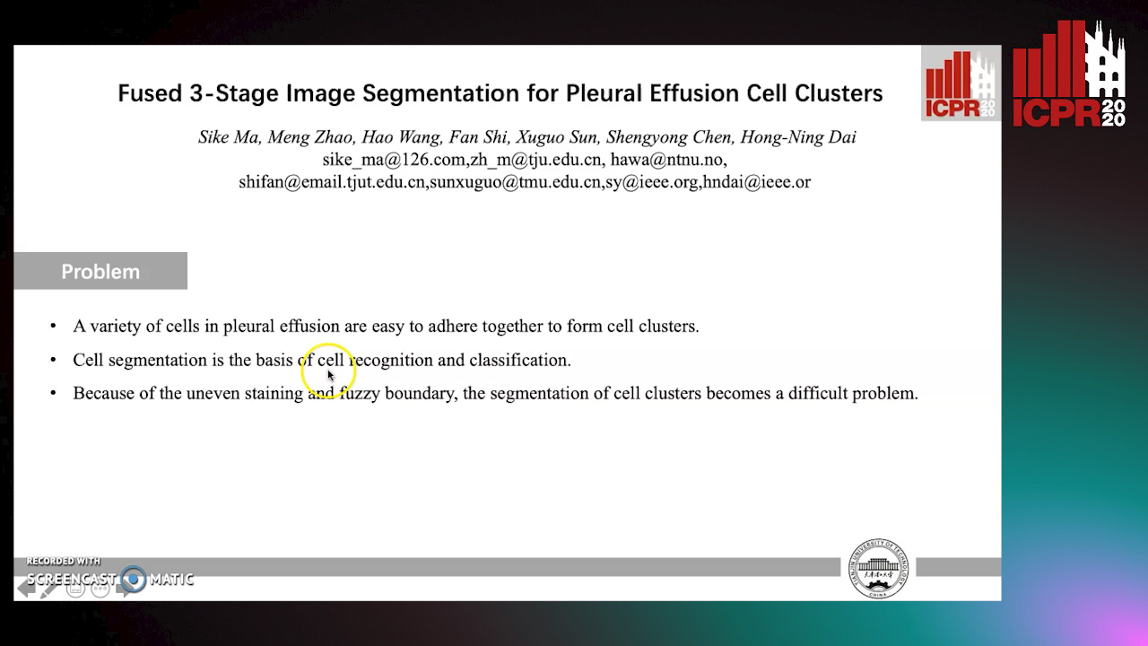
Auto-TLDR; Coarse Segmentation of Stained and Stained Unstained Cell Clusters in pleural effusion using 3-stage segmentation method

Slides Poster Similar

The appearance of tumor cell clusters in pleural effusion is usually a vital sign of cancer metastasis. Segmentation, as an indispensable basis, is of crucial importance for diagnosing, chemical treatment, and prognosis in patients. However, accurate segmentation of unstained cell clusters containing more detailed features than the fluorescent staining images remains to be a challenging problem due to the complex background and the unclear boundary. Therefore, in this paper, we propose a fused 3-stage image segmentation algorithm, namely Coarse segmentation-Mapping-Fine segmentation (CMF) to achieve unstained cell clusters from whole slide images. Firstly, we establish a tumor cell cluster dataset consisting of 107 sets of images, with each set containing one unstained image, one stained image, and one ground-truth image. Then, according to the features of the unstained and stained cell clusters, we propose a three-stage segmentation method: 1) Coarse segmentation on stained images to extract suspicious cell regions-Region of Interest (ROI); 2) Mapping this ROI to the corresponding unstained image to get the ROI of the unstained image (UI-ROI); 3) Fine Segmentation using improved automatic fuzzy clustering framework (AFCF) on the UI-ROI to get precise cell cluster boundaries. Experimental results on 107 sets of images demonstrate that the proposed algorithm can achieve better performance on unstained cell clusters with an F1 score of 90.40%.

Semantic Segmentation of Breast Ultrasound Image with Pyramid Fuzzy Uncertainty Reduction and Direction Connectedness Feature

Kuan Huang, Yingtao Zhang, Heng-Da Cheng, Ping Xing, Boyu Zhang

Responsive image

Auto-TLDR; Uncertainty-Based Deep Learning for Breast Ultrasound Image Segmentation

Slides Poster Similar

Deep learning approaches have achieved impressive results in breast ultrasound (BUS) image segmentation. However, these methods did not solve uncertainty and noise in BUS images well. To address this issue, we present a novel deep learning structure for BUS image semantic segmentation by analyzing the uncertainty using a pyramid fuzzy block and generating a novel feature based on connectedness. Firstly, feature maps in the proposed network are down-sampled to different resolutions. Fuzzy transformation and uncertainty representation are applied to each resolution to obtain the uncertainty degree on different scales. Meanwhile, the BUS images contain layer structures. From top to bottom, there are skin layer, fat layer, mammary layer, muscle layer, and background area. A spatial recurrent neural network (RNN) is utilized to calculate the connectedness between each pixel and the pixels on the four boundaries in horizontal and vertical lines. The spatial-wise context feature can introduce the characteristic of layer structure to deep neural network. Finally, the original convolutional features are combined with connectedness feature according to the uncertainty degrees. The proposed methods are applied to two datasets: a BUS image benchmark with two categories (background and tumor) and a five-category BUS image dataset with fat layer, mammary layer, muscle layer, background, and tumor. The proposed method achieves the best results on both datasets compared with eight state-of-the-art deep learning-based approaches.

Deep Learning Based Sepsis Intervention: The Modelling and Prediction of Severe Sepsis Onset

Gavin Tsang, Xianghua Xie

Responsive image

Auto-TLDR; Predicting Sepsis onset by up to six hours prior using a boosted cascading training methodology and adjustable margin hinge loss function

Slides Poster Similar

Sepsis presents a significant challenge to healthcare providers during critical care scenarios such as within an intensive care unit. The prognosis of the onset of severe septic shock results in significant increases in mortality rate, length of stay and readmission rates. Continual advancements in health informatics data allows for applications within the machine learning field to predict sepsis onset in a timely manner, allowing for effective preventative intervention of severe septic shock. A novel deep learning application is proposed to provide effective prediction of sepsis onset by up to six hours prior, involving the use of novel concepts such as a boosted cascading training methodology and adjustable margin hinge loss function. The proposed methodology provides statistically significant improvements to that of current machine learning based modelling applications based off the Physionet Computing in Cardiology 2019 challenge. Results show test F1 scores of 0.420, a significant improvement of 0.281 as compared to the next best challenger results.

Deep Recurrent-Convolutional Model for AutomatedSegmentation of Craniomaxillofacial CT Scans

Francesca Murabito, Simone Palazzo, Federica Salanitri Proietto, Francesco Rundo, Ulas Bagci, Daniela Giordano, Rosalia Leonardi, Concetto Spampinato

Responsive image

Auto-TLDR; Automated Segmentation of Anatomical Structures in Craniomaxillofacial CT Scans using Fully Convolutional Deep Networks

Slides Poster Similar

In this paper we define a deep learning architecture for automated segmentation of anatomical structures in Craniomaxillofacial (CMF) CT scans that leverages the recent success of encoder-decoder models for semantic segmentation of natural images. In particular, we propose a fully convolutional deep network that combines the advantages of recent fully convolutional models, such as Tiramisu, with squeeze-and-excitation blocks for feature recalibration, integrated with convolutional LSTMs to model spatio-temporal correlations between consecutive slices. The proposed segmentation network shows superior performance and generalization capabilities (to different structures and imaging modalities) than state of the art methods on automated segmentation of CMF structures (e.g., mandibles and airways) in several standard benchmarks (e.g., MICCAI datasets) and on new datasets proposed herein, effectively facing shape variability.