Deep Multi-Stage Model for Automated Landmarking of Craniomaxillofacial CT Scans

Simone Palazzo, Giovanni Bellitto, Luca Prezzavento, Francesco Rundo, Ulas Bagci, Daniela Giordano, Rosalia Leonardi, Concetto Spampinato

Responsive image

Auto-TLDR; Automated Landmarking of Craniomaxillofacial CT Images Using Deep Multi-Stage Architecture

Slides

In this paper we define a deep multi-stage architecture for automated landmarking of craniomaxillofacial (CMF) CT images. Our model is composed of three subnetworks that first localize, on reduced-resolution images, areas where land-marks may be found and then refine the search, at full-resolution scale, through a hierarchical structure aiming at increasing the granularity of the investigated region. The multi-stage pipeline is designed to deal with full resolution data and does not require any additional pre-processing step to reduce search space, as opposed to existing methods that can be only adopted for searching landmarks located in well-defined anatomical structures (e.g.,mandibles). The automated landmarking system is tested on identifying landmarks located in several CMF regions, achieving an average error of 0.8 mm, significantly lower than expert readings. The proposed model also outperforms baselines and is on par with existing models that employ additional upstream segmentation, on state-of-the-art benchmarks.

Similar papers

Deep Recurrent-Convolutional Model for AutomatedSegmentation of Craniomaxillofacial CT Scans

Francesca Murabito, Simone Palazzo, Federica Salanitri Proietto, Francesco Rundo, Ulas Bagci, Daniela Giordano, Rosalia Leonardi, Concetto Spampinato

Responsive image

Auto-TLDR; Automated Segmentation of Anatomical Structures in Craniomaxillofacial CT Scans using Fully Convolutional Deep Networks

Slides Poster Similar

In this paper we define a deep learning architecture for automated segmentation of anatomical structures in Craniomaxillofacial (CMF) CT scans that leverages the recent success of encoder-decoder models for semantic segmentation of natural images. In particular, we propose a fully convolutional deep network that combines the advantages of recent fully convolutional models, such as Tiramisu, with squeeze-and-excitation blocks for feature recalibration, integrated with convolutional LSTMs to model spatio-temporal correlations between consecutive slices. The proposed segmentation network shows superior performance and generalization capabilities (to different structures and imaging modalities) than state of the art methods on automated segmentation of CMF structures (e.g., mandibles and airways) in several standard benchmarks (e.g., MICCAI datasets) and on new datasets proposed herein, effectively facing shape variability.

One-Stage Multi-Task Detector for 3D Cardiac MR Imaging

Weizeng Lu, Xi Jia, Wei Chen, Nicolò Savioli, Antonio De Marvao, Linlin Shen, Declan O'Regan, Jinming Duan

Responsive image

Auto-TLDR; Multi-task Learning for Real-Time, simultaneous landmark location and bounding box detection in 3D space

Slides Poster Similar

Fast and accurate landmark location and bounding box detection are important steps in 3D medical imaging. In this paper, we propose a novel multi-task learning framework, for real-time, simultaneous landmark location and bounding box detection in 3D space. Our method extends the famous single-shot multibox detector (SSD) from single-task learning to multi-task learning and from 2D to 3D. Furthermore, we propose a post-processing approach to refine the network landmark output, by averaging the candidate landmarks. Owing to these settings, the proposed framework is fast and accurate. For 3D cardiac magnetic resonance (MR) images with size 224 × 224 × 64, our framework runs about 128 volumes per second (VPS) on GPU and achieves 6.75mm average point-to-point distance error for landmark location, which outperforms both state-of-the-art and baseline methods. We also show that segmenting the 3D image cropped with the bounding box results in both improved performance and efficiency.

A Benchmark Dataset for Segmenting Liver, Vasculature and Lesions from Large-Scale Computed Tomography Data

Bo Wang, Zhengqing Xu, Wei Xu, Qingsen Yan, Liang Zhang, Zheng You

Responsive image

Auto-TLDR; The Biggest Treatment-Oriented Liver Cancer Dataset for Segmentation

Slides Poster Similar

How to build a high-performance liver-related computer assisted diagnosis system is an open question of great interest. However, the performance of the state-of-art algorithm is always limited by the amount of data and quality of the label. To address this problem, we propose the biggest treatment-oriented liver cancer dataset for liver surgery and treatment planning. This dataset provides 216 cases (totally about 268K frames) scanned images in contrast-enhanced computed tomography (CT). We labeled all the CT images with the liver, liver vasculature and liver tumor segmentation ground truth for train and tune segmentation algorithms in advance. Based on that, we evaluate several recent and state-of-the-art segmentation algorithms, including 7 deep learning methods, on CT sequences. All results are compared to reference segmentations five error metrics that highlight different aspects of segmentation accuracy. In general, compared with previous datasets, our dataset is really a challenging dataset. To our knowledge, the proposed dataset and benchmark allow for the first time systematic exploration of such issues, and will be made available to allow for further research in this field.

A Transformer-Based Network for Anisotropic 3D Medical Image Segmentation

Guo Danfeng, Demetri Terzopoulos

Responsive image

Auto-TLDR; A transformer-based model to tackle the anisotropy problem in 3D medical image analysis

Slides Poster Similar

A critical challenge of applying neural networks to 3D medical image analysis is to deal with the anisotropy problem. The inter-slice contextual information contained in medical images is important, especially when the structural information of lesions is needed. However, such information often varies with cases because of variable slice spacing. Image anisotropy downgrades model performance especially when slice spacing varies significantly among training and testing datasets. ExsiWe proposed a transformer-based model to tackle the anisotropy problem. It is adaptable to different levels of anisotropy and is computationally efficient. Experiments are conducted on 3D lung cancer segmentation task. Our model achieves an average Dice score of approximately 0.87, which generally outperforms baseline models.

Planar 3D Transfer Learning for End to End Unimodal MRI Unbalanced Data Segmentation

Martin Kolarik, Radim Burget, Carlos M. Travieso-Gonzalez, Jan Kocica

Responsive image

Auto-TLDR; Planar 3D Res-U-Net Network for Unbalanced 3D Image Segmentation using Fluid Attenuation Inversion Recover

Slides Similar

We present a novel approach of 2D to 3D transfer learning based on mapping pre-trained 2D convolutional neural network weights into planar 3D kernels. The method is validated by proposed planar 3D res-u-net network with encoder transferred from the 2D VGG-16 which is applied for a single-stage unbalanced 3D image data segmentation. In particular, we evaluate the method on the MICCAI 2016 MS lesion segmentation challenge dataset utilizing solely Fluid Attenuation Inversion Recover (FLAIR) sequence without brain extraction for training and inference to simulate real medical praxis. The planar 3D res-u-net network performed the best both in sensitivity and Dice score amongst end to end methods processing raw MRI scans and achieved comparable Dice score to a state-of-the-art unimodal not end to end approach. Complete source code was released under the open-source license and this paper is in compliance with the Machine learning Reproducibility Checklist. By implementing practical transfer learning for 3D data representation we were able to successfully segment heavily unbalanced data without selective sampling and achieved more reliable results using less training data in single modality. From medical perspective, the unimodal approach gives an advantage in real praxis as it does not require co-registration nor additional scanning time during examination. Although modern medical imaging methods capture high resolution 3D anatomy scans suitable for computer aided detection system processing, deployment of automatic systems for interpretation of radiology imaging is still rather theoretical in many medical areas. Our work aims to bridge the gap offering solution for partial research questions.

DARN: Deep Attentive Refinement Network for Liver Tumor Segmentation from 3D CT Volume

Yao Zhang, Jiang Tian, Cheng Zhong, Yang Zhang, Zhongchao Shi, Zhiqiang He

Responsive image

Auto-TLDR; Deep Attentive Refinement Network for Liver Tumor Segmentation from 3D Computed Tomography Using Multi-Level Features

Slides Poster Similar

Automatic liver tumor segmentation from 3D Computed Tomography (CT) is a necessary prerequisite in the interventions of hepatic abnormalities and surgery planning. However, accurate liver tumor segmentation remains challenging due to the large variability of tumor sizes and inhomogeneous texture. Recent advances based on Fully Convolutional Network (FCN) in liver tumor segmentation draw on success of learning discriminative multi-level features. In this paper, we propose a Deep Attentive Refinement Network (DARN) for improved liver tumor segmentation from CT volumes by fully exploiting both low and high level features embedded in different layers of FCN. Different from existing works, we exploit attention mechanism to leverage the relation of different levels of features encoded in different layers of FCN. Specifically, we introduce a Semantic Attention Refinement (SemRef) module to selectively emphasize global semantic information in low level features with the guidance of high level ones, and a Spatial Attention Refinement (SpaRef) module to adaptively enhance spatial details in high level features with the guidance of low level ones. We evaluate our network on the public MICCAI 2017 Liver Tumor Segmentation Challenge dataset (LiTS dataset) and it achieves state-of-the-art performance. The proposed refinement modules are an effective strategy to exploit multi-level features and has great potential to generalize to other medical image segmentation tasks.

Automatic Semantic Segmentation of Structural Elements related to the Spinal Cord in the Lumbar Region by Using Convolutional Neural Networks

Jhon Jairo Sáenz Gamboa, Maria De La Iglesia-Vaya, Jon Ander Gómez

Responsive image

Auto-TLDR; Semantic Segmentation of Lumbar Spine Using Convolutional Neural Networks

Slides Poster Similar

This work addresses the problem of automatically segmenting the MR images corresponding to the lumbar spine. The purpose is to detect and delimit the different structural elements like vertebrae, intervertebral discs, nerves, blood vessels, etc. This task is known as semantic segmentation. The approach proposed in this work is based on convolutional neural networks whose output is a mask where each pixel from the input image is classified into one of the possible classes. Classes were defined by radiologists and correspond to structural elements and tissues. The proposed network architectures are variants of the U-Net. Several complementary blocks were used to define the variants: spatial attention models, deep supervision and multi-kernels at input, this last block type is based on the idea of inception. Those architectures which got the best results are described in this paper, and their results are discussed. Two of the proposed architectures outperform the standard U-Net used as baseline.

BiLuNet: A Multi-Path Network for Semantic Segmentation on X-Ray Images

Van Luan Tran, Huei-Yung Lin, Rachel Liu, Chun-Han Tseng, Chun-Han Tseng

Responsive image

Auto-TLDR; BiLuNet: Multi-path Convolutional Neural Network for Semantic Segmentation of Lumbar vertebrae, sacrum,

Similar

Semantic segmentation and shape detection of lumbar vertebrae, sacrum, and femoral heads from clinical X-ray images are important and challenging tasks. In this paper, we propose a new multi-path convolutional neural network, BiLuNet, for semantic segmentation on X-ray images. The network is capable of medical image segmentation with very limited training data. With the shape fitting of the bones, we can identify the location of the target regions very accurately for lumbar vertebra inspection. We collected our dataset and annotated by doctors for model training and performance evaluation. Compared to the state-of-the-art methods, the proposed technique provides better mIoUs and higher success rates with the same training data. The experimental results have demonstrated the feasibility of our network to perform semantic segmentation for lumbar vertebrae, sacrum, and femoral heads.

BG-Net: Boundary-Guided Network for Lung Segmentation on Clinical CT Images

Rui Xu, Yi Wang, Tiantian Liu, Xinchen Ye, Lin Lin, Yen-Wei Chen, Shoji Kido, Noriyuki Tomiyama

Responsive image

Auto-TLDR; Boundary-Guided Network for Lung Segmentation on CT Images

Slides Poster Similar

Lung segmentation on CT images is a crucial step for a computer-aided diagnosis system of lung diseases. The existing deep learning based lung segmentation methods are less efficient to segment lungs on clinical CT images, especially that the segmentation on lung boundaries is not accurate enough due to complex pulmonary opacities in practical clinics. In this paper, we propose a boundary-guided network (BG-Net) to address this problem. It contains two auxiliary branches that separately segment lungs and extract the lung boundaries, and an aggregation branch that efficiently exploits lung boundary cues to guide the network for more accurate lung segmentation on clinical CT images. We evaluate the proposed method on a private dataset collected from the Osaka university hospital and four public datasets including StructSeg, HUG, VESSEL12, and a Novel Coronavirus 2019 (COVID-19) dataset. Experimental results show that the proposed method can segment lungs more accurately and outperform several other deep learning based methods.

Segmentation of Axillary and Supraclavicular Tumoral Lymph Nodes in PET/CT: A Hybrid CNN/Component-Tree Approach

Diana Lucia Farfan Cabrera, Nicolas Gogin, David Morland, Benoît Naegel, Dimitri Papathanassiou, Nicolas Passat

Responsive image

Auto-TLDR; Coupling Convolutional Neural Networks and Component-Trees for Lymph node Segmentation from PET/CT Images

Slides Similar

The analysis of axillary and supraclavicular lymph nodes is a primary prognostic factor for the staging of breast cancer. However, due to the size of lymph nodes and the low resolution of PET data, their segmentation is challenging. We investigate the relevance of considering axillary and supraclavicular lymph node segmentation from PET/CT images by coupling Convolutional Neural Networks (CNNs) and Component-Trees (C-Trees). Building upon the U-Net architecture, we propose a framework that couples a multi-modal U-Net fed with PET and CT, coupled with a hierarchical model obtained from the PET that provides additional high-level region-based features as input channels. Our working hypotheses are twofold. First, we take advantage of both anatomical information from CT for detecting the nodes, and from functional information from PET for detecting the pathological ones. Second, we consider region-based attributes extracted from C-Tree analysis of 3D PET/CT images to improve the CNN segmentation. We carried out experiments on a dataset of 240 pathological lymph nodes from 52 patients scans, and compared our outputs with human expert-defined ground-truth, leading to promising results.

Weakly Supervised Geodesic Segmentation of Egyptian Mummy CT Scans

Avik Hati, Matteo Bustreo, Diego Sona, Vittorio Murino, Alessio Del Bue

Responsive image

Auto-TLDR; A Weakly Supervised and Efficient Interactive Segmentation of Ancient Egyptian Mummies CT Scans Using Geodesic Distance Measure and GrabCut

Slides Poster Similar

In this paper, we tackle the task of automatically analyzing 3D volumetric scans obtained from computed tomography (CT) devices. In particular, we address a particular task for which data is very limited: the segmentation of ancient Egyptian mummies CT scans. We aim at digitally unwrapping the mummy and identify different segments such as body, bandages and jewelry. The problem is complex because of the lack of annotated data for the different semantic regions to segment, thus discouraging the use of strongly supervised approaches. We, therefore, propose a weakly supervised and efficient interactive segmentation method to solve this challenging problem. After segmenting the wrapped mummy from its exterior region using histogram analysis and template matching, we first design a voxel distance measure to find an approximate solution for the body and bandage segments. Here, we use geodesic distances since voxel features as well as spatial relationship among voxels is incorporated in this measure. Next, we refine the solution using a GrabCut based segmentation together with a tracking method on the slices of the scan that assigns labels to different regions in the volume, using limited supervision in the form of scribbles drawn by the user. The efficiency of the proposed method is demonstrated using visualizations and validated through quantitative measures and qualitative unwrapping of the mummy.

Segmenting Kidney on Multiple Phase CT Images Using ULBNet

Yanling Chi, Yuyu Xu, Gang Feng, Jiawei Mao, Sihua Wu, Guibin Xu, Weimin Huang

Responsive image

Auto-TLDR; A ULBNet network for kidney segmentation on multiple phase CT images

Poster Similar

Abstract—Segmentation of kidney on CT images is critical to computer-assisted surgical planning for kidney interventional therapy. Segmenting kidney manually is impractical in clinical, automatic segmentation is desirable. U-Net has been successful in medical image segmentation and is a promising candidate for the task. However, semantic gap still exists, especially when multiple phase images or multiple center images are involved. In this paper, we proposed an ULBNet to reduce the semantic gap and to improve segmentation performance. The proposed architecture includes new skip connections of local binary convolution (LBC). We also proposed a novel strategy of fast retraining a model for a new task without manually labelling required. We evaluated the network for kidney segmentation on multiple phase CT images. ULBNet resulted in an overall accuracy of 98.0% with comparison to Resunet 97.5%. Specifically, on the plain phase CT images, 98.1% resulted from ULBNet and 97.6% from Resunet; on the corticomedullay phase images, 97.8% from ULBNet and 97.2% from Resunet; on the nephrographic phase images, 97.6% from ULBNet and 97.4% from Resunet; on the excretory phase images, 98.1% from ULBNet and 97.4% from Resunet. The proposed network architecture performs better than Resunet on generalizing to multiple phase images.

MTGAN: Mask and Texture-Driven Generative Adversarial Network for Lung Nodule Segmentation

Wei Chen, Qiuli Wang, Kun Wang, Dan Yang, Xiaohong Zhang, Chen Liu, Yucong Li

Responsive image

Auto-TLDR; Mask and Texture-driven Generative Adversarial Network for Lung Nodule Segmentation

Slides Poster Similar

Accurate segmentation for lung nodules in lung computed tomography (CT) scans plays a key role in the early diagnosis of lung cancer. Many existing methods, especially UNet, have made significant progress in lung nodule segmentation. However, due to the complex shapes of lung nodules and the similarity of visual characteristics between nodules and lung tissues, an accurate segmentation with few false positives of lung nodules is still a challenging problem. Considering the fact that both boundary and texture information of lung nodules are important for obtaining an accurate segmentation result, we propose a novel Mask and Texture-driven Generative Adversarial Network (MTGAN) with a joint multi-scale L1 loss for lung nodule segmentation, which takes full advantages of U-Net and adversarial training. The proposed MTGAN leverages adversarial learning strategy guided by the boundary and texture information of lung nodules to generate more accurate segmentation results with lesser false positives. We validate our model with the LIDC–IDRI dataset, and experimental results show that our method achieves excellent segmentation results for a variety of lung nodules, especially for juxtapleural nodules and low-dense nodules. Without any bells and whistles, the proposed MTGAN achieves significant segmentation performance with the Dice similarity coefficient (DSC) of 85.24% on the LIDC–IDRI dataset.

BCAU-Net: A Novel Architecture with Binary Channel Attention Module for MRI Brain Segmentation

Yongpei Zhu, Zicong Zhou, Guojun Liao, Kehong Yuan

Responsive image

Auto-TLDR; BCAU-Net: Binary Channel Attention U-Net for MRI brain segmentation

Slides Poster Similar

Recently deep learning-based networks have achieved advanced performance in medical image segmentation. However, the development of deep learning is slow in magnetic resonance image (MRI) segmentation of normal brain tissues. In this paper, inspired by channel attention module, we propose a new architecture, Binary Channel Attention U-Net (BCAU-Net), by introducing a novel Binary Channel Attention Module (BCAM) into skip connection of U-Net, which can take full advantages of the channel information extracted from the encoding path and corresponding decoding path. To better aggregate multi-scale spatial information of the feature map, spatial pyramid pooling (SPP) modules with different pooling operations are used in BCAM instead of original average-pooling and max-pooling operations. We verify this model on two datasets including IBSR and MRBrainS18, and obtain better performance on MRI brain segmentation compared with other methods. We believe the proposed method can advance the performance in brain segmentation and clinical diagnosis.

Segmentation of Intracranial Aneurysm Remnant in MRA Using Dual-Attention Atrous Net

Subhashis Banerjee, Ashis Kumar Dhara, Johan Wikström, Robin Strand

Responsive image

Auto-TLDR; Dual-Attention Atrous Net for Segmentation of Intracranial Aneurysm Remnant from MRA Images

Slides Poster Similar

Due to the advancement of non-invasive medical imaging modalities like Magnetic Resonance Angiography (MRA), an increasing number of Intracranial Aneurysm (IA) cases are being reported in recent years. The IAs are typically treated by so-called endovascular coiling, where blood flow in the IA is prevented by embolization with a platinum coil. Accurate quantification of the IA Remnant (IAR), i.e. the volume with blood flow present post treatment is the utmost important factor in choosing the right treatment planning. This is typically done by manually segmenting the aneurysm remnant from the MRA volume. Since manual segmentation of volumetric images is a labour-intensive and error-prone process, development of an automatic volumetric segmentation method is required. Segmentation of small structures such as IA, that may largely vary in size, shape, and location is considered extremely difficult. Similar intensity distribution of IAs and surrounding blood vessels makes it more challenging and susceptible to false positive. In this paper we propose a novel 3D CNN architecture called Dual-Attention Atrous Net (DAtt-ANet), which can efficiently segment IAR volumes from MRA images by reconciling features at different scales using the proposed Parallel Atrous Unit (PAU) along with the use of self-attention mechanism for extracting fine-grained features and intra-class correlation. The proposed DAtt-ANet model is trained and evaluated on a clinical MRA image dataset (prospective research project, approved by the local ethical committee) of IAR consisting of 46 subjects, annotated by an expert radiologist from our group. We compared the proposed DAtt-ANet with five state-of-the-art CNN models based on their segmentation performance. The proposed DAtt-ANet outperformed all other methods and was able to achieve a five-fold cross-validation DICE score of $0.73\pm0.06$.

FOANet: A Focus of Attention Network with Application to Myocardium Segmentation

Zhou Zhao, Elodie Puybareau, Nicolas Boutry, Thierry Geraud

Responsive image

Auto-TLDR; FOANet: A Hybrid Loss Function for Myocardium Segmentation of Cardiac Magnetic Resonance Images

Slides Poster Similar

In myocardium segmentation of cardiac magnetic resonance images, ambiguities often appear near the boundaries of the target domains due to tissue similarities. To address this issue, we propose a new architecture, called FOANet, which can be decomposed in three main steps: a localization step, a Gaussian-based contrast enhancement step, and a segmentation step. This architecture is supplied with a hybrid loss function that guides the FOANet to study the transformation relationship between the input image and the corresponding label in a threelevel hierarchy (pixel-, patch- and map-level), which is helpful to improve segmentation and recovery of the boundaries. We demonstrate the efficiency of our approach on two public datasets in terms of regional and boundary segmentations.

End-To-End Multi-Task Learning for Lung Nodule Segmentation and Diagnosis

Wei Chen, Qiuli Wang, Dan Yang, Xiaohong Zhang, Chen Liu, Yucong Li

Responsive image

Auto-TLDR; A novel multi-task framework for lung nodule diagnosis based on deep learning and medical features

Slides Similar

Computer-Aided Diagnosis (CAD) systems for lung nodule diagnosis based on deep learning have attracted much attention in recent years. However, most existing methods ignore the relationships between the segmentation and classification tasks, which leads to unstable performances. To address this problem, we propose a novel multi-task framework, which can provide lung nodule segmentation mask, malignancy prediction, and medical features for interpretable diagnosis at the same time. Our framework mainly contains two sub-network: (1) Multi-Channel Segmentation Sub-network (MSN) for lung nodule segmentation, and (2) Joint Classification Sub-network (JCN) for interpretable lung nodule diagnosis. In the proposed framework, we use U-Net down-sampling processes for extracting low-level deep learning features, which are shared by two sub-networks. The JCN forces the down-sampling processes to learn better lowlevel deep features, which lead to a better construct of segmentation masks. Meanwhile, two additional channels constructed by OTSU and super-pixel (SLIC) methods, are utilized as the guideline of the feature extraction. The proposed framework takes advantages of deep learning methods and classical methods, which can significantly improve the performances of all tasks. We evaluate the proposed framework on public dataset LIDCIDRI. Our framework achieves a promising Dice score of 86.43% in segmentation, 87.07% in malignancy level prediction, and convincing results in interpretable medical feature predictions.

3D Medical Multi-Modal Segmentation Network Guided by Multi-Source Correlation Constraint

Tongxue Zhou, Stéphane Canu, Pierre Vera, Su Ruan

Responsive image

Auto-TLDR; Multi-modality Segmentation with Correlation Constrained Network

Slides Poster Similar

In the field of multimodal segmentation, the correlation between different modalities can be considered for improving the segmentation results. In this paper, we propose a multi-modality segmentation network with a correlation constraint. Our network includes N model-independent encoding paths with N image sources, a correlation constrain block, a feature fusion block, and a decoding path. The model-independent encoding path can capture modality-specific features from the N modalities. Since there exists a strong correlation between different modalities, we first propose a linear correlation block to learn the correlation between modalities, then a loss function is used to guide the network to learn the correlated features based on the correlation representation block. This block forces the network to learn the latent correlated features which are more relevant for segmentation. Considering that not all the features extracted from the encoders are useful for segmentation, we propose to use dual attention based fusion block to recalibrate the features along the modality and spatial paths, which can suppress less informative features and emphasize the useful ones. The fused feature representation is finally projected by the decoder to obtain the segmentation result. Our experiment results tested on BraTS-2018 dataset for brain tumor segmentation demonstrate the effectiveness of our proposed method.

A Multi-Task Contextual Atrous Residual Network for Brain Tumor Detection & Segmentation

Ngan Le, Kashu Yamazaki, Quach Kha Gia, Thanh-Dat Truong, Marios Savvides

Responsive image

Auto-TLDR; Contextual Brain Tumor Segmentation Using 3D atrous Residual Networks and Cascaded Structures

Poster Similar

In recent years, deep neural networks have achieved state-of-the-art performance in a variety of recognition and segmentation tasks in medical imaging including brain tumor segmentation. We investigate that segmenting brain tumor is facing to the imbalanced data problem where the number of pixels belonging to background class (non tumor pixel) is much larger than the number of pixels belonging to foreground class (tumor pixel). To address this problem, we propose a multi-task network which is formed as a cascaded structure and designed to share the feature maps. Our model consists of two targets, i.e., (i) effectively differentiating brain tumor regions and (ii) estimating brain tumor masks. The first task is performed by our proposed contextual brain tumor detection network, which plays the role of an attention gate and focuses on the region around brain tumor only while ignore the background (non tumor area). Instead of processing every pixel, our contextual brain tumor detection network only processes contextual regions around ground-truth instances and this strategy helps to produce meaningful regions proposals. The second task is built upon a 3D atrous residual network and under an encode-decode network in order to effectively segment both large and small objects (brain tumor). Our 3D atrous residual network is designed with a skip connection to enables the gradient from the deep layers to be directly propagated to shallow layers, thus, features of different depths are preserved and used for refining each other. In order to incorporate larger contextual information in volume MRI data, our network is designed by 3D atrous convolution with various kernel sizes, which enlarges the receptive field of filters. Our proposed network has been evaluated on various datasets including BRATS2015, BRATS2017 and BRATS2018 datasets with both validation set and testing set. Our performance has been benchmarked by both region-based metrics and surface-based metrics. We also have conducted comparisons against state-of-the-art approaches.

Do Not Treat Boundaries and Regions Differently: An Example on Heart Left Atrial Segmentation

Zhou Zhao, Elodie Puybareau, Nicolas Boutry, Thierry Geraud

Responsive image

Auto-TLDR; Attention Full Convolutional Network for Atrial Segmentation using ResNet-101 Architecture

Slides Similar

Atrial fibrillation is the most common heart rhythm disease. Due to a lack of understanding in matter of underlying atrial structures, current treatments are still not satisfying. Recently, with the popularity of deep learning, many segmentation methods based on fully convolutional networks have been proposed to analyze atrial structures, especially from late gadolinium-enhanced magnetic resonance imaging. However, two problems still occur: 1) segmentation results include the atrial-like background; 2) boundaries are very hard to segment. Most segmentation approaches design a specific network that mainly focuses on the regions, to the detriment of the boundaries. Therefore, this paper proposes an attention full convolutional network framework based on the ResNet-101 architecture, which focuses on boundaries as much as on regions. The additional attention module is added to have the network pay more attention on regions and then to reduce the impact of the misleading similarity of neighboring tissues. We also use a hybrid loss composed of a region loss and a boundary loss to treat boundaries and regions at the same time. We demonstrate the efficiency of the proposed approach on the MICCAI 2018 Atrial Segmentation Challenge public dataset.

CAggNet: Crossing Aggregation Network for Medical Image Segmentation

Xu Cao, Yanghao Lin

Responsive image

Auto-TLDR; Crossing Aggregation Network for Medical Image Segmentation

Slides Poster Similar

In this paper, we present Crossing Aggregation Network (CAggNet), a novel densely connected semantic segmentation method for medical image analysis. The crossing aggregation network absorbs the idea of deep layer aggregation and makes significant innovations in layer connection and semantic information fusion. In this architecture, the traditional skip-connection structure of general U-Net is replaced by aggregations of multi-level down-sampling and up-sampling layers. This enables the network to fuse information interactively flows at different levels of layers in semantic segmentation. It also introduces weighted aggregation module to aggregate multi-scale output information. We have evaluated and compared our CAggNet with several advanced U-Net based methods in two public medical image datasets, including the 2018 Data Science Bowl nuclei detection dataset and the 2015 MICCAI gland segmentation competition dataset. Experimental results indicate that CAggNet improves medical object recognition and achieves a more accurate and efficient segmentation compared to existing improved U-Net and UNet++ structure.

Bridging the Gap between Natural and Medical Images through Deep Colorization

Lia Morra, Luca Piano, Fabrizio Lamberti, Tatiana Tommasi

Responsive image

Auto-TLDR; Transfer Learning for Diagnosis on X-ray Images Using Color Adaptation

Slides Poster Similar

Deep learning has thrived by training on large-scale datasets. However, in many applications, as for medical image diagnosis, getting massive amount of data is still prohibitive due to privacy, lack of acquisition homogeneity and annotation cost. In this scenario transfer learning from natural image collections is a standard practice that attempts to tackle shape, texture and color discrepancy all at once through pretrained model fine-tuning. In this work we propose to disentangle those challenges and design a dedicated network module that focuses on color adaptation. We combine learning from scratch of the color module with transfer learning of different classification backbones obtaining an end-to-end, easy-to-train architecture for diagnostic image recognition on X-ray images. Extensive experiments show how our approach is particularly efficient in case of data scarcity and provides a new path for further transferring the learned color information across multiple medical datasets.

OCT Image Segmentation Using NeuralArchitecture Search and SRGAN

Saba Heidari, Omid Dehzangi, Nasser M. Nasarabadi, Ali Rezai

Responsive image

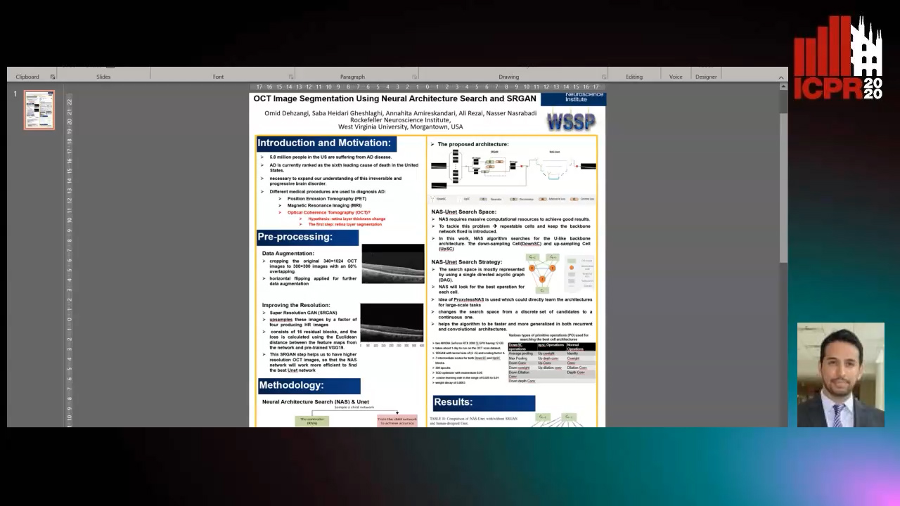
Auto-TLDR; Automatic Segmentation of Retinal Layers in Optical Coherence Tomography using Neural Architecture Search

Poster Similar

Alzheimer’s disease (AD) diagnosis is one of the major research areas in computational medicine. Optical coherence tomography (OCT) is a non-invasive, inexpensive, and timely efficient method that scans the human’s retina with depth. It has been hypothesized that the thickness of the retinal layers extracted from OCTs could be an efficient and effective biomarker for early diagnosis of AD. In this work, we aim to design a self-training model architecture for the task of segmenting the retinal layers in OCT scans. Neural architecture search (NAS) is a subfield of AutoML domain, which has a significant impact on improving the accuracy of machine vision tasks. We integrate the NAS algorithm with a Unet auto-encoder architecture as its backbone. Then, we employ our proposed model to segment the retinal nerve fiber layer in our preprocessed OCT images with the aim of AD diagnosis. In this work, we trained a super-resolution generative adversarial network on the raw OCT scans to improve the quality of the images before the modeling stage. In our architecture search strategy, different primitive operations suggested to find down- \& up-sampling Unet cell blocks and the binary gate method has been applied to make the search strategy more practical. Our architecture search method is empirically evaluated by training on the Unet and NAS-Unet from scratch. Specifically, the proposed NAS-Unet training significantly outperforms the baseline human-designed architecture by achieving 95.1\% in the mean Intersection over Union metric and 79.1\% in the Dice similarity coefficient.

Unsupervised Detection of Pulmonary Opacities for Computer-Aided Diagnosis of COVID-19 on CT Images

Rui Xu, Xiao Cao, Yufeng Wang, Yen-Wei Chen, Xinchen Ye, Lin Lin, Wenchao Zhu, Chao Chen, Fangyi Xu, Yong Zhou, Hongjie Hu, Shoji Kido, Noriyuki Tomiyama

Responsive image

Auto-TLDR; A computer-aided diagnosis of COVID-19 from CT images using unsupervised pulmonary opacity detection

Slides Poster Similar

COVID-19 emerged towards the end of 2019 which was identified as a global pandemic by the world heath organization (WHO). With the rapid spread of COVID-19, the number of infected and suspected patients has increased dramatically. Chest computed tomography (CT) has been recognized as an efficient tool for the diagnosis of COVID-19. However, the huge CT data make it difficult for radiologist to fully exploit them on the diagnosis. In this paper, we propose a computer-aided diagnosis system that can automatically analyze CT images to distinguish the COVID-19 against to community-acquired pneumonia (CAP). The proposed system is based on an unsupervised pulmonary opacity detection method that locates opacity regions by a detector unsupervisedly trained from CT images with normal lung tissues. Radiomics based features are extracted insides the opacity regions, and fed into classifiers for classification. We evaluate the proposed CAD system by using 200 CT images collected from different patients in several hospitals. The accuracy, precision, recall, f1-score and AUC achieved are 95.5%, 100%, 91%, 95.1% and 95.9% respectively, exhibiting the promising capacity on the differential diagnosis of COVID-19 from CT images.

DA-RefineNet: Dual-Inputs Attention RefineNet for Whole Slide Image Segmentation

Ziqiang Li, Rentuo Tao, Qianrun Wu, Bin Li

Responsive image

Auto-TLDR; DA-RefineNet: A dual-inputs attention network for whole slide image segmentation

Slides Poster Similar

Automatic medical image segmentation techniques have wide applications for disease diagnosing, however, its much more challenging than natural optical image segmentation tasks due to the high-resolution of medical images and the corresponding huge computation cost. Sliding window was a commonly used technique for whole slide image (WSI) segmentation, however, for these methods that based on sliding window, the main drawback was lacking of global contextual information for supervision. In this paper, we proposed a dual-inputs attention network (denoted as DA-RefineNet) for WSI segmentation, where both local fine-grained information and global coarse information can be efficiently utilized. Sufficient comparative experiments were conducted to evaluate the effectiveness of the proposed method, the results proved that the proposed method can achieve better performance on WSI segmentation tasks compared to methods rely on single-input.

A Systematic Investigation on Deep Architectures for Automatic Skin Lesions Classification

Pierluigi Carcagni, Marco Leo, Andrea Cuna, Giuseppe Celeste, Cosimo Distante

Responsive image

Auto-TLDR; RegNet: Deep Investigation of Convolutional Neural Networks for Automatic Classification of Skin Lesions

Slides Poster Similar

Computer vision-based techniques are more and more employed in healthcare and medical fields nowadays in order, principally, to be as a support to the experienced medical staff to help them to make a quick and correct diagnosis. One of the hot topics in this arena concerns the automatic classification of skin lesions. Several promising works exist about it, mainly leveraging Convolutional Neural Networks (CNN), but proposed pipeline mainly rely on complex data preprocessing and there is no systematic investigation about how available deep models can actually reach the accuracy needed for real applications. In order to overcome these drawbacks, in this work, an end-to-end pipeline is introduced and some of the most recent Convolutional Neural Networks (CNNs) architectures are included in it and compared on the largest common benchmark dataset recently introduced. To this aim, for the first time in this application context, a new network design paradigm, namely RegNet, has been exploited to get the best models among a population of configurations. The paper introduces a threefold level of contribution and novelty with respect the previous literature: the deep investigation of several CNN architectures driving to a consistent improvement of the lesions recognition accuracy, the exploitation of a new network design paradigm able to study the behavior of populations of models and a deep discussion about pro and cons of each analyzed method paving the path towards new research lines.

NephCNN: A Deep-Learning Framework for Vessel Segmentation in Nephrectomy Laparoscopic Videos

Alessandro Casella, Sara Moccia, Chiara Carlini, Emanuele Frontoni, Elena De Momi, Leonardo Mattos

Responsive image

Auto-TLDR; Adversarial Fully Convolutional Neural Networks for kidney vessel segmentation from nephrectomy laparoscopic videos

Slides Poster Similar

Objective: In the last years, Robot-assisted partial nephrectomy (RAPN) is establishing as elected treatment for renal cell carcinoma (RCC). Reduced field of view, field occlusions by surgical tools, and reduced maneuverability may potentially cause accidents, such as unwanted vessel resection with consequent bleeding. Surgical Data Science (SDS) can provide effective context-aware tools for supporting surgeons. However, currently no tools have been exploited for automatic vessels segmentation from nephrectomy laparoscopic videos. Herein, we propose a new approach based on adversarial Fully Convolutional Neural Networks (FCNNs) to kidney vessel segmentation from nephrectomy laparoscopic vision. Methods: The proposed approach enhances existing segmentation framework by (i) encoding 3D kernels for spatio-temporal features extraction to enforce pixel connectivity in time, and (ii) perform training in adversarial fashion, which constrains vessels shape. Results: We performed a preliminary study using 8 different RAPN videos (1871 frames), the first in the field, achieving a median Dice Similarity Coefficient of 71.76%. Conclusions: Results showed that the proposed approach could be a valuable solution with a view to assist surgeon during RAPN.

A Deep Learning-Based Method for Predicting Volumes of Nasopharyngeal Carcinoma for Adaptive Radiation Therapy Treatment

Bilel Daoud, Ken'Ichi Morooka, Shoko Miyauchi, Ryo Kurazume, Wafa Mnejja, Leila Farhat, Jamel Daoud

Responsive image

Auto-TLDR; TEP-Net: Tumor Evolution Prediction of Nasopharyngeal Carcinoma and Organ-at-risks Using CT Images

Slides Poster Similar

This paper presents a new system for predicting the spatial change of Nasopharyngeal carcinoma(NPC) and organ-at-risks (OARs) volumes over the course of the radiation therapy (RT) treatment for facilitating the workflow of adaptive radiotherapy. The proposed system, called " Tumor Evolution Prediction (TEP-Net)", predicts the spatial distributions of NPC and 5 OARs, separately, in response to RT in the coming week, week n. Here, TEP-Net has (n-1)-inputs that are week 1 to week n-1 of CT axial, coronal or sagittal images acquired once the patient complete the planned RT treatment of the corresponding week. As a result, three predicted results of each target region are obtained from the three-view CT images. To determine the final prediction of NPC and 5 OARs, two integration methods, weighted fully connected layers and weighted voting methods, are introduced. From the experiments using weekly CT images of 140 NPC patients, our proposed system achieves the best performance for predicting NPC and OARs compared with conventional methods.

A Deep Learning Approach for the Segmentation of Myocardial Diseases

Khawala Brahim, Abdull Qayyum, Alain Lalande, Arnaud Boucher, Anis Sakly, Fabrice Meriaudeau

Responsive image

Auto-TLDR; Segmentation of Myocardium Infarction Using Late GADEMRI and SegU-Net

Slides Poster Similar

Cardiac left ventricular (LV) segmentation is of paramount essential step for both diagnosis and treatment of cardiac pathologies such as ischemia, myocardial infarction, arrhythmia and myocarditis. However, this segmentation is challenging due to high variability across patients and the potential lack of contrast between structures. In this work, we propose and evaluate a (2.5D) SegU-Net model based on the fusion of two deep learning techniques (U-Net and Seg-Net) for automated LGEMRI (Late gadolinium enhanced magnetic resonance imaging) myocardial disease (infarct core and no reflow region) quantification in a new multifield expert annotated dataset. Given that the scar tissue represents a small part of the whole MRI slices, we focused on myocardium area. Segmentation results show that this preprocessing step facilitate the learning procedure. In order to solve the class imbalance problem, we propose to apply the Jaccard loss and the Focal Loss as optimization loss function and to integrate a class weights strategy into the objective function. Late combination has been used to merge the output of the best trained models on a different set of hyperparameters. The final network segmentation performances will be useful for future comparison of new method to the current related work for this task. A total number of 2237 of slices (320 cases) were used for training/validation and 210 slices (35 cases) were used for testing. Experiments over our proposed dataset, using several evaluation metrics such Jaccard distance (IOU), Accuracy and Dice similarity coefficient (DSC), demonstrate efficiency performance in quantifying different zones of myocardium infarction across various patients. As compared to the second intra-observer study, our testing results showed that the SegUNet prediction model leads to these average dice coefficients over all segmented tissue classes, respectively : 'Background': 0.99999, 'Myocardium': 0.99434, 'Infarctus': 0.95587, 'Noreflow': 0.78187.

A Lumen Segmentation Method in Ureteroscopy Images Based on a Deep Residual U-Net Architecture

Jorge Lazo, Marzullo Aldo, Sara Moccia, Michele Catellani, Benoit Rosa, Elena De Momi, Michel De Mathelin, Francesco Calimeri

Responsive image

Auto-TLDR; A Deep Neural Network for Ureteroscopy with Residual Units

Slides Poster Similar

Ureteroscopy is becoming the first surgical treatment option for the majority of urinary affections. This procedure is carried out using an endoscope which provides the surgeon with the visual and spatial information necessary to navigate inside the urinary tract. Having in mind the development of surgical assistance systems, that could enhance the performance of surgeon, the task of lumen segmentation is a fundamental part since this is the visual reference which marks the path that the endoscope should follow. This is something that has not been analyzed in ureteroscopy data before. However, this task presents several challenges given the image quality and the conditions itself of ureteroscopy procedures. In this paper, we study the implementation of a Deep Neural Network which exploits the advantage of residual units in an architecture based on U-Net. For the training of these networks, we analyze the use of two different color spaces: gray-scale and RGB data images. We found that training on gray-scale images gives the best results obtaining mean values of Dice Score, Precision, and Recall of 0.73, 0.58, and 0.92 respectively. The results obtained show that the use of residual U-Net could be a suitable model for further development for a computer-aided system for navigation and guidance through the urinary system.

Inferring Functional Properties from Fluid Dynamics Features

Andrea Schillaci, Maurizio Quadrio, Carlotta Pipolo, Marcello Restelli, Giacomo Boracchi

Responsive image

Auto-TLDR; Exploiting Convective Properties of Computational Fluid Dynamics for Medical Diagnosis

Slides Poster Similar

In a wide range of applied problems involving fluid flows, Computational Fluid Dynamics (CFD) provides detailed quantitative information on the flow field, at various levels of fidelity and computational cost. However, CFD alone cannot predict high-level functional properties of the system that are not easily obtained from the equations of fluid motion. In this work, we present a data-driven framework to extract additional information, such as medical diagnostic output, from CFD solutions. The task is made difficult by the huge data dimensionality of CFD, together with the limited amount of training data implied by its high computational cost. By pursuing a traditional ML pipeline of pre-processing, feature extraction, and model training, we demonstrate that informative features can be extracted from CFD data. Two experiments, pertaining to different application domains, support the claim that the convective properties implicit into a CFD solution can be leveraged to retrieve functional information for which an analytical definition is missing. Despite the preliminary nature of our study and the relative simplicity of both the geometrical and CFD models, for the first time we demonstrate that the combination of ML and CFD can diagnose a complex system in terms of high-level functional information.

Hybrid Approach for 3D Head Reconstruction: Using Neural Networks and Visual Geometry

Oussema Bouafif, Bogdan Khomutenko, Mohammed Daoudi

Responsive image

Auto-TLDR; Recovering 3D Head Geometry from a Single Image using Deep Learning and Geometric Techniques

Slides Poster Similar

Recovering the 3D geometric structure of a face from a single input image is a challenging active research area in computer vision. In this paper, we present a novel method for reconstructing 3D heads from a single or multiple image(s) using a hybrid approach based on deep learning and geometric techniques. We propose an encoder-decoder network based on the U-net architecture and trained on synthetic data only. It predicts both pixel-wise normal vectors and landmarks maps from a single input photo. Landmarks are used for the pose computation and the initialization of the optimization problem, which, in turn, reconstructs the 3D head geometry by using a parametric morphable model and normal vector fields. State-of-the-art results are achieved through qualitative and quantitative evaluation tests on both single and multi-view settings. Despite the fact that the model was trained only on synthetic data, it successfully recovers 3D geometry and precise poses for real-world images.

A Novel Computer-Aided Diagnostic System for Early Assessment of Hepatocellular Carcinoma

Ahmed Alksas, Mohamed Shehata, Gehad Saleh, Ahmed Shaffie, Ahmed Soliman, Mohammed Ghazal, Hadil Abukhalifeh, Abdel Razek Ahmed, Ayman El-Baz

Responsive image

Auto-TLDR; Classification of Liver Tumor Lesions from CE-MRI Using Structured Structural Features and Functional Features

Slides Poster Similar

Early assessment of liver cancer patients with hepatocellular carcinoma (HCC) is of immense importance to provide the proper treatment plan. In this paper, we have developed a two-stage classification computer-aided diagnostic (CAD) system that has the ability to detect and grade the liver observations from multiphase contrast enhanced magnetic resonance imaging (CE-MRI). The proposed approach consists of three main steps. First, a pre-processing is applied to the CE-MRI scans to delineate the tumor lesions that will be used as an ROI across the four different phases of the CE-MRI, (namely, the pre-contrast, late-arterial, portal-venous, and delayed-contrast). Second, a group of three features are modeled to provide a quantitative discrimination between the tumor lesions; namely: i) the tumor appearance that is modeled using a set of texture features, (namely; the first-order histogram, second-order gray-level co-occurrence matrix, and second-order gray-level run-length matrix), to capture any discrimination that may appear in the lesion texture, ii) the spherical harmonics (SH) based shape features that have the ability to describe the shape complexity of the liver tumors, and iii) the functional features that are based on the calculation of the wash-in/wash-out through that evaluate the intensity changes across the post-contrast phases. Finally, the aforementioned individual features were then integrated together to obtain the combined features to be fed to a machine learning classifier towards getting the final diagnostic decision. The proposed CAD system has been tested using hepatic observations that was obtained from 85 participating patients, 34 patients with benign tumors, 34 patients with intermediate tumors and 34 with malignant tumors. Using a random forests based classifier with a leave-one-subject-out (LOSO) cross-validation, the developed CAD system achieved an 87.1% accuracy in distinguishing the malignant, intermediate and benign tumors. The classification performance is then evaluated using k-fold (5/10-fold) cross-validation approach to examine the robustness of the system. The LR-1 lesions were classified from LR-2 benign lesions with 91.2% accuracy, while 85.3% accuracy was achieved differentiating between LR-4 and LR-5 malignant tumors. The obtained results hold a promise of the proposed framework to be reliably used as a noninvasive diagnostic tool for the early detection and grading of liver cancer tumors.

Inner Eye Canthus Localization for Human Body Temperature Screening

Claudio Ferrari, Lorenzo Berlincioni, Marco Bertini, Alberto Del Bimbo

Responsive image

Auto-TLDR; Automatic Localization of the Inner Eye Canthus in Thermal Face Images using 3D Morphable Face Model

Slides Poster Similar

In this paper, we propose an automatic approach for localizing the inner eye canthus in thermal face images. We first coarsely detect 5 facial keypoints corresponding to the center of the eyes, the nosetip and the ears. Then we compute a sparse 2D-3D points correspondence using a 3D Morphable Face Model (3DMM). This correspondence is used to project the entire 3D face onto the image, and subsequently locate the inner eye canthus. Detecting this location allows to obtain the most precise body temperature measurement for a person using a thermal camera. We evaluated the approach on a thermal face dataset provided with manually annotated landmarks. However, such manual annotations are normally conceived to identify facial parts such as eyes, nose and mouth, and are not specifically tailored for localizing the eye canthus region. As additional contribution, we enrich the original dataset by using the annotated landmarks to deform and project the 3DMM onto the images. Then, by manually selecting a small region corresponding to the eye canthus, we enrich the dataset with additional annotations. By using the manual landmarks, we ensure the correctness of the 3DMM projection, which can be used as ground-truth for future evaluations. Moreover, we supply the dataset with the 3D head poses and per-point visibility masks for detecting self-occlusions. The data will be publicly released.

Robust Skeletonization for Plant Root Structure Reconstruction from MRI

Jannis Horn

Responsive image

Auto-TLDR; Structural reconstruction of plant roots from MRI using semantic root vs shoot segmentation and 3D skeletonization

Slides Poster Similar

Structural reconstruction of plant roots from MRI is challenging, because of low resolution and low signal-to-noise ratio of the 3D measurements which may lead to disconnectivities and wrongly connected roots. We propose a two-stage approach for this task. The first stage is based on semantic root vs. soil segmentation and finds lowest-cost paths from any root voxel to the shoot. The second stage takes the largest fully connected component generated in the first stage and uses 3D skeletonization to extract a graph structure. We evaluate our method on 22 MRI scans and compare to human expert reconstructions.

MedZip: 3D Medical Images Lossless Compressor Using Recurrent Neural Network (LSTM)

Omniah Nagoor, Joss Whittle, Jingjing Deng, Benjamin Mora, Mark W. Jones

Responsive image

Auto-TLDR; Recurrent Neural Network for Lossless Medical Image Compression using Long Short-Term Memory

Poster Similar

As scanners produce higher-resolution and more densely sampled images, this raises the challenge of data storage, transmission and communication within healthcare systems. Since the quality of medical images plays a crucial role in diagnosis accuracy, medical imaging compression techniques are desired to reduce scan bitrate while guaranteeing lossless reconstruction. This paper presents a lossless compression method that integrates a Recurrent Neural Network (RNN) as a 3D sequence prediction model. The aim is to learn the long dependencies of the voxel's neighbourhood in 3D using Long Short-Term Memory (LSTM) network then compress the residual error using arithmetic coding. Experiential results reveal that our method obtains a higher compression ratio achieving 15% saving compared to the state-of-the-art lossless compression standards, including JPEG-LS, JPEG2000, JP3D, HEVC, and PPMd. Our evaluation demonstrates that the proposed method generalizes well to unseen modalities CT and MRI for the lossless compression scheme. To the best of our knowledge, this is the first lossless compression method that uses LSTM neural network for 16-bit volumetric medical image compression.

Vehicle Classification from Profile Measures

Marco Patanè, Andrea Fusiello

Responsive image

Auto-TLDR; SliceNets: Convolutional Neural Networks for 3D Object Classification of Planar Slices

Slides Similar

This paper proposes two novel convolutional neural networks for 3D object classification, tailored to process point clouds that are composed of planar slices (profiles). In particular, the application that we are targeting is the classification of vehicles by scanning them along planes perpendicular to the driving direction, within the context of Electronic Toll Collection. Depending on sensors configurations, the distance between slices can be measured or not, thus resulting in two types of point clouds, namely metric and non-metric. In the latter case, two coordinates are indeed metric but the third one is merely a temporal index. Our networks, named SliceNets, extract metric information from the spatial coordinates and neighborhood information from the third one (either metric or temporal), thus being able to handle both types of point clouds. Experiments on two datasets collected in the field show the effectiveness of our networks in comparison with state-of-the-art ones.

Vesselness Filters: A Survey with Benchmarks Applied to Liver Imaging

Jonas Lamy, Odyssée Merveille, Bertrand Kerautret, Nicolas Passat, Antoine Vacavant

Responsive image

Auto-TLDR; Comparison of Vessel Enhancement Filters for Liver Vascular Network Segmentation

Slides Poster Similar

The accurate knowledge of vascular network geometry is crucial for many clinical applications such as cardiovascular disease diagnosis and surgery planning. Vessel enhancement algorithms are often a key step to improve the robustness of vessel segmentation. A wide variety of enhancement filters exists in the literature, but they are often difficult to compare as the applications and datasets differ from a paper to another and the code is rarely available. In this article, we compare seven vessel enhancement filters covering the last twenty years literature in a unique common framework. We focus our study on the liver vascular network which is under-represented in the literature. The evaluation is made from three points of view: in the whole liver, in the vessel neighborhood and near the bifurcations. The study is performed on two publicly available datasets: the Ircad dataset (CT images) and the VascuSynth dataset adapted for MRI simulation. We discuss the strengths and weaknesses of each method in the hepatic context. In addition, the benchmark framework including a C++ implementation of each compared method is provided. An online demonstration ensures the reproducibility of the results without requiring any additional software.

Deep Learning-Based Type Identification of Volumetric MRI Sequences

Jean Pablo De Mello, Thiago Paixão, Rodrigo Berriel, Mauricio Reyes, Alberto F. De Souza, Claudine Badue, Thiago Oliveira-Santos

Responsive image

Auto-TLDR; Deep Learning for Brain MRI Sequences Identification Using Convolutional Neural Network

Slides Poster Similar

The analysis of Magnetic Resonance Imaging (MRI) sequences enables clinical professionals to monitor the progression of a brain tumor. As the interest for automatizing brain volume MRI analysis increases, it becomes convenient to have each sequence well identified. However, the unstandardized naming of MRI sequences make their identification difficult for automated systems, as well as make it difficult for researches to generate or use datasets for machine learning research. In face of that, we propose a system for identifying types of brain MRI sequences based on deep learning. By training a Convolutional Neural Network (CNN) based on 18-layer ResNet architecture, our system is able to classify a volumetric brain MRI as a T1, T1c, T2 or FLAIR sequence, or whether it does not belong to any of these classes. The network was trained with both pre-processed (BraTS dataset) and non-pre-processed (TCGA-GBM dataset) images with diverse acquisition protocols, requiring only a few layers of the volume for training. Our system is able to classify among sequence types with an accuracy of 96.27%.

RefiNet: 3D Human Pose Refinement with Depth Maps

Andrea D'Eusanio, Stefano Pini, Guido Borghi, Roberto Vezzani, Rita Cucchiara

Responsive image

Auto-TLDR; RefiNet: A Multi-stage Framework for 3D Human Pose Estimation

Slides Similar

Human Pose Estimation is a fundamental task for many applications in the Computer Vision community and it has been widely investigated in the 2D domain, i.e. intensity images. Therefore, most of the available methods for this task are mainly based on 2D Convolutional Neural Networks and huge manually-annotated RGB datasets, achieving stunning results. In this paper, we propose RefiNet, a multi-stage framework that regresses an extremely-precise 3D human pose estimation from a given 2D pose and a depth map. The framework consists of three different modules, each one specialized in a particular refinement and data representation, i.e. depth patches, 3D skeleton and point clouds. Moreover, we collect a new dataset, namely Baracca, acquired with RGB, depth and thermal cameras and specifically created for the automotive context. Experimental results confirm the quality of the refinement procedure that largely improves the human pose estimations of off-the-shelf 2D methods.

Triplet-Path Dilated Network for Detection and Segmentation of General Pathological Images

Jiaqi Luo, Zhicheng Zhao, Fei Su, Limei Guo

Responsive image

Auto-TLDR; Triplet-path Network for One-Stage Object Detection and Segmentation in Pathological Images

Slides Similar

Deep learning has been widely applied in the field of medical image processing. However, compared with flourishing visual tasks in natural images, the progress achieved in pathological images is not remarkable, and detection and segmentation, which are among basic tasks of computer vision, are regarded as two independent tasks. In this paper, we make full use of existing datasets and construct a triplet-path network using dilated convolutions to cooperatively accomplish one-stage object detection and nuclei segmentation for general pathological images. First, in order to meet the requirement of detection and segmentation, a novel structure called triplet feature generation (TFG) is designed to extract high-resolution and multiscale features, where features from different layers can be properly integrated. Second, considering that pathological datasets are usually small, a location-aware and partially truncated loss function is proposed to improve the classification accuracy of datasets with few images and widely varying targets. We compare the performance of both object detection and instance segmentation with state-of-the-art methods. Experimental results demonstrate the effectiveness and efficiency of the proposed network on two datasets collected from multiple organs.

Human Embryo Cell Centroid Localization and Counting in Time-Lapse Sequences

Lisette Lockhart, Parvaneh Saeedi, Jason Au, Jon Havelock

Responsive image

Auto-TLDR; Automated Time-Lapse Estimation of Embryo Cell Stage in Time-lapse Sequences

Slides Poster Similar

Couples suffering from infertility issues often use In Vitro Fertilization (IVF) treatment to give birth. Continuous embryo monitoring with time-lapse imaging enables time-based development metrics alongside visual features to assess an embryo’s quality before transfer. Tracking embryonic cell development provides valuable information about its likelihood of leading to a positive pregnancy. Automating this task is challenging due to cell overlap, occlusion, and variation. In this paper, cell stage is identified by counting detected cell centroids in early embryo time-lapse sequences. A convolutional regression network is trained on Gaussian-annotated centroid maps to localize cell centroids. Added network attention blocks encode spatio-temporal relationship in time-lapse sequences to emphasize relevant features in the current frame based on previous frame and cell (i.e. blastomere) movement. The proposed approach was applied to 108 embryo sequences including 1- to 4-cell stage, achieving cell centroid localization distance error of 3.98 pixels, cell detection rate 80.9%, and cell counting accuracy of 80.2%.

Joint Face Alignment and 3D Face Reconstruction with Efficient Convolution Neural Networks

Keqiang Li, Huaiyu Wu, Xiuqin Shang, Zhen Shen, Gang Xiong, Xisong Dong, Bin Hu, Fei-Yue Wang

Responsive image

Auto-TLDR; Mobile-FRNet: Efficient 3D Morphable Model Alignment and 3D Face Reconstruction from a Single 2D Facial Image

Slides Poster Similar

3D face reconstruction from a single 2D facial image is a challenging and concerned problem. Recent methods based on CNN typically aim to learn parameters of 3D Morphable Model (3DMM) from 2D images to render face alignment and 3D face reconstruction. Most algorithms are designed for faces with small, medium yaw angles, which is extremely challenging to align faces in large poses. At the same time, they are not efficient usually. The main challenge is that it takes time to determine the parameters accurately. In order to address this challenge with the goal of improving performance, this paper proposes a novel and efficient end-to-end framework. We design an efficient and lightweight network model combined with Depthwise Separable Convolution and Muti-scale Representation, Lightweight Attention Mechanism, named Mobile-FRNet. Simultaneously, different loss functions are used to constrain and optimize 3DMM parameters and 3D vertices during training to improve the performance of the network. Meanwhile, extensive experiments on the challenging datasets show that our method significantly improves the accuracy of face alignment and 3D face reconstruction. The model parameters and complexity of our method are also improved greatly.

PS^2-Net: A Locally and Globally Aware Network for Point-Based Semantic Segmentation

Na Zhao, Tat Seng Chua, Gim Hee Lee

Responsive image

Auto-TLDR; PS2-Net: A Local and Globally Aware Deep Learning Framework for Semantic Segmentation on 3D Point Clouds

Slides Poster Similar

In this paper, we present the PS^2-Net - a locally and globally aware deep learning framework for semantic segmentation on 3D scene-level point clouds. In order to deeply incorporate local structures and global context to support 3D scene segmentation, our network is built on four repeatedly stacked encoders, where each encoder has two basic components: EdgeConv that captures local structures and NetVLAD that models global context. Different from existing start-of-the-art methods for point-based scene semantic segmentation that either violate or do not achieve permutation invariance, our PS2-Net is designed to be permutation invariant which is an essential property of any deep network used to process unordered point clouds. We further provide theoretical proof to guarantee the permutation invariance property of our network. We perform extensive experiments on two large-scale 3D indoor scene datasets and demonstrate that our PS2-Net is able to achieve state-of-the-art performances as compared to existing approaches.

A New Geodesic-Based Feature for Characterization of 3D Shapes: Application to Soft Tissue Organ Temporal Deformations

Karim Makki, Amine Bohi, Augustin Ogier, Marc-Emmanuel Bellemare

Responsive image

Auto-TLDR; Spatio-Temporal Feature Descriptors for 3D Shape Characterization from Point Clouds

Slides Poster Similar

Spatio-temporal feature descriptors are of great importance for characterizing the local changes of 3D deformable shapes. In this study, we propose a method for characterizing 3D shapes from point clouds and we show a direct application on a study of organ temporal deformations. As an example, we characterize the behavior of the bladder during forced respiratory motion with a reduced number of 3D surface points: first, a set of equidistant points representing the vertices of quadrilateral mesh for the organ surface are tracked throughout a long dynamic MRI sequence using a large deformation diffeomorphic metric mapping (LDDMM) framework. Second, a novel 3D shape descriptor invariant to translation, scale and rotation is proposed for characterizing the temporal organ deformations by employing an Eulerian Partial Differential Equations (PDEs) methodology. We demonstrate the robustness of our feature on both synthetic 3D shapes and realistic dynamic Magnetic Resonance Imaging (MRI) data sequences portraying the bladder deformation during a forced breathing exercise. Promising results are obtained, showing that the proposed feature may be useful for several computer vision applications such as medical imaging, aerodynamics and robotics.

Fast and Accurate Real-Time Semantic Segmentation with Dilated Asymmetric Convolutions

Leonel Rosas-Arias, Gibran Benitez-Garcia, Jose Portillo-Portillo, Gabriel Sanchez-Perez, Keiji Yanai

Responsive image

Auto-TLDR; FASSD-Net: Dilated Asymmetric Pyramidal Fusion for Real-Time Semantic Segmentation

Slides Poster Similar

Recent works have shown promising results applied to real-time semantic segmentation tasks. To maintain fast inference speed, most of the existing networks make use of light decoders, or they simply do not use them at all. This strategy helps to maintain a fast inference speed; however, their accuracy performance is significantly lower in comparison to non-real-time semantic segmentation networks. In this paper, we introduce two key modules aimed to design a high-performance decoder for real-time semantic segmentation for reducing the accuracy gap between real-time and non-real-time segmentation networks. Our first module, Dilated Asymmetric Pyramidal Fusion (DAPF), is designed to substantially increase the receptive field on the top of the last stage of the encoder, obtaining richer contextual features. Our second module, Multi-resolution Dilated Asymmetric (MDA) module, fuses and refines detail and contextual information from multi-scale feature maps coming from early and deeper stages of the network. Both modules exploit contextual information without excessively increasing the computational complexity by using asymmetric convolutions. Our proposed network entitled “FASSD-Net” reaches 78.8% of mIoU accuracy on the Cityscapes validation dataset at 41.1 FPS on full resolution images (1024x2048). Besides, with a light version of our network, we reach 74.1% of mIoU at 133.1 FPS (full resolution) on a single NVIDIA GTX 1080Ti card with no additional acceleration techniques. The source code and pre-trained models are available at https://github.com/GibranBenitez/FASSD-Net.

RescueNet: Joint Building Segmentation and Damage Assessment from Satellite Imagery

Rohit Gupta, Mubarak Shah

Responsive image

Auto-TLDR; RescueNet: End-to-End Building Segmentation and Damage Classification for Humanitarian Aid and Disaster Response

Slides Poster Similar

Accurate and fine-grained information about the extent of damage to buildings is essential for directing Humanitarian Aid and Disaster Response (HADR) operations in the immediate aftermath of any natural calamity. In recent years, satellite and UAV (drone) imagery has been used for this purpose, sometimes aided by computer vision algorithms. Existing Computer Vision approaches for building damage assessment typically rely on a two stage approach, consisting of building detection using an object detection model, followed by damage assessment through classification of the detected building tiles. These multi-stage methods are not end-to-end trainable, and suffer from poor overall results. We propose RescueNet, a unified model that can simultaneously segment buildings and assess the damage levels to individual buildings and can be trained end-to end. In order to to model the composite nature of this problem, we propose a novel localization aware loss function, which consists of a Binary Cross Entropy loss for building segmentation, and a foreground only selective Categorical Cross-Entropy loss for damage classification, and show significant improvement over the widely used Cross-Entropy loss. RescueNet is tested on the large scale and diverse xBD dataset and achieves significantly better building segmentation and damage classification performance than previous methods and achieves generalization across varied geographical regions and disaster types.

DE-Net: Dilated Encoder Network for Automated Tongue Segmentation

Hui Tang, Bin Wang, Jun Zhou, Yongsheng Gao

Responsive image

Auto-TLDR; Automated Tongue Image Segmentation using De-Net

Slides Poster Similar

Automated tongue recognition is a growing research field due to global demand for personal health care. Using mobile devices to take tongue pictures is convenient and of low cost for tongue recognition. It is particularly suitable for self-health evaluation of the public. However, images taken by mobile devices are easily affected by various imaging environment, which makes fine segmentation a more challenging task compared with those taken by specialized acquisition devices. Deep learning approaches are promising for tongue image segmentation because they have powerful feature learning and representation capability. However, the successive pooling operations in these methods lead to loss of information on image details, making them fail when segmenting low-quality images captured by mobile devices. To address this issue, we propose a dilated encoder network (DE-Net) to capture more high-level features and get high-resolution output for automated tongue image segmentation. In addition, we construct two tongue image datasets which contain images taken by specialized devices and mobile devices, respectively, to verify the effectiveness of the proposed method. Experimental results on both datasets demonstrate that the proposed method outperforms the state-of-the-art methods in tongue image segmentation.