Robust Skeletonization for Plant Root Structure Reconstruction from MRI

Jannis Horn

Responsive image

Auto-TLDR; Structural reconstruction of plant roots from MRI using semantic root vs shoot segmentation and 3D skeletonization

Slides Poster

Structural reconstruction of plant roots from MRI is challenging, because of low resolution and low signal-to-noise ratio of the 3D measurements which may lead to disconnectivities and wrongly connected roots. We propose a two-stage approach for this task. The first stage is based on semantic root vs. soil segmentation and finds lowest-cost paths from any root voxel to the shoot. The second stage takes the largest fully connected component generated in the first stage and uses 3D skeletonization to extract a graph structure. We evaluate our method on 22 MRI scans and compare to human expert reconstructions.

Similar papers

A Heuristic-Based Decision Tree for Connected Components Labeling of 3D Volumes

Maximilian Söchting, Stefano Allegretti, Federico Bolelli, Costantino Grana

Responsive image

Auto-TLDR; Entropy Partitioning Decision Tree for Connected Components Labeling

Slides Poster Similar

Connected Components Labeling represents a fundamental step for many Computer Vision and Image Processing pipelines. Since the first appearance of the task in the sixties, many algorithmic solutions to optimize the computational load needed to label an image have been proposed. Among them, block-based scan approaches and decision trees revealed to be some of the most valuable strategies. However, due to the cost of the manual construction of optimal decision trees and the computational limitations of automatic strategies employed in the past, the application of blocks and decision trees has been restricted to small masks, and thus to 2D algorithms. With this paper we present a novel heuristic algorithm based on decision tree learning methodology, called Entropy Partitioning Decision Tree (EPDT). It allows to compute near-optimal decision trees for large scan masks. Experimental results demonstrate that algorithms based on the generated decision trees outperform state-of-the-art competitors.

Weakly Supervised Geodesic Segmentation of Egyptian Mummy CT Scans

Avik Hati, Matteo Bustreo, Diego Sona, Vittorio Murino, Alessio Del Bue

Responsive image

Auto-TLDR; A Weakly Supervised and Efficient Interactive Segmentation of Ancient Egyptian Mummies CT Scans Using Geodesic Distance Measure and GrabCut

Slides Poster Similar

In this paper, we tackle the task of automatically analyzing 3D volumetric scans obtained from computed tomography (CT) devices. In particular, we address a particular task for which data is very limited: the segmentation of ancient Egyptian mummies CT scans. We aim at digitally unwrapping the mummy and identify different segments such as body, bandages and jewelry. The problem is complex because of the lack of annotated data for the different semantic regions to segment, thus discouraging the use of strongly supervised approaches. We, therefore, propose a weakly supervised and efficient interactive segmentation method to solve this challenging problem. After segmenting the wrapped mummy from its exterior region using histogram analysis and template matching, we first design a voxel distance measure to find an approximate solution for the body and bandage segments. Here, we use geodesic distances since voxel features as well as spatial relationship among voxels is incorporated in this measure. Next, we refine the solution using a GrabCut based segmentation together with a tracking method on the slices of the scan that assigns labels to different regions in the volume, using limited supervision in the form of scribbles drawn by the user. The efficiency of the proposed method is demonstrated using visualizations and validated through quantitative measures and qualitative unwrapping of the mummy.

A New Geodesic-Based Feature for Characterization of 3D Shapes: Application to Soft Tissue Organ Temporal Deformations

Karim Makki, Amine Bohi, Augustin Ogier, Marc-Emmanuel Bellemare

Responsive image

Auto-TLDR; Spatio-Temporal Feature Descriptors for 3D Shape Characterization from Point Clouds

Slides Poster Similar

Spatio-temporal feature descriptors are of great importance for characterizing the local changes of 3D deformable shapes. In this study, we propose a method for characterizing 3D shapes from point clouds and we show a direct application on a study of organ temporal deformations. As an example, we characterize the behavior of the bladder during forced respiratory motion with a reduced number of 3D surface points: first, a set of equidistant points representing the vertices of quadrilateral mesh for the organ surface are tracked throughout a long dynamic MRI sequence using a large deformation diffeomorphic metric mapping (LDDMM) framework. Second, a novel 3D shape descriptor invariant to translation, scale and rotation is proposed for characterizing the temporal organ deformations by employing an Eulerian Partial Differential Equations (PDEs) methodology. We demonstrate the robustness of our feature on both synthetic 3D shapes and realistic dynamic Magnetic Resonance Imaging (MRI) data sequences portraying the bladder deformation during a forced breathing exercise. Promising results are obtained, showing that the proposed feature may be useful for several computer vision applications such as medical imaging, aerodynamics and robotics.

Segmentation of Axillary and Supraclavicular Tumoral Lymph Nodes in PET/CT: A Hybrid CNN/Component-Tree Approach

Diana Lucia Farfan Cabrera, Nicolas Gogin, David Morland, Benoît Naegel, Dimitri Papathanassiou, Nicolas Passat

Responsive image

Auto-TLDR; Coupling Convolutional Neural Networks and Component-Trees for Lymph node Segmentation from PET/CT Images

Slides Similar

The analysis of axillary and supraclavicular lymph nodes is a primary prognostic factor for the staging of breast cancer. However, due to the size of lymph nodes and the low resolution of PET data, their segmentation is challenging. We investigate the relevance of considering axillary and supraclavicular lymph node segmentation from PET/CT images by coupling Convolutional Neural Networks (CNNs) and Component-Trees (C-Trees). Building upon the U-Net architecture, we propose a framework that couples a multi-modal U-Net fed with PET and CT, coupled with a hierarchical model obtained from the PET that provides additional high-level region-based features as input channels. Our working hypotheses are twofold. First, we take advantage of both anatomical information from CT for detecting the nodes, and from functional information from PET for detecting the pathological ones. Second, we consider region-based attributes extracted from C-Tree analysis of 3D PET/CT images to improve the CNN segmentation. We carried out experiments on a dataset of 240 pathological lymph nodes from 52 patients scans, and compared our outputs with human expert-defined ground-truth, leading to promising results.

A Hierarchical Framework for Leaf Instance Segmentation: Application to Plant Phenotyping

Swati Bhugra, Kanish Garg, Santanu Chaudhury, Brejesh Lall

Responsive image

Auto-TLDR; Under-segmentation of plant image using a graph based formulation to extract leaf shape knowledge for the task of leaf instance segmentation

Slides Poster Similar

Image based analysis of plants is a high-throughput and non-invasive approach to study plant traits. Based on plant image data, the quantitative estimation of many plant traits (leaf area index, biomass etc.) is associated with accurate segmentation of individual leaves. However, this task is challenging due to the presence of overlapped leaves and lack of discernible boundaries between them. In addition, variability in leaf shapes and arrangement among different plant species limits the broad utilisation of current leaf instance segmentation algorithms. In this paper, we propose a novel framework that relies on under-segmentation of plant image using a graph based formulation to extract leaf shape knowledge for the task of leaf instance segmentation. These shape priors are generated based on leaf shape characteristics independent of plant species. We demonstrate the performance of the proposed framework across multiple plant dataset i.e. Arabidopsis, Komatsuna and Salad. Experimental results indicate its broad utility.

Planar 3D Transfer Learning for End to End Unimodal MRI Unbalanced Data Segmentation

Martin Kolarik, Radim Burget, Carlos M. Travieso-Gonzalez, Jan Kocica

Responsive image

Auto-TLDR; Planar 3D Res-U-Net Network for Unbalanced 3D Image Segmentation using Fluid Attenuation Inversion Recover

Slides Similar

We present a novel approach of 2D to 3D transfer learning based on mapping pre-trained 2D convolutional neural network weights into planar 3D kernels. The method is validated by proposed planar 3D res-u-net network with encoder transferred from the 2D VGG-16 which is applied for a single-stage unbalanced 3D image data segmentation. In particular, we evaluate the method on the MICCAI 2016 MS lesion segmentation challenge dataset utilizing solely Fluid Attenuation Inversion Recover (FLAIR) sequence without brain extraction for training and inference to simulate real medical praxis. The planar 3D res-u-net network performed the best both in sensitivity and Dice score amongst end to end methods processing raw MRI scans and achieved comparable Dice score to a state-of-the-art unimodal not end to end approach. Complete source code was released under the open-source license and this paper is in compliance with the Machine learning Reproducibility Checklist. By implementing practical transfer learning for 3D data representation we were able to successfully segment heavily unbalanced data without selective sampling and achieved more reliable results using less training data in single modality. From medical perspective, the unimodal approach gives an advantage in real praxis as it does not require co-registration nor additional scanning time during examination. Although modern medical imaging methods capture high resolution 3D anatomy scans suitable for computer aided detection system processing, deployment of automatic systems for interpretation of radiology imaging is still rather theoretical in many medical areas. Our work aims to bridge the gap offering solution for partial research questions.

On Morphological Hierarchies for Image Sequences

Caglayan Tuna, Alain Giros, François Merciol, Sébastien Lefèvre

Responsive image

Auto-TLDR; Comparison of Hierarchies for Image Sequences

Slides Poster Similar

Morphological hierarchies form a popular framework aiming at emphasizing the multiscale structure of digital image by performing an unsupervised spatial partitioning of the data. These hierarchies have been recently extended to cope with image sequences, and different strategies have been proposed to allow their construction from spatio-temporal data. In this paper, we compare these hierarchical representation strategies for image sequences according to their structural properties. We introduce a projection method to make these representations comparable. Furthermore, we extend one of these recent strategies in order to obtain more efficient hierarchical representations for image sequences. Experiments were conducted on both synthetic and real datasets, the latter being made of satellite image time series. We show that building one hierarchy by using spatial and temporal information together is more efficient comparing to other existing strategies.

Recovery of 2D and 3D Layout Information through an Advanced Image Stitching Algorithm Using Scanning Electron Microscope Images

Aayush Singla, Bernhard Lippmann, Helmut Graeb

Responsive image

Auto-TLDR; Image Stitching for True Geometrical Layout Recovery in Nanoscale Dimension

Slides Poster Similar

Image stitching describes the process of reconstruction of a high resolution image from combining multiple images. Using a scanning electron microscope as the image source, individual images will show patterns in a nm dimension whereas the combined image may cover an area of several mm2. The recovery of the physical layout of modern semiconductor products manufactured in advanced technologies nodes down to 22 nm requires a perfect stitching process with no deviation with respect to the original design data, as any stitching error will result in failures during the reconstruction of the electrical design. In addition, the recovery of the complete design requires the acquisition of all individual layers of a semiconductor device which represent a 3D structure with interconnections defining error limits on the stitching error for each individual scanned image mosaic. An advanced stitching and alignment process is presented enabling a true geometrical layout recovery in nanoscale dimensions which is also applied and evaluated on other use cases from biological applications.

Vesselness Filters: A Survey with Benchmarks Applied to Liver Imaging

Jonas Lamy, Odyssée Merveille, Bertrand Kerautret, Nicolas Passat, Antoine Vacavant

Responsive image

Auto-TLDR; Comparison of Vessel Enhancement Filters for Liver Vascular Network Segmentation

Slides Poster Similar

The accurate knowledge of vascular network geometry is crucial for many clinical applications such as cardiovascular disease diagnosis and surgery planning. Vessel enhancement algorithms are often a key step to improve the robustness of vessel segmentation. A wide variety of enhancement filters exists in the literature, but they are often difficult to compare as the applications and datasets differ from a paper to another and the code is rarely available. In this article, we compare seven vessel enhancement filters covering the last twenty years literature in a unique common framework. We focus our study on the liver vascular network which is under-represented in the literature. The evaluation is made from three points of view: in the whole liver, in the vessel neighborhood and near the bifurcations. The study is performed on two publicly available datasets: the Ircad dataset (CT images) and the VascuSynth dataset adapted for MRI simulation. We discuss the strengths and weaknesses of each method in the hepatic context. In addition, the benchmark framework including a C++ implementation of each compared method is provided. An online demonstration ensures the reproducibility of the results without requiring any additional software.

One Step Clustering Based on A-Contrario Framework for Detection of Alterations in Historical Violins

Alireza Rezaei, Sylvie Le Hégarat-Mascle, Emanuel Aldea, Piercarlo Dondi, Marco Malagodi

Responsive image

Auto-TLDR; A-Contrario Clustering for the Detection of Altered Violins using UVIFL Images

Slides Poster Similar

Preventive conservation is an important practice in Cultural Heritage. The constant monitoring of the state of conservation of an artwork helps us reduce the risk of damage and number of interventions necessary. In this work, we propose a probabilistic approach for the detection of alterations on the surface of historical violins based on an a-contrario framework. Our method is a one step NFA clustering solution which considers grey-level and spatial density information in one background model. The proposed method is robust to noise and avoids parameter tuning and any assumption about the quantity of the worn out areas. We have used as input UV induced fluorescence (UVIFL) images for considering details not perceivable with visible light. Tests were conducted on image sequences included in the ``Violins UVIFL imagery'' dataset. Results illustrate the ability of the algorithm to distinguish the worn area from the surrounding regions. Comparisons with the state of the art clustering methods shows improved overall precision and recall.

Graph-Based Image Decoding for Multiplexed in Situ RNA Detection

Gabriele Partel, Carolina Wahlby

Responsive image

Auto-TLDR; A Graph-based Decoding Approach for Multiplexed In situ RNA Detection

Poster Similar

Image-based multiplexed in situ RNA detection makes it possible to map the spatial gene expression of hundreds to thousands of genes in parallel, and thus discern at the same time a large numbers of different cell types to better understand tissue development, heterogeneity, and disease. Fluorescent signals are detected over multiple fluorescent channels and imaging rounds and decoded in order to identify RNA molecules in their morphological context. Here we present a graph-based decoding approach that models the decoding process as a network flow problem jointly optimizing observation likelihoods and distances of signal detections, thus achieving robustness with respect to noise and spatial jitter of the fluorescent signals. We evaluated our method on synthetic data generated at different experimental conditions, and on real data of in situ RNA sequencing, comparing results with respect to alternative and gold standard image decoding pipelines.

Segmentation of Intracranial Aneurysm Remnant in MRA Using Dual-Attention Atrous Net

Subhashis Banerjee, Ashis Kumar Dhara, Johan Wikström, Robin Strand

Responsive image

Auto-TLDR; Dual-Attention Atrous Net for Segmentation of Intracranial Aneurysm Remnant from MRA Images

Slides Poster Similar

Due to the advancement of non-invasive medical imaging modalities like Magnetic Resonance Angiography (MRA), an increasing number of Intracranial Aneurysm (IA) cases are being reported in recent years. The IAs are typically treated by so-called endovascular coiling, where blood flow in the IA is prevented by embolization with a platinum coil. Accurate quantification of the IA Remnant (IAR), i.e. the volume with blood flow present post treatment is the utmost important factor in choosing the right treatment planning. This is typically done by manually segmenting the aneurysm remnant from the MRA volume. Since manual segmentation of volumetric images is a labour-intensive and error-prone process, development of an automatic volumetric segmentation method is required. Segmentation of small structures such as IA, that may largely vary in size, shape, and location is considered extremely difficult. Similar intensity distribution of IAs and surrounding blood vessels makes it more challenging and susceptible to false positive. In this paper we propose a novel 3D CNN architecture called Dual-Attention Atrous Net (DAtt-ANet), which can efficiently segment IAR volumes from MRA images by reconciling features at different scales using the proposed Parallel Atrous Unit (PAU) along with the use of self-attention mechanism for extracting fine-grained features and intra-class correlation. The proposed DAtt-ANet model is trained and evaluated on a clinical MRA image dataset (prospective research project, approved by the local ethical committee) of IAR consisting of 46 subjects, annotated by an expert radiologist from our group. We compared the proposed DAtt-ANet with five state-of-the-art CNN models based on their segmentation performance. The proposed DAtt-ANet outperformed all other methods and was able to achieve a five-fold cross-validation DICE score of $0.73\pm0.06$.

Deep Multi-Stage Model for Automated Landmarking of Craniomaxillofacial CT Scans

Simone Palazzo, Giovanni Bellitto, Luca Prezzavento, Francesco Rundo, Ulas Bagci, Daniela Giordano, Rosalia Leonardi, Concetto Spampinato

Responsive image

Auto-TLDR; Automated Landmarking of Craniomaxillofacial CT Images Using Deep Multi-Stage Architecture

Slides Similar

In this paper we define a deep multi-stage architecture for automated landmarking of craniomaxillofacial (CMF) CT images. Our model is composed of three subnetworks that first localize, on reduced-resolution images, areas where land-marks may be found and then refine the search, at full-resolution scale, through a hierarchical structure aiming at increasing the granularity of the investigated region. The multi-stage pipeline is designed to deal with full resolution data and does not require any additional pre-processing step to reduce search space, as opposed to existing methods that can be only adopted for searching landmarks located in well-defined anatomical structures (e.g.,mandibles). The automated landmarking system is tested on identifying landmarks located in several CMF regions, achieving an average error of 0.8 mm, significantly lower than expert readings. The proposed model also outperforms baselines and is on par with existing models that employ additional upstream segmentation, on state-of-the-art benchmarks.

Automatic Semantic Segmentation of Structural Elements related to the Spinal Cord in the Lumbar Region by Using Convolutional Neural Networks

Jhon Jairo Sáenz Gamboa, Maria De La Iglesia-Vaya, Jon Ander Gómez

Responsive image

Auto-TLDR; Semantic Segmentation of Lumbar Spine Using Convolutional Neural Networks

Slides Poster Similar

This work addresses the problem of automatically segmenting the MR images corresponding to the lumbar spine. The purpose is to detect and delimit the different structural elements like vertebrae, intervertebral discs, nerves, blood vessels, etc. This task is known as semantic segmentation. The approach proposed in this work is based on convolutional neural networks whose output is a mask where each pixel from the input image is classified into one of the possible classes. Classes were defined by radiologists and correspond to structural elements and tissues. The proposed network architectures are variants of the U-Net. Several complementary blocks were used to define the variants: spatial attention models, deep supervision and multi-kernels at input, this last block type is based on the idea of inception. Those architectures which got the best results are described in this paper, and their results are discussed. Two of the proposed architectures outperform the standard U-Net used as baseline.

A Benchmark Dataset for Segmenting Liver, Vasculature and Lesions from Large-Scale Computed Tomography Data

Bo Wang, Zhengqing Xu, Wei Xu, Qingsen Yan, Liang Zhang, Zheng You

Responsive image

Auto-TLDR; The Biggest Treatment-Oriented Liver Cancer Dataset for Segmentation

Slides Poster Similar

How to build a high-performance liver-related computer assisted diagnosis system is an open question of great interest. However, the performance of the state-of-art algorithm is always limited by the amount of data and quality of the label. To address this problem, we propose the biggest treatment-oriented liver cancer dataset for liver surgery and treatment planning. This dataset provides 216 cases (totally about 268K frames) scanned images in contrast-enhanced computed tomography (CT). We labeled all the CT images with the liver, liver vasculature and liver tumor segmentation ground truth for train and tune segmentation algorithms in advance. Based on that, we evaluate several recent and state-of-the-art segmentation algorithms, including 7 deep learning methods, on CT sequences. All results are compared to reference segmentations five error metrics that highlight different aspects of segmentation accuracy. In general, compared with previous datasets, our dataset is really a challenging dataset. To our knowledge, the proposed dataset and benchmark allow for the first time systematic exploration of such issues, and will be made available to allow for further research in this field.

Deep Recurrent-Convolutional Model for AutomatedSegmentation of Craniomaxillofacial CT Scans

Francesca Murabito, Simone Palazzo, Federica Salanitri Proietto, Francesco Rundo, Ulas Bagci, Daniela Giordano, Rosalia Leonardi, Concetto Spampinato

Responsive image

Auto-TLDR; Automated Segmentation of Anatomical Structures in Craniomaxillofacial CT Scans using Fully Convolutional Deep Networks

Slides Poster Similar

In this paper we define a deep learning architecture for automated segmentation of anatomical structures in Craniomaxillofacial (CMF) CT scans that leverages the recent success of encoder-decoder models for semantic segmentation of natural images. In particular, we propose a fully convolutional deep network that combines the advantages of recent fully convolutional models, such as Tiramisu, with squeeze-and-excitation blocks for feature recalibration, integrated with convolutional LSTMs to model spatio-temporal correlations between consecutive slices. The proposed segmentation network shows superior performance and generalization capabilities (to different structures and imaging modalities) than state of the art methods on automated segmentation of CMF structures (e.g., mandibles and airways) in several standard benchmarks (e.g., MICCAI datasets) and on new datasets proposed herein, effectively facing shape variability.

A Bayesian Deep CNN Framework for Reconstructing K-T-Undersampled Resting-fMRI

Karan Taneja, Prachi Kulkarni, Shabbir Merchant, Suyash Awate

Responsive image

Auto-TLDR; K-t undersampled R-fMRI Reconstruction using Deep Convolutional Neural Networks

Slides Poster Similar

Undersampled reconstruction in resting functional magnetic resonance imaging (R-fMRI) holds the potential to enable higher spatial resolution in brain R-fMRI without increasing scan duration. We propose a novel framework to reconstruct k-t undersampled R-fMRI relying on a deep convolutional neural network (CNN) framework that leverages the insight that R-fMRI measurements are in k-space (frequency domain) and explicitly models the Fourier transformation from the frequency domain to the spatial domain. The architecture of our CNN framework comprises a multi-stage scheme that jointly learns two multilayer CNN components for (i)~filling in missing k-space data using acquired data in frequency-temporal neighborhoods and (ii)~image quality enhancement in the spatiotemporal domain. We propose four methods within our framework, including a Bayesian CNN that produces uncertainty maps indicating the per-voxel (and per-timepoint) confidence in the blood oxygenation level dependent (BOLD) time-series reconstruction. Results on brain R-fMRI show that our CNN framework improves over the state of the art, quantitatively and qualitatively, in terms of the connectivity maps for three cerebral functional networks.

A Multi-Task Contextual Atrous Residual Network for Brain Tumor Detection & Segmentation

Ngan Le, Kashu Yamazaki, Quach Kha Gia, Thanh-Dat Truong, Marios Savvides

Responsive image

Auto-TLDR; Contextual Brain Tumor Segmentation Using 3D atrous Residual Networks and Cascaded Structures

Poster Similar

In recent years, deep neural networks have achieved state-of-the-art performance in a variety of recognition and segmentation tasks in medical imaging including brain tumor segmentation. We investigate that segmenting brain tumor is facing to the imbalanced data problem where the number of pixels belonging to background class (non tumor pixel) is much larger than the number of pixels belonging to foreground class (tumor pixel). To address this problem, we propose a multi-task network which is formed as a cascaded structure and designed to share the feature maps. Our model consists of two targets, i.e., (i) effectively differentiating brain tumor regions and (ii) estimating brain tumor masks. The first task is performed by our proposed contextual brain tumor detection network, which plays the role of an attention gate and focuses on the region around brain tumor only while ignore the background (non tumor area). Instead of processing every pixel, our contextual brain tumor detection network only processes contextual regions around ground-truth instances and this strategy helps to produce meaningful regions proposals. The second task is built upon a 3D atrous residual network and under an encode-decode network in order to effectively segment both large and small objects (brain tumor). Our 3D atrous residual network is designed with a skip connection to enables the gradient from the deep layers to be directly propagated to shallow layers, thus, features of different depths are preserved and used for refining each other. In order to incorporate larger contextual information in volume MRI data, our network is designed by 3D atrous convolution with various kernel sizes, which enlarges the receptive field of filters. Our proposed network has been evaluated on various datasets including BRATS2015, BRATS2017 and BRATS2018 datasets with both validation set and testing set. Our performance has been benchmarked by both region-based metrics and surface-based metrics. We also have conducted comparisons against state-of-the-art approaches.

A Plane-Based Approach for Indoor Point Clouds Registration

Ketty Favre, Muriel Pressigout, Luce Morin, Eric Marchand

Responsive image

Auto-TLDR; A plane-based registration approach for indoor environments based on LiDAR data

Slides Poster Similar

Iterative Closest Point (ICP) is one of the mostly used algorithms for 3D point clouds registration. This classical approach can be impacted by the large number of points contained in a point cloud. Planar structures, which are less numerous than points, can be used in well-structured man-made environment. In this paper we propose a registration method inspired by the ICP algorithm in a plane-based registration approach for indoor environments. This method is based solely on data acquired with a LiDAR sensor. A new metric based on plane characteristics is introduced to find the best plane correspondences. The optimal transformation is estimated through a two-step minimization approach, successively performing robust plane-to-plane minimization and non-linear robust point-to-plane registration. Experiments on the Autonomous Systems Lab (ASL) dataset show that the proposed method enables to successfully register 100% of the scans from the three indoor sequences. Experiments also show that the proposed method is more robust in large motion scenarios than other state-of-the-art algorithms.

Generic Document Image Dewarping by Probabilistic Discretization of Vanishing Points

Gilles Simon, Salvatore Tabbone

Responsive image

Auto-TLDR; Robust Document Dewarping using vanishing points

Slides Poster Similar

Document images dewarping is still a challenge especially when documents are captured with one camera in an uncontrolled environment. In this paper we propose a generic approach based on vanishing points (VP) to reconstruct the 3D shape of document pages. Unlike previous methods we do not need to segment the text included in the documents. Therefore, our approach is less sensitive to pre-processing and segmentation errors. The computation of the VPs is robust and relies on the a-contrario framework, which has only one parameter whose setting is based on probabilistic reasoning instead of experimental tuning. Thus, our method can be applied to any kind of document including text and non-text blocks and extended to other kind of images. Experimental results show that the proposed method is robust to a variety of distortions.

A Novel Computer-Aided Diagnostic System for Early Assessment of Hepatocellular Carcinoma

Ahmed Alksas, Mohamed Shehata, Gehad Saleh, Ahmed Shaffie, Ahmed Soliman, Mohammed Ghazal, Hadil Abukhalifeh, Abdel Razek Ahmed, Ayman El-Baz

Responsive image

Auto-TLDR; Classification of Liver Tumor Lesions from CE-MRI Using Structured Structural Features and Functional Features

Slides Poster Similar

Early assessment of liver cancer patients with hepatocellular carcinoma (HCC) is of immense importance to provide the proper treatment plan. In this paper, we have developed a two-stage classification computer-aided diagnostic (CAD) system that has the ability to detect and grade the liver observations from multiphase contrast enhanced magnetic resonance imaging (CE-MRI). The proposed approach consists of three main steps. First, a pre-processing is applied to the CE-MRI scans to delineate the tumor lesions that will be used as an ROI across the four different phases of the CE-MRI, (namely, the pre-contrast, late-arterial, portal-venous, and delayed-contrast). Second, a group of three features are modeled to provide a quantitative discrimination between the tumor lesions; namely: i) the tumor appearance that is modeled using a set of texture features, (namely; the first-order histogram, second-order gray-level co-occurrence matrix, and second-order gray-level run-length matrix), to capture any discrimination that may appear in the lesion texture, ii) the spherical harmonics (SH) based shape features that have the ability to describe the shape complexity of the liver tumors, and iii) the functional features that are based on the calculation of the wash-in/wash-out through that evaluate the intensity changes across the post-contrast phases. Finally, the aforementioned individual features were then integrated together to obtain the combined features to be fed to a machine learning classifier towards getting the final diagnostic decision. The proposed CAD system has been tested using hepatic observations that was obtained from 85 participating patients, 34 patients with benign tumors, 34 patients with intermediate tumors and 34 with malignant tumors. Using a random forests based classifier with a leave-one-subject-out (LOSO) cross-validation, the developed CAD system achieved an 87.1% accuracy in distinguishing the malignant, intermediate and benign tumors. The classification performance is then evaluated using k-fold (5/10-fold) cross-validation approach to examine the robustness of the system. The LR-1 lesions were classified from LR-2 benign lesions with 91.2% accuracy, while 85.3% accuracy was achieved differentiating between LR-4 and LR-5 malignant tumors. The obtained results hold a promise of the proposed framework to be reliably used as a noninvasive diagnostic tool for the early detection and grading of liver cancer tumors.

Inferring Functional Properties from Fluid Dynamics Features

Andrea Schillaci, Maurizio Quadrio, Carlotta Pipolo, Marcello Restelli, Giacomo Boracchi

Responsive image

Auto-TLDR; Exploiting Convective Properties of Computational Fluid Dynamics for Medical Diagnosis

Slides Poster Similar

In a wide range of applied problems involving fluid flows, Computational Fluid Dynamics (CFD) provides detailed quantitative information on the flow field, at various levels of fidelity and computational cost. However, CFD alone cannot predict high-level functional properties of the system that are not easily obtained from the equations of fluid motion. In this work, we present a data-driven framework to extract additional information, such as medical diagnostic output, from CFD solutions. The task is made difficult by the huge data dimensionality of CFD, together with the limited amount of training data implied by its high computational cost. By pursuing a traditional ML pipeline of pre-processing, feature extraction, and model training, we demonstrate that informative features can be extracted from CFD data. Two experiments, pertaining to different application domains, support the claim that the convective properties implicit into a CFD solution can be leveraged to retrieve functional information for which an analytical definition is missing. Despite the preliminary nature of our study and the relative simplicity of both the geometrical and CFD models, for the first time we demonstrate that the combination of ML and CFD can diagnose a complex system in terms of high-level functional information.

FC-DCNN: A Densely Connected Neural Network for Stereo Estimation

Dominik Hirner, Friedrich Fraundorfer

Responsive image

Auto-TLDR; FC-DCNN: A Lightweight Network for Stereo Estimation

Slides Poster Similar

We propose a novel lightweight network for stereo estimation. Our network consists of a fully-convolutional densely connected neural network (FC-DCNN) that computes matching costs between rectified image pairs. Our FC-DCNN method learns expressive features and performs some simple but effective post-processing steps. The densely connected layer structure connects the output of each layer to the input of each subsequent layer. This network structure in addition to getting rid of any fully-connected layers leads to a very lightweight network. The output of this network is used in order to calculate matching costs and create a cost-volume. Instead of using time and memory-inefficient cost-aggregation methods such as semi-global matching or conditional random fields in order to improve the result, we rely on filtering techniques, namely median filter and guided filter. By computing a left-right consistency check we get rid of inconsistent values. Afterwards we use a watershed foreground-background segmentation on the disparity image with removed inconsistencies. This mask is then used to refine the final prediction. We show that our method works well for both challenging indoor and outdoor scenes by evaluating it on the Middlebury, KITTI and ETH3D benchmarks respectively.

Generic Merging of Structure from Motion Maps with a Low Memory Footprint

Gabrielle Flood, David Gillsjö, Patrik Persson, Anders Heyden, Kalle Åström

Responsive image

Auto-TLDR; A Low-Memory Footprint Representation for Robust Map Merge

Slides Poster Similar

With the development of cheap image sensors, the amount of available image data have increased enormously, and the possibility of using crowdsourced collection methods has emerged. This calls for development of ways to handle all these data. In this paper, we present new tools that will enable efficient, flexible and robust map merging. Assuming that separate optimisations have been performed for the individual maps, we show how only relevant data can be stored in a low memory footprint representation. We use these representations to perform map merging so that the algorithm is invariant to the merging order and independent of the choice of coordinate system. The result is a robust algorithm that can be applied to several maps simultaneously. The result of a merge can also be represented with the same type of low-memory footprint format, which enables further merging and updating of the map in a hierarchical way. Furthermore, the method can perform loop closing and also detect changes in the scene between the capture of the different image sequences. Using both simulated and real data — from both a hand held mobile phone and from a drone — we verify the performance of the proposed method.

A Deep Learning Approach for the Segmentation of Myocardial Diseases

Khawala Brahim, Abdull Qayyum, Alain Lalande, Arnaud Boucher, Anis Sakly, Fabrice Meriaudeau

Responsive image

Auto-TLDR; Segmentation of Myocardium Infarction Using Late GADEMRI and SegU-Net

Slides Poster Similar

Cardiac left ventricular (LV) segmentation is of paramount essential step for both diagnosis and treatment of cardiac pathologies such as ischemia, myocardial infarction, arrhythmia and myocarditis. However, this segmentation is challenging due to high variability across patients and the potential lack of contrast between structures. In this work, we propose and evaluate a (2.5D) SegU-Net model based on the fusion of two deep learning techniques (U-Net and Seg-Net) for automated LGEMRI (Late gadolinium enhanced magnetic resonance imaging) myocardial disease (infarct core and no reflow region) quantification in a new multifield expert annotated dataset. Given that the scar tissue represents a small part of the whole MRI slices, we focused on myocardium area. Segmentation results show that this preprocessing step facilitate the learning procedure. In order to solve the class imbalance problem, we propose to apply the Jaccard loss and the Focal Loss as optimization loss function and to integrate a class weights strategy into the objective function. Late combination has been used to merge the output of the best trained models on a different set of hyperparameters. The final network segmentation performances will be useful for future comparison of new method to the current related work for this task. A total number of 2237 of slices (320 cases) were used for training/validation and 210 slices (35 cases) were used for testing. Experiments over our proposed dataset, using several evaluation metrics such Jaccard distance (IOU), Accuracy and Dice similarity coefficient (DSC), demonstrate efficiency performance in quantifying different zones of myocardium infarction across various patients. As compared to the second intra-observer study, our testing results showed that the SegUNet prediction model leads to these average dice coefficients over all segmented tissue classes, respectively : 'Background': 0.99999, 'Myocardium': 0.99434, 'Infarctus': 0.95587, 'Noreflow': 0.78187.

3D Pots Configuration System by Optimizing Over Geometric Constraints

Jae Eun Kim, Muhammad Zeeshan Arshad, Seong Jong Yoo, Je Hyeong Hong, Jinwook Kim, Young Min Kim

Responsive image

Auto-TLDR; Optimizing 3D Configurations for Stable Pottery Restoration from irregular and noisy evidence

Slides Poster Similar

While potteries are common artifacts excavated in archaeological sites, the restoration process relies on the manual cleaning and reassembling shattered pieces. Since the number of possible 3D configurations is considerably large, the exhaustive manual trial may result in an abrasion on fractured surfaces and even failure to find the correct matches. As a result, many recent works suggest virtual reassembly from 3D scans of the fragments. The problem is challenging in the view of the conventional 3D geometric analysis, as it is hard to extract reliable shape features from the thin break lines. We propose to optimize the global configuration by combining geometric constraints with information from noisy shape features. Specifically, we enforce bijection and continuity of sequence of correspondences given estimates of corners and pair-wise matching scores between multiple break lines. We demonstrate that our pipeline greatly increases the accuracy of correspondences, resulting in the stable restoration of 3D configurations from irregular and noisy evidence.

MedZip: 3D Medical Images Lossless Compressor Using Recurrent Neural Network (LSTM)

Omniah Nagoor, Joss Whittle, Jingjing Deng, Benjamin Mora, Mark W. Jones

Responsive image

Auto-TLDR; Recurrent Neural Network for Lossless Medical Image Compression using Long Short-Term Memory

Poster Similar

As scanners produce higher-resolution and more densely sampled images, this raises the challenge of data storage, transmission and communication within healthcare systems. Since the quality of medical images plays a crucial role in diagnosis accuracy, medical imaging compression techniques are desired to reduce scan bitrate while guaranteeing lossless reconstruction. This paper presents a lossless compression method that integrates a Recurrent Neural Network (RNN) as a 3D sequence prediction model. The aim is to learn the long dependencies of the voxel's neighbourhood in 3D using Long Short-Term Memory (LSTM) network then compress the residual error using arithmetic coding. Experiential results reveal that our method obtains a higher compression ratio achieving 15% saving compared to the state-of-the-art lossless compression standards, including JPEG-LS, JPEG2000, JP3D, HEVC, and PPMd. Our evaluation demonstrates that the proposed method generalizes well to unseen modalities CT and MRI for the lossless compression scheme. To the best of our knowledge, this is the first lossless compression method that uses LSTM neural network for 16-bit volumetric medical image compression.

PIF: Anomaly detection via preference embedding

Filippo Leveni, Luca Magri, Giacomo Boracchi, Cesare Alippi

Responsive image

Auto-TLDR; PIF: Anomaly Detection with Preference Embedding for Structured Patterns

Slides Poster Similar

We address the problem of detecting anomalies with respect to structured patterns. To this end, we conceive a novel anomaly detection method called PIF, that combines the advantages of adaptive isolation methods with the flexibility of preference embedding. Specifically, we propose to embed the data in a high dimensional space where an efficient tree-based method, PI-FOREST, is employed to compute an anomaly score. Experiments on synthetic and real datasets demonstrate that PIF favorably compares with state-of-the-art anomaly detection techniques, and confirm that PI-FOREST is better at measuring arbitrary distances and isolate points in the preference space.

Stochastic 3D Rock Reconstruction Using GANs

Sergio Damas, Andrea Valsecchi

Responsive image

Auto-TLDR; Generative Adversarial Neural Networks for 3D-to-3D Reconstruction of Porous Media

Slides Poster Similar

The study of the physical properties of porous media is crucial for petrophysics laboratories. Even though micro computed tomography (CT) could be useful, the appropriate evaluation of flow properties would involve the acquisition of a large number of representative images. That is often unfeasible. Stochastic reconstruction methods aim to generate novel, realistic rock images from a small sample, thus avoiding a large acquisition process. In this contribution, we improve a previous method for 3D-to-3D reconstruction of the structure of porous media by applying generative adversarial neural networks (GANs). We compare several measures of pore morphology between simulated and acquired images. Experiments include Beadpack, Berea sandstone, and Ketton limestone images. Results show that our GANs-based method can reconstruct three-dimensional images of porous media at different scales that are representative of the morphology of the original images. Furthermore, the generation of multiple images is much faster than classical image reconstruction methods.

Tensor Factorization of Brain Structural Graph for Unsupervised Classification in Multiple Sclerosis

Berardino Barile, Marzullo Aldo, Claudio Stamile, Françoise Durand-Dubief, Dominique Sappey-Marinier

Responsive image

Auto-TLDR; A Fully Automated Tensor-based Algorithm for Multiple Sclerosis Classification based on Structural Connectivity Graph of the White Matter Network

Slides Poster Similar

Analysis of longitudinal changes in brain diseases is essential for a better characterization of pathological processes and evaluation of the prognosis. This is particularly important in Multiple Sclerosis (MS) which is the first traumatic disease in young adults, with unknown etiology and characterized by complex inflammatory and degenerative processes leading to different clinical courses. In this work, we propose a fully automated tensor-based algorithm for the classification of MS clinical forms based on the structural connectivity graph of the white matter (WM) network. Using non-negative tensor factorization (NTF), we first focused on the detection of pathological patterns of the brain WM network affected by significant longitudinal variations. Second, we performed unsupervised classification of different MS phenotypes based on these longitudinal patterns, and finally, we used the latent factors obtained by the factorization algorithm to identify the most affected brain regions.

Enhancing Deep Semantic Segmentation of RGB-D Data with Entangled Forests

Matteo Terreran, Elia Bonetto, Stefano Ghidoni

Responsive image

Auto-TLDR; FuseNet: A Lighter Deep Learning Model for Semantic Segmentation

Slides Poster Similar

Semantic segmentation is a problem which is getting more and more attention in the computer vision community. Nowadays, deep learning methods represent the state of the art to solve this problem, and the trend is to use deeper networks to get higher performance. The drawback with such models is a higher computational cost, which makes it difficult to integrate them on mobile robot platforms. In this work we want to explore how to obtain lighter deep learning models without compromising performance. To do so we will consider the features used in the Entangled Random Forest algorithm and we will study the best strategies to integrate these within FuseNet deep network. Such new features allow us to shrink the network size without loosing performance, obtaining hence a lighter model which achieves state-of-the-art performance on the semantic segmentation task and represents an interesting alternative for mobile robotics applications, where computational power and energy are limited.

Automated Whiteboard Lecture Video Summarization by Content Region Detection and Representation

Bhargava Urala Kota, Alexander Stone, Kenny Davila, Srirangaraj Setlur, Venu Govindaraju

Responsive image

Auto-TLDR; A Framework for Summarizing Whiteboard Lecture Videos Using Feature Representations of Handwritten Content Regions

Poster Similar

Lecture videos are rapidly becoming an invaluable source of information for students across the globe. Given the large number of online courses currently available, it is important to condense the information within these videos into a compact yet representative summary that can be used for search-based applications. We propose a framework to summarize whiteboard lecture videos by finding feature representations of detected handwritten content regions to determine unique content. We investigate multi-scale histogram of gradients and embeddings from deep metric learning for feature representation. We explicitly handle occluded, growing and disappearing handwritten content. Our method is capable of producing two kinds of lecture video summaries - the unique regions themselves or so-called key content and keyframes (which contain all unique content in a video segment). We use weighted spatio-temporal conflict minimization to segment the lecture and produce keyframes from detected regions and features. We evaluate both types of summaries and find that we obtain state-of-the-art peformance in terms of number of summary keyframes while our unique content recall and precision are comparable to state-of-the-art.

BAT Optimized CNN Model Identifies Water Stress in Chickpea Plant Shoot Images

Shiva Azimi, Taranjit Kaur, Tapan Gandhi

Responsive image

Auto-TLDR; BAT Optimized ResNet-18 for Stress Classification of chickpea shoot images under water deficiency

Slides Poster Similar

Stress due to water deficiency in plants can significantly lower the agricultural yield. It can affect many visible plant traits such as size and surface area, the number of leaves and their color, etc. In recent years, computer vision-based plant phenomics has emerged as a promising tool for plant research and management. Such techniques have the advantage of being non-destructive, non-evasive, fast, and offer high levels of automation. Pulses like chickpeas play an important role in ensuring food security in poor countries owing to their high protein and nutrition content. In the present work, we have built a dataset comprising of two varieties of chickpea plant shoot images under different moisture stress conditions. Specifically, we propose a BAT optimized ResNet-18 model for classifying stress induced by water deficiency using chickpea shoot images. BAT algorithm identifies the optimal value of the mini-batch size to be used for training rather than employing the traditional manual approach of trial and error. Experimentation on two crop varieties (JG and Pusa) reveals that BAT optimized approach achieves an accuracy of 96% and 91% for JG and Pusa varieties that is better than the traditional method by 4%. The experimental results are also compared with state of the art CNN models like Alexnet, GoogleNet, and ResNet-50. The comparison results demonstrate that the proposed BAT optimized ResNet-18 model achieves higher performance than the comparison counterparts.

Point In: Counting Trees with Weakly Supervised Segmentation Network

Pinmo Tong, Shuhui Bu, Pengcheng Han

Responsive image

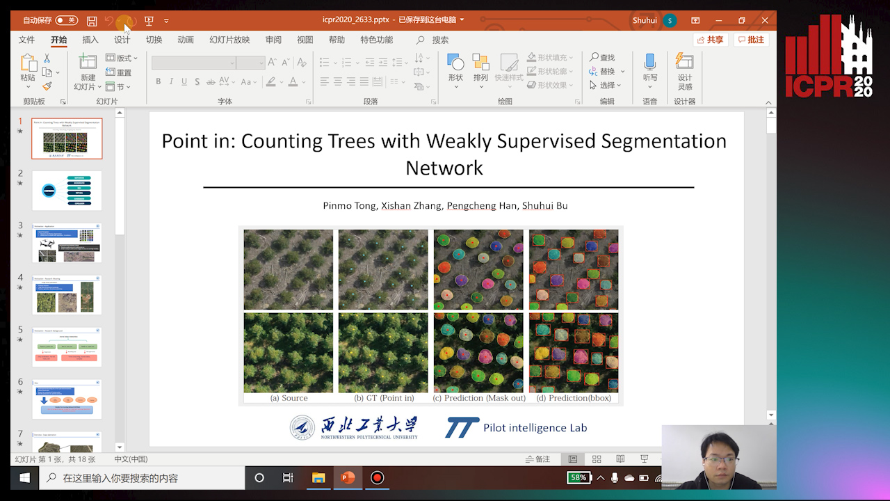
Auto-TLDR; Weakly Tree counting using Deep Segmentation Network with Localization and Mask Prediction

Slides Poster Similar

For tree counting tasks, since traditional image processing methods require expensive feature engineering and are not end-to-end frameworks, this will cause additional noise and cannot be optimized overall, so this method has not been widely used in recent trends of tree counting application. Recently, many deep learning based approaches are designed for this task because of the powerful feature extracting ability. The representative way is bounding box based supervised method, but time-consuming annotations are indispensable for them. Moreover, these methods are difficult to overcome the occlusion or overlap. To solve this problem, we propose a weakly tree counting network (WTCNet) based on deep segmentation network with only point supervision. It can simultaneously complete tree counting with localization and output mask of each tree at the same time. We first adopt a novel feature extractor network (FENet) to get features of input images, and then an effective strategy is introduced to deal with different mask predictions. In the end, we propose a basic localization guidance accompany with rectification guidance to train the network. We create two different datasets and select an existing challenging plant dataset to evaluate our method on three different tasks. Experimental results show the good performance improvement of our method compared with other existing methods. Further study shows that our method has great potential to reduce human labor and provide effective ground-truth masks and the results show the superiority of our method over the advanced methods.

Expectation-Maximization for Scheduling Problems in Satellite Communication

Werner Bailer, Martin Winter, Johannes Ebert, Joel Flavio, Karin Plimon

Responsive image

Auto-TLDR; Unsupervised Machine Learning for Satellite Communication Using Expectation-Maximization

Slides Poster Similar

In this paper we address unsupervised machine learning for two use cases in satellite communication, which are scheduling problems: (i) Ka-band frequency plan optimization and (ii) dynamic configuration of an active antenna array satellite. We apply approaches based on the Expectation-Maximization (EM) framework to both of them. We compare against baselines of currently deployed solutions, and show that they can be significantly outperformed by the EM-based approach. In addition, the approaches can be applied incrementally, thus supporting fast adaptation to small changes in the input configuration.

Estimation of Abundance and Distribution of SaltMarsh Plants from Images Using Deep Learning

Jayant Parashar, Suchendra Bhandarkar, Jacob Simon, Brian Hopkinson, Steven Pennings

Responsive image

Auto-TLDR; CNN-based approaches to automated plant identification and localization in salt marsh images

Poster Similar

Recent advances in computer vision and machine learning, most notably deep convolutional neural networks (CNNs), are exploited to identify and localize various plant species in salt marsh images. Three different approaches are explored that provide estimations of abundance and spatial distribution at varying levels of granularity in terms of spatial resolution. In the coarsest-grained approach, CNNs are tasked with identifying which of six plant species are present/absent in large patches within the salt marsh images. CNNs with diverse topological properties and attention mechanisms are shown capable of providing accurate estimations with >90 % precision and recall in the case of the more abundant plant species whereas the performance declines for less common plant species. Estimation of percent cover of each plant species is performed at a finer spatial resolution, where smaller image patches are extracted and the CNNs tasked with identifying the plant species or substrate at the center of the image patch. For the percent cover estimation task, the CNNs are observed to exhibit a performance profile similar to that for the presence/absence estimation task, but with an ~ 5-10% reduction in precision and recall. Finally, fine-grained estimation of the spatial distribution of the various plant species is performed via semantic segmentation. The Deeplab-V3 semantic segmentation architecture is observed to provide very accurate estimations for abundant plant species; however,a significant degradation in performance is observed in the case of less abundant plant species and, in extreme cases, rare plant classes are seen to be ignored entirely. Overall, a clear trade-off is observed between the CNN estimation quality and the spatial resolution of the underlying estimation thereby offering guidance for ecological applications of CNN-based approaches to automated plant identification and localization in salt marsh images.

Few Shot Learning Framework to Reduce Inter-Observer Variability in Medical Images

Sohini Roychowdhury

Responsive image

Auto-TLDR; Few-Shot Learning for Quality Image Annotation

Slides Poster Similar

Most computer aided pathology detection systems rely on large volumes of quality annotated data to aid diagnostics and follow up procedures. However, quality assuring large volumes of annotated medical image data can be subjective and expensive. In this work we present a novel standardization framework that implements three few-shot learning (FSL) models that can be iteratively trained by atmost 5 images per 3D stack to generate multiple regional proposals (RPs) per test image. These FSL models include a novel parallel echo state network framework and an augmented U-net model. Additionally, we propose a novel target label selection algorithm (TLSA) that measures relative agreeability between RPs and the manually annotated target labels to detect the ``best" quality annotation per image. Using the FSL models, our system achieves 0.28-0.64 Dice coefficient across vendor image stacks for intra-retinal cyst segmentation. Additionally, the TLSA is capable of correctly classifying high quality target labels from their noisy counterparts with 70-97% accuracy. The proposed system significantly automates high quality annotation selection on an image level while minimizing manual quality checking to 12-28% of the images only. Thus, the proposed framework is flexible to extensions for quality image annotation curation of other image stacks as well.

Learn to Segment Retinal Lesions and Beyond

Qijie Wei, Xirong Li, Weihong Yu, Xiao Zhang, Yongpeng Zhang, Bojie Hu, Bin Mo, Di Gong, Ning Chen, Dayong Ding, Youxin Chen

Responsive image

Auto-TLDR; Multi-task Lesion Segmentation and Disease Classification for Diabetic Retinopathy Grading

Poster Similar

Towards automated retinal screening, this paper makes an endeavor to simultaneously achieve pixel-level retinal lesion segmentation and image-level disease classification. Such a multi-task approach is crucial for accurate and clinically interpretable disease diagnosis. Prior art is insufficient due to three challenges, i.e., lesions lacking objective boundaries, clinical importance of lesions irrelevant to their size, and the lack of one-to-one correspondence between lesion and disease classes. This paper attacks the three challenges in the context of diabetic retinopathy (DR) grading. We propose Lesion-Net, a new variant of fully convolutional networks, with its expansive path re- designed to tackle the first challenge. A dual Dice loss that leverages both semantic segmentation and image classification losses is introduced to resolve the second challenge. Lastly, we build a multi-task network that employs Lesion-Net as a side- attention branch for both DR grading and result interpretation. A set of 12K fundus images is manually segmented by 45 ophthalmologists for 8 DR-related lesions, resulting in 290K manual segments in total. Extensive experiments on this large- scale dataset show that our proposed approach surpasses the prior art for multiple tasks including lesion segmentation, lesion classification and DR grading.

Multi-Scale Keypoint Matching

Sina Lotfian, Hassan Foroosh

Responsive image

Auto-TLDR; Multi-Scale Keypoint Matching Using Multi-Scale Information

Slides Poster Similar

We propose a new hierarchical method to match keypoints by exploiting information across multiple scales. Traditionally, for each keypoint a single scale is detected and the matching process is done in the specific scale. We replace this approach with matching across scale-space. The holistic information from higher scales are used for early rejection of candidates that are far away in the feature space. The more localized and finer details of lower scale are then used to decide between remaining possible points. The proposed multi-scale solution is more consistent with the multi-scale processing that is present in the human visual system and is therefore biologically plausible. We evaluate our method on several datasets and achieve state of the art accuracy, while significantly outperforming others in extraction time.

BiLuNet: A Multi-Path Network for Semantic Segmentation on X-Ray Images

Van Luan Tran, Huei-Yung Lin, Rachel Liu, Chun-Han Tseng, Chun-Han Tseng

Responsive image

Auto-TLDR; BiLuNet: Multi-path Convolutional Neural Network for Semantic Segmentation of Lumbar vertebrae, sacrum,

Similar

Semantic segmentation and shape detection of lumbar vertebrae, sacrum, and femoral heads from clinical X-ray images are important and challenging tasks. In this paper, we propose a new multi-path convolutional neural network, BiLuNet, for semantic segmentation on X-ray images. The network is capable of medical image segmentation with very limited training data. With the shape fitting of the bones, we can identify the location of the target regions very accurately for lumbar vertebra inspection. We collected our dataset and annotated by doctors for model training and performance evaluation. Compared to the state-of-the-art methods, the proposed technique provides better mIoUs and higher success rates with the same training data. The experimental results have demonstrated the feasibility of our network to perform semantic segmentation for lumbar vertebrae, sacrum, and femoral heads.

BCAU-Net: A Novel Architecture with Binary Channel Attention Module for MRI Brain Segmentation

Yongpei Zhu, Zicong Zhou, Guojun Liao, Kehong Yuan

Responsive image

Auto-TLDR; BCAU-Net: Binary Channel Attention U-Net for MRI brain segmentation

Slides Poster Similar

Recently deep learning-based networks have achieved advanced performance in medical image segmentation. However, the development of deep learning is slow in magnetic resonance image (MRI) segmentation of normal brain tissues. In this paper, inspired by channel attention module, we propose a new architecture, Binary Channel Attention U-Net (BCAU-Net), by introducing a novel Binary Channel Attention Module (BCAM) into skip connection of U-Net, which can take full advantages of the channel information extracted from the encoding path and corresponding decoding path. To better aggregate multi-scale spatial information of the feature map, spatial pyramid pooling (SPP) modules with different pooling operations are used in BCAM instead of original average-pooling and max-pooling operations. We verify this model on two datasets including IBSR and MRBrainS18, and obtain better performance on MRI brain segmentation compared with other methods. We believe the proposed method can advance the performance in brain segmentation and clinical diagnosis.

Joint Supervised and Self-Supervised Learning for 3D Real World Challenges

Antonio Alliegro, Davide Boscaini, Tatiana Tommasi

Responsive image

Auto-TLDR; Self-supervision for 3D Shape Classification and Segmentation in Point Clouds

Slides Similar

Point cloud processing and 3D shape understanding are very challenging tasks for which deep learning techniques have demonstrated great potentials. Still further progresses are essential to allow artificial intelligent agents to interact with the real world. In many practical conditions the amount of annotated data may be limited and integrating new sources of knowledge becomes crucial to support autonomous learning. Here we consider several scenarios involving synthetic and real world point clouds where supervised learning fails due to data scarcity and large domain gaps. We propose to enrich standard feature representations by leveraging self-supervision through a multi-task model that can solve a 3D puzzle while learning the main task of shape classification or part segmentation. An extensive analysis investigating few-shot, transfer learning and cross-domain settings shows the effectiveness of our approach with state-of-the-art results for 3D shape classification and part segmentation.

Neural Machine Registration for Motion Correction in Breast DCE-MRI

Federica Aprea, Stefano Marrone, Carlo Sansone

Responsive image

Auto-TLDR; A Neural Registration Network for Dynamic Contrast Enhanced-Magnetic Resonance Imaging

Slides Poster Similar

Cancer is one of the leading causes of death in the western world, with medical imaging playing a key role for early diagnosis. Focusing on breast cancer, one of the emerging imaging methodologies is Dynamic Contrast Enhanced-Magnetic Resonance Imaging (DCE-MRI). The flip side of using DCE-MRI is in its long acquisition times, often causing the patient to move, resulting in motion artefacts, namely distortions in the acquired image that can affect DCE-MRI analysis. A possible solution consists in the use of Motion Correction Techniques (MCTs), i.e. procedures intended to re-align the post-contrast image to the corresponding pre-contrast (reference) one. This task is particularly critic in DCE-MRI, due to brightness variations introduced in post-contrast images by the contrast-agent flowing. To face this problem, in this work we introduce a new MCT for breast DCE-MRI leveraging Physiologically Based PharmacoKinetic (PBPK) modelling and Artificial Neural Networks (ANN) to determine the most suitable physiologically-compliant transformation. To this aim, we propose a Neural Registration Network relying on a very task-specific loss function explicitly designed to take into account the contrast agent flowing while enforcing a correct re-alignment. We compared the obtained results against some conventional motion correction techniques, evaluating the performance on a patient-by-patient basis. Results clearly show the effectiveness of the proposed approach, resulting as the best performing even when compares against other techniques designed to take into account for brightness variations.

Holistic Grid Fusion Based Stop Line Estimation

Runsheng Xu, Faezeh Tafazzoli, Li Zhang, Timo Rehfeld, Gunther Krehl, Arunava Seal

Responsive image

Auto-TLDR; Fused Multi-Sensory Data for Stop Lines Detection in Intersection Scenarios

Slides Similar

Intersection scenarios provide the most complex traffic situations in Autonomous Driving and Driving Assistance Systems. Knowing where to stop in advance in an intersection is an essential parameter in controlling the longitudinal velocity of the vehicle. Most of the existing methods in literature solely use cameras to detect stop lines, which is typically not sufficient in terms of detection range. To address this issue, we propose a method that takes advantage of fused multi-sensory data including stereo camera and lidar as input and utilizes a carefully designed convolutional neural network architecture to detect stop lines. Our experiments show that the proposed approach can improve detection range compared to camera data alone, works under heavy occlusion without observing the ground markings explicitly, is able to predict stop lines for all lanes and allows detection at a distance up to 50 meters.

Uncertainty Guided Recognition of Tiny Craters on the Moon

Thorsten Wilhelm, Christian Wöhler

Responsive image

Auto-TLDR; Accurately Detecting Tiny Craters in Remote Sensed Images Using Deep Neural Networks

Slides Poster Similar

Accurately detecting craters in remotely sensed images is an important task when analysing the properties of planetary bodies. Commonly, only large craters in the range of several kilometres are detected. In this work we provide the first example of automatically detecting tiny craters in the range of several meters with the help of a deep neural network by using only a small set of annotated craters. Additionally, we propose a novel way to group overlapping detections and replace the commonly used non-maximum suppression with a probabilistic treatment. As a result, we receive valuable uncertainty estimates of the detections and the aggregated detections are shown to be vastly superior.

A Riemannian Framework for Detecting Stimulus-Relevant Fiber Pathways

Jingyong Su, Linlin Tang, Zhipeng Yang, Mengmeng Guo

Responsive image

Auto-TLDR; Clustering Task-Specific Fiber Pathways in Functional MRI using BOLD Signals

Poster Similar

Functional MRI based on blood oxygenation level-dependent (BOLD) contrast is well established as a neuro-imaging technique for detecting neural activity in the cortex of the human brain. Recent studies have shown that variations of BOLD signals in white matter are also related to neural activities both in resting state and under functional loading. We develop a comprehensive framework of detecting task-specific fiber pathways. We not only study fiber tracts as open curves with different physical features (shape, scale, orientation and position), but also incorporate the BOLD signals transmitted along them to find stimulus-relevant pathways. Specifically, we propose a novel Riemannian metric, which is a weighted sum of distances in product space of shapes and functions. This metric provides both a cost function for registration and a proper distance for comparison. Experimental results on real data have shown that we can cluster fiber pathways correctly by evaluating correlations between BOLD signals and stimuli, temporal variations and power spectra of them. The proposed framework can also be easily generalized to various applications where multi-modality data exist.

Deep Learning on Active Sonar Data Using Bayesian Optimization for Hyperparameter Tuning

Henrik Berg, Karl Thomas Hjelmervik

Responsive image

Auto-TLDR; Bayesian Optimization for Sonar Operations in Littoral Environments

Slides Poster Similar

Sonar operations in littoral environments may be challenging due to an increased probability of false alarms. Machine learning can be used to train classifiers that are able to filter out most of the false alarms automatically, however, this is a time consuming process, with many hyperparameters that need to be tuned in order to yield useful results. In this paper, Bayesian optimization is used to search for good values for some of the hyperparameters, like topology and training parameters, resulting in performance superior to earlier trial-and-error based training. Additionally, we analyze some of the parameters involved in the Bayesian optimization, as well as the resulting hyperparameter values.

Anime Sketch Colorization by Component-Based Matching Using Deep Appearance Features and Graph Representation

Thien Do, Pham Van, Anh Nguyen, Trung Dang, Quoc Nguyen, Bach Hoang, Giao Nguyen

Responsive image

Auto-TLDR; Combining Deep Learning and Graph Representation for Sketch Colorization

Slides Poster Similar

Sketch colorization is usually expensive and time-consuming for artists, and automating this process can have many pragmatic applications in the animation, comic book, and video game industry. However, automatic image colorization faces many challenges, because sketches not only lack texture information but also potentially entail complicated objects that require acute coloring. These difficulties usually result in incorrect color assignments that can ruin the aesthetic appeal of the final output. In this paper, we present a novel component-based matching framework that combines deep learned features and quadratic programming {\color{red} with a new cost function} to solve this colorization problem. The proposed framework inputs a character's sketches as well as a colored image in the same cut of a movie, and outputs a high-quality sequence of colorized frames based on the color assignment in the reference colored image. To carry out this colorization task, we first utilize a pretrained ResNet-34 model to extract elementary components' features to match certain pairs of components (one component from the sketch and one from reference). Next, a graph representation is constructed in order to process and match the remaining components that could not be done in the first step. Since the first step has reduced the number of components to be matched by the graph, we can solve this graph problem in a short computing time even when there are hundreds of different components present in each sketch. We demonstrate the effectiveness of the proposed solution by conducting comprehensive experiments and producing aesthetically pleasing results. To the best of our knowledge, our framework is the first work that combines deep learning and graph representation to colorize anime and achieves a high pixel-level accuracy at a reasonable time cost.