Vesselness Filters: A Survey with Benchmarks Applied to Liver Imaging

Jonas Lamy, Odyssée Merveille, Bertrand Kerautret, Nicolas Passat, Antoine Vacavant

Responsive image

Auto-TLDR; Comparison of Vessel Enhancement Filters for Liver Vascular Network Segmentation

Slides Poster

The accurate knowledge of vascular network geometry is crucial for many clinical applications such as cardiovascular disease diagnosis and surgery planning. Vessel enhancement algorithms are often a key step to improve the robustness of vessel segmentation. A wide variety of enhancement filters exists in the literature, but they are often difficult to compare as the applications and datasets differ from a paper to another and the code is rarely available. In this article, we compare seven vessel enhancement filters covering the last twenty years literature in a unique common framework. We focus our study on the liver vascular network which is under-represented in the literature. The evaluation is made from three points of view: in the whole liver, in the vessel neighborhood and near the bifurcations. The study is performed on two publicly available datasets: the Ircad dataset (CT images) and the VascuSynth dataset adapted for MRI simulation. We discuss the strengths and weaknesses of each method in the hepatic context. In addition, the benchmark framework including a C++ implementation of each compared method is provided. An online demonstration ensures the reproducibility of the results without requiring any additional software.

Similar papers

A Benchmark Dataset for Segmenting Liver, Vasculature and Lesions from Large-Scale Computed Tomography Data

Bo Wang, Zhengqing Xu, Wei Xu, Qingsen Yan, Liang Zhang, Zheng You

Responsive image

Auto-TLDR; The Biggest Treatment-Oriented Liver Cancer Dataset for Segmentation

Slides Poster Similar

How to build a high-performance liver-related computer assisted diagnosis system is an open question of great interest. However, the performance of the state-of-art algorithm is always limited by the amount of data and quality of the label. To address this problem, we propose the biggest treatment-oriented liver cancer dataset for liver surgery and treatment planning. This dataset provides 216 cases (totally about 268K frames) scanned images in contrast-enhanced computed tomography (CT). We labeled all the CT images with the liver, liver vasculature and liver tumor segmentation ground truth for train and tune segmentation algorithms in advance. Based on that, we evaluate several recent and state-of-the-art segmentation algorithms, including 7 deep learning methods, on CT sequences. All results are compared to reference segmentations five error metrics that highlight different aspects of segmentation accuracy. In general, compared with previous datasets, our dataset is really a challenging dataset. To our knowledge, the proposed dataset and benchmark allow for the first time systematic exploration of such issues, and will be made available to allow for further research in this field.

Segmentation of Axillary and Supraclavicular Tumoral Lymph Nodes in PET/CT: A Hybrid CNN/Component-Tree Approach

Diana Lucia Farfan Cabrera, Nicolas Gogin, David Morland, Benoît Naegel, Dimitri Papathanassiou, Nicolas Passat

Responsive image

Auto-TLDR; Coupling Convolutional Neural Networks and Component-Trees for Lymph node Segmentation from PET/CT Images

Slides Similar

The analysis of axillary and supraclavicular lymph nodes is a primary prognostic factor for the staging of breast cancer. However, due to the size of lymph nodes and the low resolution of PET data, their segmentation is challenging. We investigate the relevance of considering axillary and supraclavicular lymph node segmentation from PET/CT images by coupling Convolutional Neural Networks (CNNs) and Component-Trees (C-Trees). Building upon the U-Net architecture, we propose a framework that couples a multi-modal U-Net fed with PET and CT, coupled with a hierarchical model obtained from the PET that provides additional high-level region-based features as input channels. Our working hypotheses are twofold. First, we take advantage of both anatomical information from CT for detecting the nodes, and from functional information from PET for detecting the pathological ones. Second, we consider region-based attributes extracted from C-Tree analysis of 3D PET/CT images to improve the CNN segmentation. We carried out experiments on a dataset of 240 pathological lymph nodes from 52 patients scans, and compared our outputs with human expert-defined ground-truth, leading to promising results.

Segmentation of Intracranial Aneurysm Remnant in MRA Using Dual-Attention Atrous Net

Subhashis Banerjee, Ashis Kumar Dhara, Johan Wikström, Robin Strand

Responsive image

Auto-TLDR; Dual-Attention Atrous Net for Segmentation of Intracranial Aneurysm Remnant from MRA Images

Slides Poster Similar

Due to the advancement of non-invasive medical imaging modalities like Magnetic Resonance Angiography (MRA), an increasing number of Intracranial Aneurysm (IA) cases are being reported in recent years. The IAs are typically treated by so-called endovascular coiling, where blood flow in the IA is prevented by embolization with a platinum coil. Accurate quantification of the IA Remnant (IAR), i.e. the volume with blood flow present post treatment is the utmost important factor in choosing the right treatment planning. This is typically done by manually segmenting the aneurysm remnant from the MRA volume. Since manual segmentation of volumetric images is a labour-intensive and error-prone process, development of an automatic volumetric segmentation method is required. Segmentation of small structures such as IA, that may largely vary in size, shape, and location is considered extremely difficult. Similar intensity distribution of IAs and surrounding blood vessels makes it more challenging and susceptible to false positive. In this paper we propose a novel 3D CNN architecture called Dual-Attention Atrous Net (DAtt-ANet), which can efficiently segment IAR volumes from MRA images by reconciling features at different scales using the proposed Parallel Atrous Unit (PAU) along with the use of self-attention mechanism for extracting fine-grained features and intra-class correlation. The proposed DAtt-ANet model is trained and evaluated on a clinical MRA image dataset (prospective research project, approved by the local ethical committee) of IAR consisting of 46 subjects, annotated by an expert radiologist from our group. We compared the proposed DAtt-ANet with five state-of-the-art CNN models based on their segmentation performance. The proposed DAtt-ANet outperformed all other methods and was able to achieve a five-fold cross-validation DICE score of $0.73\pm0.06$.

Transfer Learning through Weighted Loss Function and Group Normalization for Vessel Segmentation from Retinal Images

Abdullah Sarhan, Jon Rokne, Reda Alhajj, Andrew Crichton

Responsive image

Auto-TLDR; Deep Learning for Segmentation of Blood Vessels in Retinal Images

Slides Poster Similar

The vascular structure of blood vessels is important in diagnosing retinal conditions such as glaucoma and diabetic retinopathy. Accurate segmentation of these vessels can help in detecting retinal objects such as the optic disc and optic cup and hence determine if there are damages to these areas. Moreover, the structure of the vessels can help in diagnosing glaucoma. The rapid development of digital imaging and computer-vision techniques has increased the potential for developing approaches for segmenting retinal vessels. In this paper, we propose an approach for segmenting retinal vessels that uses deep learning along with transfer learning. We adapted the U-Net structure to use a customized InceptionV3 as the encoder and used multiple skip connections to form the decoder. Moreover, we used a weighted loss function to handle the issue of class imbalance in retinal images. Furthermore, we contributed a new dataset to this field. We tested our approach on six publicly available datasets and a newly created dataset. We achieved an average accuracy of 95.60\% and a Dice coefficient of 80.98\%. The results obtained from comprehensive experiments demonstrate the robustness of our approach to the segmentation of blood vessels in retinal images obtained from different sources. Our approach results in greater segmentation accuracy than other approaches.

A Novel Computer-Aided Diagnostic System for Early Assessment of Hepatocellular Carcinoma

Ahmed Alksas, Mohamed Shehata, Gehad Saleh, Ahmed Shaffie, Ahmed Soliman, Mohammed Ghazal, Hadil Abukhalifeh, Abdel Razek Ahmed, Ayman El-Baz

Responsive image

Auto-TLDR; Classification of Liver Tumor Lesions from CE-MRI Using Structured Structural Features and Functional Features

Slides Poster Similar

Early assessment of liver cancer patients with hepatocellular carcinoma (HCC) is of immense importance to provide the proper treatment plan. In this paper, we have developed a two-stage classification computer-aided diagnostic (CAD) system that has the ability to detect and grade the liver observations from multiphase contrast enhanced magnetic resonance imaging (CE-MRI). The proposed approach consists of three main steps. First, a pre-processing is applied to the CE-MRI scans to delineate the tumor lesions that will be used as an ROI across the four different phases of the CE-MRI, (namely, the pre-contrast, late-arterial, portal-venous, and delayed-contrast). Second, a group of three features are modeled to provide a quantitative discrimination between the tumor lesions; namely: i) the tumor appearance that is modeled using a set of texture features, (namely; the first-order histogram, second-order gray-level co-occurrence matrix, and second-order gray-level run-length matrix), to capture any discrimination that may appear in the lesion texture, ii) the spherical harmonics (SH) based shape features that have the ability to describe the shape complexity of the liver tumors, and iii) the functional features that are based on the calculation of the wash-in/wash-out through that evaluate the intensity changes across the post-contrast phases. Finally, the aforementioned individual features were then integrated together to obtain the combined features to be fed to a machine learning classifier towards getting the final diagnostic decision. The proposed CAD system has been tested using hepatic observations that was obtained from 85 participating patients, 34 patients with benign tumors, 34 patients with intermediate tumors and 34 with malignant tumors. Using a random forests based classifier with a leave-one-subject-out (LOSO) cross-validation, the developed CAD system achieved an 87.1% accuracy in distinguishing the malignant, intermediate and benign tumors. The classification performance is then evaluated using k-fold (5/10-fold) cross-validation approach to examine the robustness of the system. The LR-1 lesions were classified from LR-2 benign lesions with 91.2% accuracy, while 85.3% accuracy was achieved differentiating between LR-4 and LR-5 malignant tumors. The obtained results hold a promise of the proposed framework to be reliably used as a noninvasive diagnostic tool for the early detection and grading of liver cancer tumors.

Automatic Semantic Segmentation of Structural Elements related to the Spinal Cord in the Lumbar Region by Using Convolutional Neural Networks

Jhon Jairo Sáenz Gamboa, Maria De La Iglesia-Vaya, Jon Ander Gómez

Responsive image

Auto-TLDR; Semantic Segmentation of Lumbar Spine Using Convolutional Neural Networks

Slides Poster Similar

This work addresses the problem of automatically segmenting the MR images corresponding to the lumbar spine. The purpose is to detect and delimit the different structural elements like vertebrae, intervertebral discs, nerves, blood vessels, etc. This task is known as semantic segmentation. The approach proposed in this work is based on convolutional neural networks whose output is a mask where each pixel from the input image is classified into one of the possible classes. Classes were defined by radiologists and correspond to structural elements and tissues. The proposed network architectures are variants of the U-Net. Several complementary blocks were used to define the variants: spatial attention models, deep supervision and multi-kernels at input, this last block type is based on the idea of inception. Those architectures which got the best results are described in this paper, and their results are discussed. Two of the proposed architectures outperform the standard U-Net used as baseline.

Multi-focus Image Fusion for Confocal Microscopy Using U-Net Regression Map

Md Maruf Hossain Shuvo, Yasmin M. Kassim, Filiz Bunyak, Olga V. Glinskii, Leike Xie, Vladislav V Glinsky, Virginia H. Huxley, Kannappan Palaniappan

Responsive image

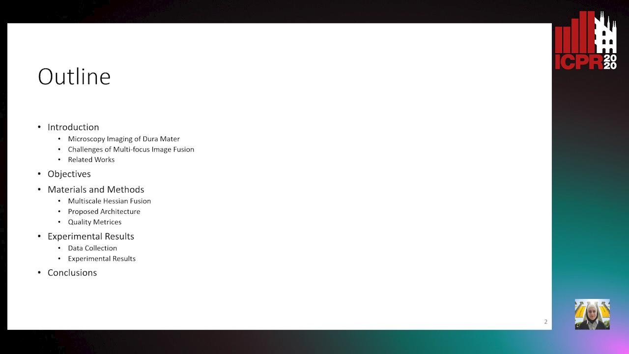
Auto-TLDR; Independent Single Channel U-Net Fusion for Multi-focus Microscopy Images

Slides Poster Similar

Multi-focus image fusion plays an important role to better visualize the detailed information and anatomical structures of microscopy images. We propose a new approach to fuse all single-focus microscopy images in each Z-stack. As the structures are different in different channels, input images are separated into red and green channels. Red for blood vessels, and green for lymphatics like structures . Taking the maximum likelihood of U-Net regression likelihood map along Z, we obtain the focus selection map for each channel. We named this approach as Independent Single Channel U-Net (ISCU) fusion. We combined each channel fusion result to get the final dual channel composite RGB image. The dataset used is extremely challenging with complex microscopy images of mice dura mater attached to bone. We compared our results with one of the popular and widely used derivative based fusion method [7] using multiscale Hessian. We found that multiscale Hessian-based approach produces banding effects with nonhomogeneous background lacking detailed anatomical structures. So, we took the advantages of Convolutional Neural Network (CNN), and used the U-Net regression likelihood map to fuse the images. Perception based no-reference image quality assessment parameters like PIQUE, NIQE, and BRISQUE confirms the effectiveness of the proposed method.

PCANet: Pyramid Context-Aware Network for Retinal Vessel Segmentation

Yi Zhang, Yixuan Chen, Kai Zhang

Responsive image

Auto-TLDR; PCANet: Adaptive Context-Aware Network for Automated Retinal Vessel Segmentation

Slides Poster Similar

Automated retinal vessel segmentation plays an important role in the diagnosis of some diseases such as diabetes, arteriosclerosis and hypertension. Recent works attempt to improve segmentation performance by exploring either global or local contexts. However, the context demands are varying from regions in each image and different levels of network. To address these problems, we propose Pyramid Context-aware Network (PCANet), which can adaptively capture multi-scale context representations. Specifically, PCANet is composed of multiple Adaptive Context-aware (ACA) blocks arranged in parallel, each of which can adaptively obtain the context-aware features by estimating affinity coefficients at a specific scale under the guidance of global contextual dependencies. Meanwhile, we import ACA blocks with specific scales in different levels of the network to obtain a coarse-to-fine result. Furthermore, an integrated test-time augmentation method is developed to further boost the performance of PCANet. Finally, extensive experiments demonstrate the effectiveness of the proposed PCANet, and state-of-the-art performances are achieved with AUCs of 0.9866, 0.9886 and F1 Scores of 0.8274, 0.8371 on two public datasets, DRIVE and STARE, respectively.

A Deep Learning Approach for the Segmentation of Myocardial Diseases

Khawala Brahim, Abdull Qayyum, Alain Lalande, Arnaud Boucher, Anis Sakly, Fabrice Meriaudeau

Responsive image

Auto-TLDR; Segmentation of Myocardium Infarction Using Late GADEMRI and SegU-Net

Slides Poster Similar

Cardiac left ventricular (LV) segmentation is of paramount essential step for both diagnosis and treatment of cardiac pathologies such as ischemia, myocardial infarction, arrhythmia and myocarditis. However, this segmentation is challenging due to high variability across patients and the potential lack of contrast between structures. In this work, we propose and evaluate a (2.5D) SegU-Net model based on the fusion of two deep learning techniques (U-Net and Seg-Net) for automated LGEMRI (Late gadolinium enhanced magnetic resonance imaging) myocardial disease (infarct core and no reflow region) quantification in a new multifield expert annotated dataset. Given that the scar tissue represents a small part of the whole MRI slices, we focused on myocardium area. Segmentation results show that this preprocessing step facilitate the learning procedure. In order to solve the class imbalance problem, we propose to apply the Jaccard loss and the Focal Loss as optimization loss function and to integrate a class weights strategy into the objective function. Late combination has been used to merge the output of the best trained models on a different set of hyperparameters. The final network segmentation performances will be useful for future comparison of new method to the current related work for this task. A total number of 2237 of slices (320 cases) were used for training/validation and 210 slices (35 cases) were used for testing. Experiments over our proposed dataset, using several evaluation metrics such Jaccard distance (IOU), Accuracy and Dice similarity coefficient (DSC), demonstrate efficiency performance in quantifying different zones of myocardium infarction across various patients. As compared to the second intra-observer study, our testing results showed that the SegUNet prediction model leads to these average dice coefficients over all segmented tissue classes, respectively : 'Background': 0.99999, 'Myocardium': 0.99434, 'Infarctus': 0.95587, 'Noreflow': 0.78187.

NephCNN: A Deep-Learning Framework for Vessel Segmentation in Nephrectomy Laparoscopic Videos

Alessandro Casella, Sara Moccia, Chiara Carlini, Emanuele Frontoni, Elena De Momi, Leonardo Mattos

Responsive image

Auto-TLDR; Adversarial Fully Convolutional Neural Networks for kidney vessel segmentation from nephrectomy laparoscopic videos

Slides Poster Similar

Objective: In the last years, Robot-assisted partial nephrectomy (RAPN) is establishing as elected treatment for renal cell carcinoma (RCC). Reduced field of view, field occlusions by surgical tools, and reduced maneuverability may potentially cause accidents, such as unwanted vessel resection with consequent bleeding. Surgical Data Science (SDS) can provide effective context-aware tools for supporting surgeons. However, currently no tools have been exploited for automatic vessels segmentation from nephrectomy laparoscopic videos. Herein, we propose a new approach based on adversarial Fully Convolutional Neural Networks (FCNNs) to kidney vessel segmentation from nephrectomy laparoscopic vision. Methods: The proposed approach enhances existing segmentation framework by (i) encoding 3D kernels for spatio-temporal features extraction to enforce pixel connectivity in time, and (ii) perform training in adversarial fashion, which constrains vessels shape. Results: We performed a preliminary study using 8 different RAPN videos (1871 frames), the first in the field, achieving a median Dice Similarity Coefficient of 71.76%. Conclusions: Results showed that the proposed approach could be a valuable solution with a view to assist surgeon during RAPN.

FOANet: A Focus of Attention Network with Application to Myocardium Segmentation

Zhou Zhao, Elodie Puybareau, Nicolas Boutry, Thierry Geraud

Responsive image

Auto-TLDR; FOANet: A Hybrid Loss Function for Myocardium Segmentation of Cardiac Magnetic Resonance Images

Slides Poster Similar

In myocardium segmentation of cardiac magnetic resonance images, ambiguities often appear near the boundaries of the target domains due to tissue similarities. To address this issue, we propose a new architecture, called FOANet, which can be decomposed in three main steps: a localization step, a Gaussian-based contrast enhancement step, and a segmentation step. This architecture is supplied with a hybrid loss function that guides the FOANet to study the transformation relationship between the input image and the corresponding label in a threelevel hierarchy (pixel-, patch- and map-level), which is helpful to improve segmentation and recovery of the boundaries. We demonstrate the efficiency of our approach on two public datasets in terms of regional and boundary segmentations.

3D Medical Multi-Modal Segmentation Network Guided by Multi-Source Correlation Constraint

Tongxue Zhou, Stéphane Canu, Pierre Vera, Su Ruan

Responsive image

Auto-TLDR; Multi-modality Segmentation with Correlation Constrained Network

Slides Poster Similar

In the field of multimodal segmentation, the correlation between different modalities can be considered for improving the segmentation results. In this paper, we propose a multi-modality segmentation network with a correlation constraint. Our network includes N model-independent encoding paths with N image sources, a correlation constrain block, a feature fusion block, and a decoding path. The model-independent encoding path can capture modality-specific features from the N modalities. Since there exists a strong correlation between different modalities, we first propose a linear correlation block to learn the correlation between modalities, then a loss function is used to guide the network to learn the correlated features based on the correlation representation block. This block forces the network to learn the latent correlated features which are more relevant for segmentation. Considering that not all the features extracted from the encoders are useful for segmentation, we propose to use dual attention based fusion block to recalibrate the features along the modality and spatial paths, which can suppress less informative features and emphasize the useful ones. The fused feature representation is finally projected by the decoder to obtain the segmentation result. Our experiment results tested on BraTS-2018 dataset for brain tumor segmentation demonstrate the effectiveness of our proposed method.

DARN: Deep Attentive Refinement Network for Liver Tumor Segmentation from 3D CT Volume

Yao Zhang, Jiang Tian, Cheng Zhong, Yang Zhang, Zhongchao Shi, Zhiqiang He

Responsive image

Auto-TLDR; Deep Attentive Refinement Network for Liver Tumor Segmentation from 3D Computed Tomography Using Multi-Level Features

Slides Poster Similar

Automatic liver tumor segmentation from 3D Computed Tomography (CT) is a necessary prerequisite in the interventions of hepatic abnormalities and surgery planning. However, accurate liver tumor segmentation remains challenging due to the large variability of tumor sizes and inhomogeneous texture. Recent advances based on Fully Convolutional Network (FCN) in liver tumor segmentation draw on success of learning discriminative multi-level features. In this paper, we propose a Deep Attentive Refinement Network (DARN) for improved liver tumor segmentation from CT volumes by fully exploiting both low and high level features embedded in different layers of FCN. Different from existing works, we exploit attention mechanism to leverage the relation of different levels of features encoded in different layers of FCN. Specifically, we introduce a Semantic Attention Refinement (SemRef) module to selectively emphasize global semantic information in low level features with the guidance of high level ones, and a Spatial Attention Refinement (SpaRef) module to adaptively enhance spatial details in high level features with the guidance of low level ones. We evaluate our network on the public MICCAI 2017 Liver Tumor Segmentation Challenge dataset (LiTS dataset) and it achieves state-of-the-art performance. The proposed refinement modules are an effective strategy to exploit multi-level features and has great potential to generalize to other medical image segmentation tasks.

BCAU-Net: A Novel Architecture with Binary Channel Attention Module for MRI Brain Segmentation

Yongpei Zhu, Zicong Zhou, Guojun Liao, Kehong Yuan

Responsive image

Auto-TLDR; BCAU-Net: Binary Channel Attention U-Net for MRI brain segmentation

Slides Poster Similar

Recently deep learning-based networks have achieved advanced performance in medical image segmentation. However, the development of deep learning is slow in magnetic resonance image (MRI) segmentation of normal brain tissues. In this paper, inspired by channel attention module, we propose a new architecture, Binary Channel Attention U-Net (BCAU-Net), by introducing a novel Binary Channel Attention Module (BCAM) into skip connection of U-Net, which can take full advantages of the channel information extracted from the encoding path and corresponding decoding path. To better aggregate multi-scale spatial information of the feature map, spatial pyramid pooling (SPP) modules with different pooling operations are used in BCAM instead of original average-pooling and max-pooling operations. We verify this model on two datasets including IBSR and MRBrainS18, and obtain better performance on MRI brain segmentation compared with other methods. We believe the proposed method can advance the performance in brain segmentation and clinical diagnosis.

A Comparison of Neural Network Approaches for Melanoma Classification

Maria Frasca, Michele Nappi, Michele Risi, Genoveffa Tortora, Alessia Auriemma Citarella

Responsive image

Auto-TLDR; Classification of Melanoma Using Deep Neural Network Methodologies

Slides Poster Similar

Melanoma is the deadliest form of skin cancer and it is diagnosed mainly visually, starting from initial clinical screening and followed by dermoscopic analysis, biopsy and histopathological examination. A dermatologist’s recognition of melanoma may be subject to errors and may take some time to diagnose it. In this regard, deep learning can be useful in the study and classification of skin cancer. In particular, by classifying images with Deep Neural Network methodologies, it is possible to obtain comparable or even superior results compared to those of dermatologists. In this paper, we propose a methodology for the classification of melanoma by adopting different deep learning techniques applied to a common dataset, composed of images from the ISIC dataset and consisting of different types of skin diseases, including melanoma on which we applied a specific pre-processing phase. In particular, a comparison of the results is performed in order to select the best effective neural network to be applied to the problem of recognition and classification of melanoma. Moreover, we also evaluate the impact of the pre- processing phase on the final classification. Different metrics such as accuracy, sensitivity, and specificity have been selected to assess the goodness of the adopted neural networks and compare them also with the manual classification of dermatologists.

SAGE: Sequential Attribute Generator for Analyzing Glioblastomas Using Limited Dataset

Padmaja Jonnalagedda, Brent Weinberg, Jason Allen, Taejin Min, Shiv Bhanu, Bir Bhanu

Responsive image

Auto-TLDR; SAGE: Generative Adversarial Networks for Imaging Biomarker Detection and Prediction

Slides Poster Similar

While deep learning approaches have shown remarkable performance in many imaging tasks, most of these methods rely on availability of large quantities of data. Medical image data, however, is scarce and fragmented. Generative Adversarial Networks (GANs) have recently been very effective in handling such datasets by generating more data. If the datasets are very small, however, GANs cannot learn the data distribution properly, resulting in less diverse or low-quality results. One such limited dataset is that for the concurrent gain of 19/20 chromosomes (19/20 co-gain), a mutation with positive prognostic value in Glioblastomas (GBM). In this paper, we detect imaging biomarkers for the mutation to streamline the extensive and invasive prognosis pipeline. Since this mutation is relatively rare, i.e. small dataset, we propose a novel generative framework – the Sequential Attribute GEnerator (SAGE), that generates detailed tumor imaging features while learning from a limited dataset. Experiments show that not only does SAGE generate high quality tumors when compared to standard Deep Convolutional GAN (DC-GAN) and Wasserstein GAN with Gradient Penalty (WGAN-GP), it also captures the imaging biomarkers accurately.

A Multi-Task Contextual Atrous Residual Network for Brain Tumor Detection & Segmentation

Ngan Le, Kashu Yamazaki, Quach Kha Gia, Thanh-Dat Truong, Marios Savvides

Responsive image

Auto-TLDR; Contextual Brain Tumor Segmentation Using 3D atrous Residual Networks and Cascaded Structures

Poster Similar

In recent years, deep neural networks have achieved state-of-the-art performance in a variety of recognition and segmentation tasks in medical imaging including brain tumor segmentation. We investigate that segmenting brain tumor is facing to the imbalanced data problem where the number of pixels belonging to background class (non tumor pixel) is much larger than the number of pixels belonging to foreground class (tumor pixel). To address this problem, we propose a multi-task network which is formed as a cascaded structure and designed to share the feature maps. Our model consists of two targets, i.e., (i) effectively differentiating brain tumor regions and (ii) estimating brain tumor masks. The first task is performed by our proposed contextual brain tumor detection network, which plays the role of an attention gate and focuses on the region around brain tumor only while ignore the background (non tumor area). Instead of processing every pixel, our contextual brain tumor detection network only processes contextual regions around ground-truth instances and this strategy helps to produce meaningful regions proposals. The second task is built upon a 3D atrous residual network and under an encode-decode network in order to effectively segment both large and small objects (brain tumor). Our 3D atrous residual network is designed with a skip connection to enables the gradient from the deep layers to be directly propagated to shallow layers, thus, features of different depths are preserved and used for refining each other. In order to incorporate larger contextual information in volume MRI data, our network is designed by 3D atrous convolution with various kernel sizes, which enlarges the receptive field of filters. Our proposed network has been evaluated on various datasets including BRATS2015, BRATS2017 and BRATS2018 datasets with both validation set and testing set. Our performance has been benchmarked by both region-based metrics and surface-based metrics. We also have conducted comparisons against state-of-the-art approaches.

Planar 3D Transfer Learning for End to End Unimodal MRI Unbalanced Data Segmentation

Martin Kolarik, Radim Burget, Carlos M. Travieso-Gonzalez, Jan Kocica

Responsive image

Auto-TLDR; Planar 3D Res-U-Net Network for Unbalanced 3D Image Segmentation using Fluid Attenuation Inversion Recover

Slides Similar

We present a novel approach of 2D to 3D transfer learning based on mapping pre-trained 2D convolutional neural network weights into planar 3D kernels. The method is validated by proposed planar 3D res-u-net network with encoder transferred from the 2D VGG-16 which is applied for a single-stage unbalanced 3D image data segmentation. In particular, we evaluate the method on the MICCAI 2016 MS lesion segmentation challenge dataset utilizing solely Fluid Attenuation Inversion Recover (FLAIR) sequence without brain extraction for training and inference to simulate real medical praxis. The planar 3D res-u-net network performed the best both in sensitivity and Dice score amongst end to end methods processing raw MRI scans and achieved comparable Dice score to a state-of-the-art unimodal not end to end approach. Complete source code was released under the open-source license and this paper is in compliance with the Machine learning Reproducibility Checklist. By implementing practical transfer learning for 3D data representation we were able to successfully segment heavily unbalanced data without selective sampling and achieved more reliable results using less training data in single modality. From medical perspective, the unimodal approach gives an advantage in real praxis as it does not require co-registration nor additional scanning time during examination. Although modern medical imaging methods capture high resolution 3D anatomy scans suitable for computer aided detection system processing, deployment of automatic systems for interpretation of radiology imaging is still rather theoretical in many medical areas. Our work aims to bridge the gap offering solution for partial research questions.

Neural Machine Registration for Motion Correction in Breast DCE-MRI

Federica Aprea, Stefano Marrone, Carlo Sansone

Responsive image

Auto-TLDR; A Neural Registration Network for Dynamic Contrast Enhanced-Magnetic Resonance Imaging

Slides Poster Similar

Cancer is one of the leading causes of death in the western world, with medical imaging playing a key role for early diagnosis. Focusing on breast cancer, one of the emerging imaging methodologies is Dynamic Contrast Enhanced-Magnetic Resonance Imaging (DCE-MRI). The flip side of using DCE-MRI is in its long acquisition times, often causing the patient to move, resulting in motion artefacts, namely distortions in the acquired image that can affect DCE-MRI analysis. A possible solution consists in the use of Motion Correction Techniques (MCTs), i.e. procedures intended to re-align the post-contrast image to the corresponding pre-contrast (reference) one. This task is particularly critic in DCE-MRI, due to brightness variations introduced in post-contrast images by the contrast-agent flowing. To face this problem, in this work we introduce a new MCT for breast DCE-MRI leveraging Physiologically Based PharmacoKinetic (PBPK) modelling and Artificial Neural Networks (ANN) to determine the most suitable physiologically-compliant transformation. To this aim, we propose a Neural Registration Network relying on a very task-specific loss function explicitly designed to take into account the contrast agent flowing while enforcing a correct re-alignment. We compared the obtained results against some conventional motion correction techniques, evaluating the performance on a patient-by-patient basis. Results clearly show the effectiveness of the proposed approach, resulting as the best performing even when compares against other techniques designed to take into account for brightness variations.

MTGAN: Mask and Texture-Driven Generative Adversarial Network for Lung Nodule Segmentation

Wei Chen, Qiuli Wang, Kun Wang, Dan Yang, Xiaohong Zhang, Chen Liu, Yucong Li

Responsive image

Auto-TLDR; Mask and Texture-driven Generative Adversarial Network for Lung Nodule Segmentation

Slides Poster Similar

Accurate segmentation for lung nodules in lung computed tomography (CT) scans plays a key role in the early diagnosis of lung cancer. Many existing methods, especially UNet, have made significant progress in lung nodule segmentation. However, due to the complex shapes of lung nodules and the similarity of visual characteristics between nodules and lung tissues, an accurate segmentation with few false positives of lung nodules is still a challenging problem. Considering the fact that both boundary and texture information of lung nodules are important for obtaining an accurate segmentation result, we propose a novel Mask and Texture-driven Generative Adversarial Network (MTGAN) with a joint multi-scale L1 loss for lung nodule segmentation, which takes full advantages of U-Net and adversarial training. The proposed MTGAN leverages adversarial learning strategy guided by the boundary and texture information of lung nodules to generate more accurate segmentation results with lesser false positives. We validate our model with the LIDC–IDRI dataset, and experimental results show that our method achieves excellent segmentation results for a variety of lung nodules, especially for juxtapleural nodules and low-dense nodules. Without any bells and whistles, the proposed MTGAN achieves significant segmentation performance with the Dice similarity coefficient (DSC) of 85.24% on the LIDC–IDRI dataset.

SA-UNet: Spatial Attention U-Net for Retinal Vessel Segmentation

Changlu Guo, Marton Szemenyei, Yugen Yi, Wenle Wang, Buer Chen, Changqi Fan

Responsive image

Auto-TLDR; Spatial Attention U-Net for Segmentation of Retinal Blood Vessels

Slides Poster Similar

The precise segmentation of retinal blood vessels is of great significance for early diagnosis of eye-related diseases such as diabetes and hypertension. In this work, we propose a lightweight network named Spatial Attention U-Net (SA-UNet) that does not require thousands of annotated training samples and can be utilized in a data augmentation manner to use the available annotated samples more efficiently. SA-UNet introduces a spatial attention module which infers the attention map along the spatial dimension, and multiplies the attention map by the input feature map for adaptive feature refinement. In addition, the proposed network employs structured dropout convolutional blocks instead of the original convolutional blocks of U-Net to prevent the network from overfitting. We evaluate SA-UNet based on two benchmark retinal datasets: the Vascular Extraction (DRIVE) dataset and the Child Heart and Health Study (CHASE_DB1) dataset. The results show that the proposed SA-UNet achieves state-of-the-art performance on both datasets.The implementation and the trained networks are available on Github1.

Unsupervised Detection of Pulmonary Opacities for Computer-Aided Diagnosis of COVID-19 on CT Images

Rui Xu, Xiao Cao, Yufeng Wang, Yen-Wei Chen, Xinchen Ye, Lin Lin, Wenchao Zhu, Chao Chen, Fangyi Xu, Yong Zhou, Hongjie Hu, Shoji Kido, Noriyuki Tomiyama

Responsive image

Auto-TLDR; A computer-aided diagnosis of COVID-19 from CT images using unsupervised pulmonary opacity detection

Slides Poster Similar

COVID-19 emerged towards the end of 2019 which was identified as a global pandemic by the world heath organization (WHO). With the rapid spread of COVID-19, the number of infected and suspected patients has increased dramatically. Chest computed tomography (CT) has been recognized as an efficient tool for the diagnosis of COVID-19. However, the huge CT data make it difficult for radiologist to fully exploit them on the diagnosis. In this paper, we propose a computer-aided diagnosis system that can automatically analyze CT images to distinguish the COVID-19 against to community-acquired pneumonia (CAP). The proposed system is based on an unsupervised pulmonary opacity detection method that locates opacity regions by a detector unsupervisedly trained from CT images with normal lung tissues. Radiomics based features are extracted insides the opacity regions, and fed into classifiers for classification. We evaluate the proposed CAD system by using 200 CT images collected from different patients in several hospitals. The accuracy, precision, recall, f1-score and AUC achieved are 95.5%, 100%, 91%, 95.1% and 95.9% respectively, exhibiting the promising capacity on the differential diagnosis of COVID-19 from CT images.

A Lumen Segmentation Method in Ureteroscopy Images Based on a Deep Residual U-Net Architecture

Jorge Lazo, Marzullo Aldo, Sara Moccia, Michele Catellani, Benoit Rosa, Elena De Momi, Michel De Mathelin, Francesco Calimeri

Responsive image

Auto-TLDR; A Deep Neural Network for Ureteroscopy with Residual Units

Slides Poster Similar

Ureteroscopy is becoming the first surgical treatment option for the majority of urinary affections. This procedure is carried out using an endoscope which provides the surgeon with the visual and spatial information necessary to navigate inside the urinary tract. Having in mind the development of surgical assistance systems, that could enhance the performance of surgeon, the task of lumen segmentation is a fundamental part since this is the visual reference which marks the path that the endoscope should follow. This is something that has not been analyzed in ureteroscopy data before. However, this task presents several challenges given the image quality and the conditions itself of ureteroscopy procedures. In this paper, we study the implementation of a Deep Neural Network which exploits the advantage of residual units in an architecture based on U-Net. For the training of these networks, we analyze the use of two different color spaces: gray-scale and RGB data images. We found that training on gray-scale images gives the best results obtaining mean values of Dice Score, Precision, and Recall of 0.73, 0.58, and 0.92 respectively. The results obtained show that the use of residual U-Net could be a suitable model for further development for a computer-aided system for navigation and guidance through the urinary system.

Deep Recurrent-Convolutional Model for AutomatedSegmentation of Craniomaxillofacial CT Scans

Francesca Murabito, Simone Palazzo, Federica Salanitri Proietto, Francesco Rundo, Ulas Bagci, Daniela Giordano, Rosalia Leonardi, Concetto Spampinato

Responsive image

Auto-TLDR; Automated Segmentation of Anatomical Structures in Craniomaxillofacial CT Scans using Fully Convolutional Deep Networks

Slides Poster Similar

In this paper we define a deep learning architecture for automated segmentation of anatomical structures in Craniomaxillofacial (CMF) CT scans that leverages the recent success of encoder-decoder models for semantic segmentation of natural images. In particular, we propose a fully convolutional deep network that combines the advantages of recent fully convolutional models, such as Tiramisu, with squeeze-and-excitation blocks for feature recalibration, integrated with convolutional LSTMs to model spatio-temporal correlations between consecutive slices. The proposed segmentation network shows superior performance and generalization capabilities (to different structures and imaging modalities) than state of the art methods on automated segmentation of CMF structures (e.g., mandibles and airways) in several standard benchmarks (e.g., MICCAI datasets) and on new datasets proposed herein, effectively facing shape variability.

Learning Defects in Old Movies from Manually Assisted Restoration

Arthur Renaudeau, Travis Seng, Axel Carlier, Jean-Denis Durou, Fabien Pierre, Francois Lauze, Jean-François Aujol

Responsive image

Auto-TLDR; U-Net: Detecting Defects in Old Movies by Inpainting Techniques

Slides Poster Similar

We propose to detect defects in old movies, as the first step of a larger framework of old movies restoration by inpainting techniques. The specificity of our work is to learn a film restorer's expertise from a pair of sequences, composed of a movie with defects, and the same movie which was semi-automatically restored with the help of a specialized software. In order to detect those defects with minimal human interaction and further reduce the time spent for a restoration, we feed a U-Net with consecutive defective frames as input to detect the unexpected variations of pixel intensity over space and time. Since the output of the network is a mask of defect location, we first have to create the dataset of mask frames on the basis of restored frames from the software used by the film restorer, instead of classical synthetic ground truth, which is not available. These masks are estimated by computing the absolute difference between restored frames and defectuous frames, combined with thresholding and morphological closing. Our network succeeds in automatically detecting real defects with more precision than the manual selection with an all-encompassing shape, including some the expert restorer could have missed for lack of time.

A New Geodesic-Based Feature for Characterization of 3D Shapes: Application to Soft Tissue Organ Temporal Deformations

Karim Makki, Amine Bohi, Augustin Ogier, Marc-Emmanuel Bellemare

Responsive image

Auto-TLDR; Spatio-Temporal Feature Descriptors for 3D Shape Characterization from Point Clouds

Slides Poster Similar

Spatio-temporal feature descriptors are of great importance for characterizing the local changes of 3D deformable shapes. In this study, we propose a method for characterizing 3D shapes from point clouds and we show a direct application on a study of organ temporal deformations. As an example, we characterize the behavior of the bladder during forced respiratory motion with a reduced number of 3D surface points: first, a set of equidistant points representing the vertices of quadrilateral mesh for the organ surface are tracked throughout a long dynamic MRI sequence using a large deformation diffeomorphic metric mapping (LDDMM) framework. Second, a novel 3D shape descriptor invariant to translation, scale and rotation is proposed for characterizing the temporal organ deformations by employing an Eulerian Partial Differential Equations (PDEs) methodology. We demonstrate the robustness of our feature on both synthetic 3D shapes and realistic dynamic Magnetic Resonance Imaging (MRI) data sequences portraying the bladder deformation during a forced breathing exercise. Promising results are obtained, showing that the proposed feature may be useful for several computer vision applications such as medical imaging, aerodynamics and robotics.

Deep Learning-Based Type Identification of Volumetric MRI Sequences

Jean Pablo De Mello, Thiago Paixão, Rodrigo Berriel, Mauricio Reyes, Alberto F. De Souza, Claudine Badue, Thiago Oliveira-Santos

Responsive image

Auto-TLDR; Deep Learning for Brain MRI Sequences Identification Using Convolutional Neural Network

Slides Poster Similar

The analysis of Magnetic Resonance Imaging (MRI) sequences enables clinical professionals to monitor the progression of a brain tumor. As the interest for automatizing brain volume MRI analysis increases, it becomes convenient to have each sequence well identified. However, the unstandardized naming of MRI sequences make their identification difficult for automated systems, as well as make it difficult for researches to generate or use datasets for machine learning research. In face of that, we propose a system for identifying types of brain MRI sequences based on deep learning. By training a Convolutional Neural Network (CNN) based on 18-layer ResNet architecture, our system is able to classify a volumetric brain MRI as a T1, T1c, T2 or FLAIR sequence, or whether it does not belong to any of these classes. The network was trained with both pre-processed (BraTS dataset) and non-pre-processed (TCGA-GBM dataset) images with diverse acquisition protocols, requiring only a few layers of the volume for training. Our system is able to classify among sequence types with an accuracy of 96.27%.

Few Shot Learning Framework to Reduce Inter-Observer Variability in Medical Images

Sohini Roychowdhury

Responsive image

Auto-TLDR; Few-Shot Learning for Quality Image Annotation

Slides Poster Similar

Most computer aided pathology detection systems rely on large volumes of quality annotated data to aid diagnostics and follow up procedures. However, quality assuring large volumes of annotated medical image data can be subjective and expensive. In this work we present a novel standardization framework that implements three few-shot learning (FSL) models that can be iteratively trained by atmost 5 images per 3D stack to generate multiple regional proposals (RPs) per test image. These FSL models include a novel parallel echo state network framework and an augmented U-net model. Additionally, we propose a novel target label selection algorithm (TLSA) that measures relative agreeability between RPs and the manually annotated target labels to detect the ``best" quality annotation per image. Using the FSL models, our system achieves 0.28-0.64 Dice coefficient across vendor image stacks for intra-retinal cyst segmentation. Additionally, the TLSA is capable of correctly classifying high quality target labels from their noisy counterparts with 70-97% accuracy. The proposed system significantly automates high quality annotation selection on an image level while minimizing manual quality checking to 12-28% of the images only. Thus, the proposed framework is flexible to extensions for quality image annotation curation of other image stacks as well.

Robust Skeletonization for Plant Root Structure Reconstruction from MRI

Jannis Horn

Responsive image

Auto-TLDR; Structural reconstruction of plant roots from MRI using semantic root vs shoot segmentation and 3D skeletonization

Slides Poster Similar

Structural reconstruction of plant roots from MRI is challenging, because of low resolution and low signal-to-noise ratio of the 3D measurements which may lead to disconnectivities and wrongly connected roots. We propose a two-stage approach for this task. The first stage is based on semantic root vs. soil segmentation and finds lowest-cost paths from any root voxel to the shoot. The second stage takes the largest fully connected component generated in the first stage and uses 3D skeletonization to extract a graph structure. We evaluate our method on 22 MRI scans and compare to human expert reconstructions.

Force Banner for the Recognition of Spatial Relations

Robin Deléarde, Camille Kurtz, Laurent Wendling, Philippe Dejean

Responsive image

Auto-TLDR; Spatial Relation Recognition using Force Banners

Slides Similar

Studying the spatial organization of objects in images is fundamental to increase both the understanding of the sensed scene and the accuracy of the perceived similarity between images. This often leads to the problem of spatial relation recognition: given two objects depicted in an image, what is their spatial relation? In this article, we consider this as a classification problem. Instead of considering directly the original image space (or imaging features) to predict the spatial relation, we propose a novel intermediate representation (called Force Banner) modeling rich spatial information between pairs of objects composing a scene. Such a representation captures the relative position between objects using a panel of forces (attraction and repulsion), that take into account the structural shapes of the objects and their distance in a directional fashion. Force Banners are used to feed a classical 2D Convolutional Neural Network (CNN) for the recognition of spatial relations, benefiting from pre-trained models and fine-tuning. Experimental results obtained on a dataset of images with various shapes highlight the interest of this approach, and in particular its benefit to describe spatial information.

Weakly Supervised Geodesic Segmentation of Egyptian Mummy CT Scans

Avik Hati, Matteo Bustreo, Diego Sona, Vittorio Murino, Alessio Del Bue

Responsive image

Auto-TLDR; A Weakly Supervised and Efficient Interactive Segmentation of Ancient Egyptian Mummies CT Scans Using Geodesic Distance Measure and GrabCut

Slides Poster Similar

In this paper, we tackle the task of automatically analyzing 3D volumetric scans obtained from computed tomography (CT) devices. In particular, we address a particular task for which data is very limited: the segmentation of ancient Egyptian mummies CT scans. We aim at digitally unwrapping the mummy and identify different segments such as body, bandages and jewelry. The problem is complex because of the lack of annotated data for the different semantic regions to segment, thus discouraging the use of strongly supervised approaches. We, therefore, propose a weakly supervised and efficient interactive segmentation method to solve this challenging problem. After segmenting the wrapped mummy from its exterior region using histogram analysis and template matching, we first design a voxel distance measure to find an approximate solution for the body and bandage segments. Here, we use geodesic distances since voxel features as well as spatial relationship among voxels is incorporated in this measure. Next, we refine the solution using a GrabCut based segmentation together with a tracking method on the slices of the scan that assigns labels to different regions in the volume, using limited supervision in the form of scribbles drawn by the user. The efficiency of the proposed method is demonstrated using visualizations and validated through quantitative measures and qualitative unwrapping of the mummy.

Segmenting Kidney on Multiple Phase CT Images Using ULBNet

Yanling Chi, Yuyu Xu, Gang Feng, Jiawei Mao, Sihua Wu, Guibin Xu, Weimin Huang

Responsive image

Auto-TLDR; A ULBNet network for kidney segmentation on multiple phase CT images

Poster Similar

Abstract—Segmentation of kidney on CT images is critical to computer-assisted surgical planning for kidney interventional therapy. Segmenting kidney manually is impractical in clinical, automatic segmentation is desirable. U-Net has been successful in medical image segmentation and is a promising candidate for the task. However, semantic gap still exists, especially when multiple phase images or multiple center images are involved. In this paper, we proposed an ULBNet to reduce the semantic gap and to improve segmentation performance. The proposed architecture includes new skip connections of local binary convolution (LBC). We also proposed a novel strategy of fast retraining a model for a new task without manually labelling required. We evaluated the network for kidney segmentation on multiple phase CT images. ULBNet resulted in an overall accuracy of 98.0% with comparison to Resunet 97.5%. Specifically, on the plain phase CT images, 98.1% resulted from ULBNet and 97.6% from Resunet; on the corticomedullay phase images, 97.8% from ULBNet and 97.2% from Resunet; on the nephrographic phase images, 97.6% from ULBNet and 97.4% from Resunet; on the excretory phase images, 98.1% from ULBNet and 97.4% from Resunet. The proposed network architecture performs better than Resunet on generalizing to multiple phase images.

Evaluation of Anomaly Detection Algorithms for the Real-World Applications

Marija Ivanovska, Domen Tabernik, Danijel Skocaj, Janez Pers

Responsive image

Auto-TLDR; Evaluating Anomaly Detection Algorithms for Practical Applications

Slides Poster Similar

Anomaly detection in complex data structures is oneof the most challenging problems in computer vision. In manyreal-world problems, for example in the quality control in modernmanufacturing, the anomalous samples are usually rare, resultingin (highly) imbalanced datasets. However, in current researchpractice, these scenarios are rarely modeled, and as a conse-quence, evaluation of anomaly detection algorithms often do notreproduce results that are useful for practical applications. First,even in case of highly unbalanced input data, anomaly detectionalgorithms are expected to significantly reduce the proportionof anomalous samples, detecting ”almost all” anomalous samples(with exact specifications depending on the target customer). Thisplaces high importance on only the small part of the ROC curve,possibly rendering the standard metrics such as AUC (AreaUnder Curve) and AP (Average Precision) useless. Second, thetarget of automatic anomaly detection in practical applicationsis significant reduction in manual work required, and standardmetrics are poor predictor of this feature. Finally, the evaluationmay produce erratic results for different randomly initializedtraining runs of the neural network, producing evaluation resultsthat may not reproduce well in practice. In this paper, we presentan evaluation methodology that avoids these pitfalls.

Automatic Tuberculosis Detection Using Chest X-Ray Analysis with Position Enhanced Structural Information

Hermann Jepdjio Nkouanga, Szilard Vajda

Responsive image

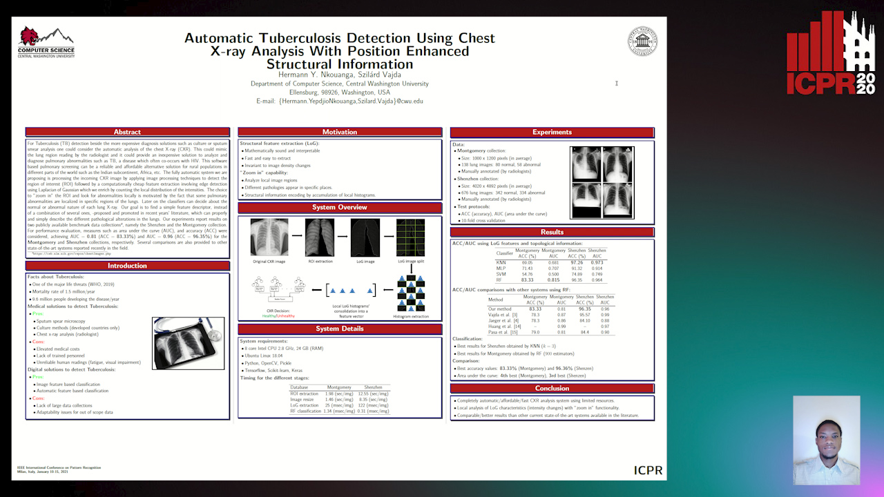
Auto-TLDR; Automatic Chest X-ray Screening for Tuberculosis in Rural Population using Localized Region on Interest

Slides Poster Similar

For Tuberculosis (TB) detection beside the more expensive diagnosis solutions such as culture or sputum smear analysis one could consider the automatic analysis of the chest X-ray (CXR). This could mimic the lung region reading by the radiologist and it could provide a cheap solution to analyze and diagnose pulmonary abnormalities such as TB which often co- occurs with HIV. This software based pulmonary screening can be a reliable and affordable solution for rural population in different parts of the world such as India, Africa, etc. Our fully automatic system is processing the incoming CXR image by applying image processing techniques to detect the region on interest (ROI) followed by a computationally cheap feature extraction involving edge detection using Laplacian of Gaussian which we enrich by counting the local distribution of the intensities. The choice to ”zoom in” the ROI and look for abnormalities locally is motivated by the fact that some pulmonary abnormalities are localized in specific regions of the lungs. Later on the classifiers can decide about the normal or abnormal nature of each lung X-ray. Our goal is to find a simple feature, instead of a combination of several ones, -proposed and promoted in recent years’ literature, which can properly describe the different pathological alterations in the lungs. Our experiments report results on two publicly available data collections1, namely the Shenzhen and the Montgomery collection. For performance evaluation, measures such as area under the curve (AUC), and accuracy (ACC) were considered, achieving AUC = 0.81 (ACC = 83.33%) and AUC = 0.96 (ACC = 96.35%) for the Montgomery and Schenzen collections, respectively. Several comparisons are also provided to other state- of-the-art systems reported recently in the field.

Offset Curves Loss for Imbalanced Problem in Medical Segmentation

Ngan Le, Duc Toan Bui, Khoa Luu, Marios Savvides

Responsive image

Auto-TLDR; Offset Curves Loss for Medical Image Segmentation

Poster Similar

Medical image segmentation has played an important role in medical analysis and widely developed for many clinical applications. Deep learning-based approaches have achieved high performance in semantic segmentation but they are limited to pixel-wise setting and imbalanced classes data problem. In this paper, we tackle those limitations by developing a new deep learning-based model which takes into account both higher feature level i.e. region inside contour, intermediate feature level i.e. offset curves around the contour and lower feature level i.e. contour. Our proposed Offset Curves (OsC) loss consists of three main fitting terms. The first fitting term focuses on pixel-wise level segmentation whereas the second fitting term acts as attention model which pays attention to the area around the boundaries (offset curves). The third terms plays a role as regularization term which takes the length of boundaries into account. We evaluate our proposed OsC loss on both 2D network and 3D network. Two common medical datasets, i.e. retina DRIVE and brain tumor BRATS 2018 datasets are used to benchmark our proposed loss performance. The experiments have showed that our proposed OsC loss function outperforms other mainstream loss functions such as Cross-Entropy, Dice, Focal on the most common segmentation networks Unet, FCN.

Deep Multi-Stage Model for Automated Landmarking of Craniomaxillofacial CT Scans

Simone Palazzo, Giovanni Bellitto, Luca Prezzavento, Francesco Rundo, Ulas Bagci, Daniela Giordano, Rosalia Leonardi, Concetto Spampinato

Responsive image

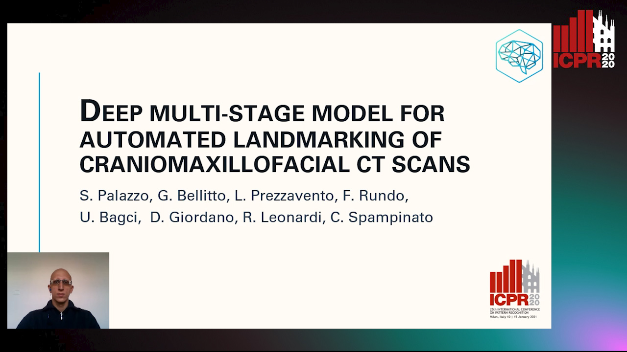
Auto-TLDR; Automated Landmarking of Craniomaxillofacial CT Images Using Deep Multi-Stage Architecture

Slides Similar

In this paper we define a deep multi-stage architecture for automated landmarking of craniomaxillofacial (CMF) CT images. Our model is composed of three subnetworks that first localize, on reduced-resolution images, areas where land-marks may be found and then refine the search, at full-resolution scale, through a hierarchical structure aiming at increasing the granularity of the investigated region. The multi-stage pipeline is designed to deal with full resolution data and does not require any additional pre-processing step to reduce search space, as opposed to existing methods that can be only adopted for searching landmarks located in well-defined anatomical structures (e.g.,mandibles). The automated landmarking system is tested on identifying landmarks located in several CMF regions, achieving an average error of 0.8 mm, significantly lower than expert readings. The proposed model also outperforms baselines and is on par with existing models that employ additional upstream segmentation, on state-of-the-art benchmarks.

One Step Clustering Based on A-Contrario Framework for Detection of Alterations in Historical Violins

Alireza Rezaei, Sylvie Le Hégarat-Mascle, Emanuel Aldea, Piercarlo Dondi, Marco Malagodi

Responsive image

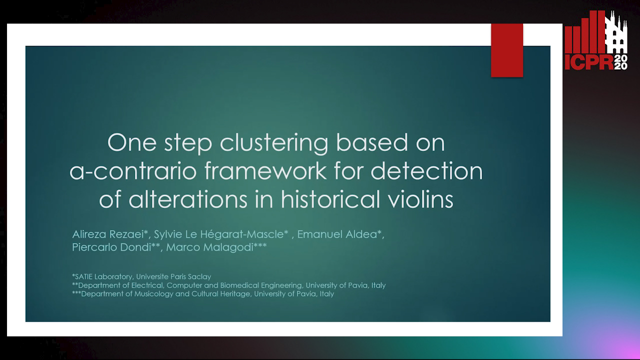
Auto-TLDR; A-Contrario Clustering for the Detection of Altered Violins using UVIFL Images

Slides Poster Similar

Preventive conservation is an important practice in Cultural Heritage. The constant monitoring of the state of conservation of an artwork helps us reduce the risk of damage and number of interventions necessary. In this work, we propose a probabilistic approach for the detection of alterations on the surface of historical violins based on an a-contrario framework. Our method is a one step NFA clustering solution which considers grey-level and spatial density information in one background model. The proposed method is robust to noise and avoids parameter tuning and any assumption about the quantity of the worn out areas. We have used as input UV induced fluorescence (UVIFL) images for considering details not perceivable with visible light. Tests were conducted on image sequences included in the ``Violins UVIFL imagery'' dataset. Results illustrate the ability of the algorithm to distinguish the worn area from the surrounding regions. Comparisons with the state of the art clustering methods shows improved overall precision and recall.

Fine-Tuning Convolutional Neural Networks: A Comprehensive Guide and Benchmark Analysis for Glaucoma Screening

Amed Mvoulana, Rostom Kachouri, Mohamed Akil

Responsive image

Auto-TLDR; Fine-tuning Convolutional Neural Networks for Glaucoma Screening

Slides Poster Similar

This work aimed at giving a comprehensive and in-detailed guide on the route to fine-tuning Convolutional Neural Networks (CNNs) for glaucoma screening. Transfer learning consists in a promising alternative to train CNNs from stratch, to avoid the huge data and resources requirements. After a thorough study of five state-of-the-art CNNs architectures, a complete and well-explained strategy for fine-tuning these networks is proposed, using hyperparameter grid-searching and two-phase training approach. Excellent performance is reached on model evaluation, with a 0.9772 AUROC validation rate, giving arise to reliable glaucoma diagosis-help systems. Also, a benchmark analysis is conducted across all fine-tuned models, studying them according to performance indices such as model complexity and size, AUROC density and inference time. This in-depth analysis allows a rigorous comparison between model characteristics, and is useful for giving practioners important trademarks for prospective applications and deployments.

End-To-End Multi-Task Learning for Lung Nodule Segmentation and Diagnosis

Wei Chen, Qiuli Wang, Dan Yang, Xiaohong Zhang, Chen Liu, Yucong Li

Responsive image

Auto-TLDR; A novel multi-task framework for lung nodule diagnosis based on deep learning and medical features

Slides Similar

Computer-Aided Diagnosis (CAD) systems for lung nodule diagnosis based on deep learning have attracted much attention in recent years. However, most existing methods ignore the relationships between the segmentation and classification tasks, which leads to unstable performances. To address this problem, we propose a novel multi-task framework, which can provide lung nodule segmentation mask, malignancy prediction, and medical features for interpretable diagnosis at the same time. Our framework mainly contains two sub-network: (1) Multi-Channel Segmentation Sub-network (MSN) for lung nodule segmentation, and (2) Joint Classification Sub-network (JCN) for interpretable lung nodule diagnosis. In the proposed framework, we use U-Net down-sampling processes for extracting low-level deep learning features, which are shared by two sub-networks. The JCN forces the down-sampling processes to learn better lowlevel deep features, which lead to a better construct of segmentation masks. Meanwhile, two additional channels constructed by OTSU and super-pixel (SLIC) methods, are utilized as the guideline of the feature extraction. The proposed framework takes advantages of deep learning methods and classical methods, which can significantly improve the performances of all tasks. We evaluate the proposed framework on public dataset LIDCIDRI. Our framework achieves a promising Dice score of 86.43% in segmentation, 87.07% in malignancy level prediction, and convincing results in interpretable medical feature predictions.

On Morphological Hierarchies for Image Sequences

Caglayan Tuna, Alain Giros, François Merciol, Sébastien Lefèvre

Responsive image

Auto-TLDR; Comparison of Hierarchies for Image Sequences

Slides Poster Similar

Morphological hierarchies form a popular framework aiming at emphasizing the multiscale structure of digital image by performing an unsupervised spatial partitioning of the data. These hierarchies have been recently extended to cope with image sequences, and different strategies have been proposed to allow their construction from spatio-temporal data. In this paper, we compare these hierarchical representation strategies for image sequences according to their structural properties. We introduce a projection method to make these representations comparable. Furthermore, we extend one of these recent strategies in order to obtain more efficient hierarchical representations for image sequences. Experiments were conducted on both synthetic and real datasets, the latter being made of satellite image time series. We show that building one hierarchy by using spatial and temporal information together is more efficient comparing to other existing strategies.

MedZip: 3D Medical Images Lossless Compressor Using Recurrent Neural Network (LSTM)

Omniah Nagoor, Joss Whittle, Jingjing Deng, Benjamin Mora, Mark W. Jones

Responsive image

Auto-TLDR; Recurrent Neural Network for Lossless Medical Image Compression using Long Short-Term Memory

Poster Similar

As scanners produce higher-resolution and more densely sampled images, this raises the challenge of data storage, transmission and communication within healthcare systems. Since the quality of medical images plays a crucial role in diagnosis accuracy, medical imaging compression techniques are desired to reduce scan bitrate while guaranteeing lossless reconstruction. This paper presents a lossless compression method that integrates a Recurrent Neural Network (RNN) as a 3D sequence prediction model. The aim is to learn the long dependencies of the voxel's neighbourhood in 3D using Long Short-Term Memory (LSTM) network then compress the residual error using arithmetic coding. Experiential results reveal that our method obtains a higher compression ratio achieving 15% saving compared to the state-of-the-art lossless compression standards, including JPEG-LS, JPEG2000, JP3D, HEVC, and PPMd. Our evaluation demonstrates that the proposed method generalizes well to unseen modalities CT and MRI for the lossless compression scheme. To the best of our knowledge, this is the first lossless compression method that uses LSTM neural network for 16-bit volumetric medical image compression.

Graph-Based Image Decoding for Multiplexed in Situ RNA Detection

Gabriele Partel, Carolina Wahlby

Responsive image

Auto-TLDR; A Graph-based Decoding Approach for Multiplexed In situ RNA Detection

Poster Similar

Image-based multiplexed in situ RNA detection makes it possible to map the spatial gene expression of hundreds to thousands of genes in parallel, and thus discern at the same time a large numbers of different cell types to better understand tissue development, heterogeneity, and disease. Fluorescent signals are detected over multiple fluorescent channels and imaging rounds and decoded in order to identify RNA molecules in their morphological context. Here we present a graph-based decoding approach that models the decoding process as a network flow problem jointly optimizing observation likelihoods and distances of signal detections, thus achieving robustness with respect to noise and spatial jitter of the fluorescent signals. We evaluated our method on synthetic data generated at different experimental conditions, and on real data of in situ RNA sequencing, comparing results with respect to alternative and gold standard image decoding pipelines.

Using Machine Learning to Refer Patients with Chronic Kidney Disease to Secondary Care

Lee Au-Yeung, Xianghua Xie, Timothy Marcus Scale, James Anthony Chess

Responsive image

Auto-TLDR; A Machine Learning Approach for Chronic Kidney Disease Prediction using Blood Test Data

Slides Poster Similar

There has been growing interest recently in using machine learning techniques as an aid in clinical medicine. Machine learning offers a range of classification algorithms which can be applied to medical data to aid in making clinical predictions. Recent studies have demonstrated the high predictive accuracy of various classification algorithms applied to clinical data. Several studies have already been conducted in diagnosing or predicting chronic kidney disease at various stages using different sets of variables. In this study we are investigating the use machine learning techniques with blood test data. Such a system could aid renal teams in making recommendations to primary care general practitioners to refer patients to secondary care where patients may benefit from earlier specialist assessment and medical intervention. We are able to achieve an overall accuracy of 88.48\% using logistic regression, 87.12\% using ANN and 85.29\% using SVM. ANNs performed with the highest sensitivity at 89.74\% compared to 86.67\% for logistic regression and 85.51\% for SVM.

One-Stage Multi-Task Detector for 3D Cardiac MR Imaging

Weizeng Lu, Xi Jia, Wei Chen, Nicolò Savioli, Antonio De Marvao, Linlin Shen, Declan O'Regan, Jinming Duan

Responsive image

Auto-TLDR; Multi-task Learning for Real-Time, simultaneous landmark location and bounding box detection in 3D space

Slides Poster Similar

Fast and accurate landmark location and bounding box detection are important steps in 3D medical imaging. In this paper, we propose a novel multi-task learning framework, for real-time, simultaneous landmark location and bounding box detection in 3D space. Our method extends the famous single-shot multibox detector (SSD) from single-task learning to multi-task learning and from 2D to 3D. Furthermore, we propose a post-processing approach to refine the network landmark output, by averaging the candidate landmarks. Owing to these settings, the proposed framework is fast and accurate. For 3D cardiac magnetic resonance (MR) images with size 224 × 224 × 64, our framework runs about 128 volumes per second (VPS) on GPU and achieves 6.75mm average point-to-point distance error for landmark location, which outperforms both state-of-the-art and baseline methods. We also show that segmenting the 3D image cropped with the bounding box results in both improved performance and efficiency.

Prediction of Obstructive Coronary Artery Disease from Myocardial Perfusion Scintigraphy using Deep Neural Networks

Ida Arvidsson, Niels Christian Overgaard, Miguel Ochoa Figueroa, Jeronimo Rose, Anette Davidsson, Kalle Åström, Anders Heyden

Responsive image

Auto-TLDR; A Deep Learning Algorithm for Multi-label Classification of Myocardial Perfusion Scintigraphy for Stable Ischemic Heart Disease

Slides Poster Similar

For diagnosis and risk assessment in patients with stable ischemic heart disease, myocardial perfusion scintigraphy is one of the most common cardiological examinations performed today. There are however many motivations for why an artificial intelligence algorithm would provide useful input to this task. For example to reduce the subjectiveness and save time for the nuclear medicine physicians working with this time consuming task. In this work we have developed a deep learning algorithm for multi-label classification based on a modified convolutional neural network to estimate probability of obstructive coronary artery disease in the left anterior artery, left circumflex artery and right coronary artery. The prediction is based on data from myocardial perfusion scintigraphy studies conducted in a dedicated Cadmium-Zinc-Telluride cardio camera (D-SPECT Spectrum Dynamics). Data from 588 patients was available, with stress images in both upright and supine position, as well as a number of auxiliary parameters such as angina symptoms and BMI. The data was used to train and evaluate the algorithm using 5-fold cross-validation. We achieve state-of-the-art results for this task with an area under the receiver operating characteristics curve of 0.89 as average on per-vessel level and 0.94 on per-patient level.

BG-Net: Boundary-Guided Network for Lung Segmentation on Clinical CT Images

Rui Xu, Yi Wang, Tiantian Liu, Xinchen Ye, Lin Lin, Yen-Wei Chen, Shoji Kido, Noriyuki Tomiyama

Responsive image

Auto-TLDR; Boundary-Guided Network for Lung Segmentation on CT Images

Slides Poster Similar

Lung segmentation on CT images is a crucial step for a computer-aided diagnosis system of lung diseases. The existing deep learning based lung segmentation methods are less efficient to segment lungs on clinical CT images, especially that the segmentation on lung boundaries is not accurate enough due to complex pulmonary opacities in practical clinics. In this paper, we propose a boundary-guided network (BG-Net) to address this problem. It contains two auxiliary branches that separately segment lungs and extract the lung boundaries, and an aggregation branch that efficiently exploits lung boundary cues to guide the network for more accurate lung segmentation on clinical CT images. We evaluate the proposed method on a private dataset collected from the Osaka university hospital and four public datasets including StructSeg, HUG, VESSEL12, and a Novel Coronavirus 2019 (COVID-19) dataset. Experimental results show that the proposed method can segment lungs more accurately and outperform several other deep learning based methods.

Extended Depth of Field Preserving Color Fidelity for Automated Digital Cytology

Alexandre Bouyssoux, Riadh Fezzani, Jean-Christophe Olivo-Marin

Responsive image

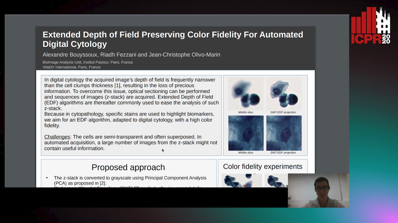
Auto-TLDR; Multi-Channel Extended Depth of Field for Digital cytology based on the stationary wavelet transform

Poster Similar

This paper presents a multi-channel Extended Depth of Field (EDF) method for digital cytology based on the stationary wavelet transform. With a coefficient selection rule adapted to a precise color recovery, a sharp image can be reconstructed even on images with transparent overlapping cells. The precision and the color fidelity of the proposed method is analyzed. Moreover, an experiment demonstrating the necessity of volume analysis in cytology to achieve precise segmentation on cell clumps is conducted, and the importance of color fidelity in this context is asserted. The proposed method was tested on pap-stained urothelial cells and gray-scale cervical cells with important overlapping.

Leveraging Unlabeled Data for Glioma Molecular Subtype and Survival Prediction

Nicholas Nuechterlein, Beibin Li, Mehmet Saygin Seyfioglu, Sachin Mehta, Patrick Cimino, Linda Shapiro

Responsive image

Auto-TLDR; Multimodal Brain Tumor Segmentation Using Unlabeled MR Data and Genomic Data for Cancer Prediction

Slides Poster Similar

In this paper, we address two long-standing challenges in neuro-oncology: (1) how to leverage large amounts of unlabeled magnetic resonance (MR) imaging data for radiogenomic tasks and (2) how to unite glioma MR imaging with genomic data. We examine multi-parametric MR data from 542 patients in the combined training, validation, and testing sets of the 2018 Multimodal Brain Tumor Segmentation Challenge and somatic copy number alteration (SCNA) data from 1090 patients in The Cancer Genome Archive's (TCGA) lower-grade glioma and glioblastoma projects. We propose a novel application of multi-task learning (MTL) that leverages unlabeled MR data by jointly learning tumor segmentation masks with glioma molecular subtype markers and allows for SCNA input when available. There are 235 patients in the intersection of these MR and SCNA datasets, which we divide into an unlabeled training set, a labeled training set, and a validation set. Our MTL model significantly outperforms comparable classification models trained only on labeled MR data for both IDH1/2 mutation and 1p/19q co-deletion glioma subtype marker prediction tasks. We also observe that models trained on genomic and imaging data improve survival prediction results achieved by models trained on either alone. We will release our source code for future research.