Few Shot Learning Framework to Reduce Inter-Observer Variability in Medical Images

Sohini Roychowdhury

Responsive image

Auto-TLDR; Few-Shot Learning for Quality Image Annotation

Slides Poster

Most computer aided pathology detection systems rely on large volumes of quality annotated data to aid diagnostics and follow up procedures. However, quality assuring large volumes of annotated medical image data can be subjective and expensive. In this work we present a novel standardization framework that implements three few-shot learning (FSL) models that can be iteratively trained by atmost 5 images per 3D stack to generate multiple regional proposals (RPs) per test image. These FSL models include a novel parallel echo state network framework and an augmented U-net model. Additionally, we propose a novel target label selection algorithm (TLSA) that measures relative agreeability between RPs and the manually annotated target labels to detect the ``best" quality annotation per image. Using the FSL models, our system achieves 0.28-0.64 Dice coefficient across vendor image stacks for intra-retinal cyst segmentation. Additionally, the TLSA is capable of correctly classifying high quality target labels from their noisy counterparts with 70-97% accuracy. The proposed system significantly automates high quality annotation selection on an image level while minimizing manual quality checking to 12-28% of the images only. Thus, the proposed framework is flexible to extensions for quality image annotation curation of other image stacks as well.

Similar papers

Transfer Learning through Weighted Loss Function and Group Normalization for Vessel Segmentation from Retinal Images

Abdullah Sarhan, Jon Rokne, Reda Alhajj, Andrew Crichton

Responsive image

Auto-TLDR; Deep Learning for Segmentation of Blood Vessels in Retinal Images

Slides Poster Similar

The vascular structure of blood vessels is important in diagnosing retinal conditions such as glaucoma and diabetic retinopathy. Accurate segmentation of these vessels can help in detecting retinal objects such as the optic disc and optic cup and hence determine if there are damages to these areas. Moreover, the structure of the vessels can help in diagnosing glaucoma. The rapid development of digital imaging and computer-vision techniques has increased the potential for developing approaches for segmenting retinal vessels. In this paper, we propose an approach for segmenting retinal vessels that uses deep learning along with transfer learning. We adapted the U-Net structure to use a customized InceptionV3 as the encoder and used multiple skip connections to form the decoder. Moreover, we used a weighted loss function to handle the issue of class imbalance in retinal images. Furthermore, we contributed a new dataset to this field. We tested our approach on six publicly available datasets and a newly created dataset. We achieved an average accuracy of 95.60\% and a Dice coefficient of 80.98\%. The results obtained from comprehensive experiments demonstrate the robustness of our approach to the segmentation of blood vessels in retinal images obtained from different sources. Our approach results in greater segmentation accuracy than other approaches.

Automatic Semantic Segmentation of Structural Elements related to the Spinal Cord in the Lumbar Region by Using Convolutional Neural Networks

Jhon Jairo Sáenz Gamboa, Maria De La Iglesia-Vaya, Jon Ander Gómez

Responsive image

Auto-TLDR; Semantic Segmentation of Lumbar Spine Using Convolutional Neural Networks

Slides Poster Similar

This work addresses the problem of automatically segmenting the MR images corresponding to the lumbar spine. The purpose is to detect and delimit the different structural elements like vertebrae, intervertebral discs, nerves, blood vessels, etc. This task is known as semantic segmentation. The approach proposed in this work is based on convolutional neural networks whose output is a mask where each pixel from the input image is classified into one of the possible classes. Classes were defined by radiologists and correspond to structural elements and tissues. The proposed network architectures are variants of the U-Net. Several complementary blocks were used to define the variants: spatial attention models, deep supervision and multi-kernels at input, this last block type is based on the idea of inception. Those architectures which got the best results are described in this paper, and their results are discussed. Two of the proposed architectures outperform the standard U-Net used as baseline.

NephCNN: A Deep-Learning Framework for Vessel Segmentation in Nephrectomy Laparoscopic Videos

Alessandro Casella, Sara Moccia, Chiara Carlini, Emanuele Frontoni, Elena De Momi, Leonardo Mattos

Responsive image

Auto-TLDR; Adversarial Fully Convolutional Neural Networks for kidney vessel segmentation from nephrectomy laparoscopic videos

Slides Poster Similar

Objective: In the last years, Robot-assisted partial nephrectomy (RAPN) is establishing as elected treatment for renal cell carcinoma (RCC). Reduced field of view, field occlusions by surgical tools, and reduced maneuverability may potentially cause accidents, such as unwanted vessel resection with consequent bleeding. Surgical Data Science (SDS) can provide effective context-aware tools for supporting surgeons. However, currently no tools have been exploited for automatic vessels segmentation from nephrectomy laparoscopic videos. Herein, we propose a new approach based on adversarial Fully Convolutional Neural Networks (FCNNs) to kidney vessel segmentation from nephrectomy laparoscopic vision. Methods: The proposed approach enhances existing segmentation framework by (i) encoding 3D kernels for spatio-temporal features extraction to enforce pixel connectivity in time, and (ii) perform training in adversarial fashion, which constrains vessels shape. Results: We performed a preliminary study using 8 different RAPN videos (1871 frames), the first in the field, achieving a median Dice Similarity Coefficient of 71.76%. Conclusions: Results showed that the proposed approach could be a valuable solution with a view to assist surgeon during RAPN.

A Benchmark Dataset for Segmenting Liver, Vasculature and Lesions from Large-Scale Computed Tomography Data

Bo Wang, Zhengqing Xu, Wei Xu, Qingsen Yan, Liang Zhang, Zheng You

Responsive image

Auto-TLDR; The Biggest Treatment-Oriented Liver Cancer Dataset for Segmentation

Slides Poster Similar

How to build a high-performance liver-related computer assisted diagnosis system is an open question of great interest. However, the performance of the state-of-art algorithm is always limited by the amount of data and quality of the label. To address this problem, we propose the biggest treatment-oriented liver cancer dataset for liver surgery and treatment planning. This dataset provides 216 cases (totally about 268K frames) scanned images in contrast-enhanced computed tomography (CT). We labeled all the CT images with the liver, liver vasculature and liver tumor segmentation ground truth for train and tune segmentation algorithms in advance. Based on that, we evaluate several recent and state-of-the-art segmentation algorithms, including 7 deep learning methods, on CT sequences. All results are compared to reference segmentations five error metrics that highlight different aspects of segmentation accuracy. In general, compared with previous datasets, our dataset is really a challenging dataset. To our knowledge, the proposed dataset and benchmark allow for the first time systematic exploration of such issues, and will be made available to allow for further research in this field.

Segmentation of Intracranial Aneurysm Remnant in MRA Using Dual-Attention Atrous Net

Subhashis Banerjee, Ashis Kumar Dhara, Johan Wikström, Robin Strand

Responsive image

Auto-TLDR; Dual-Attention Atrous Net for Segmentation of Intracranial Aneurysm Remnant from MRA Images

Slides Poster Similar

Due to the advancement of non-invasive medical imaging modalities like Magnetic Resonance Angiography (MRA), an increasing number of Intracranial Aneurysm (IA) cases are being reported in recent years. The IAs are typically treated by so-called endovascular coiling, where blood flow in the IA is prevented by embolization with a platinum coil. Accurate quantification of the IA Remnant (IAR), i.e. the volume with blood flow present post treatment is the utmost important factor in choosing the right treatment planning. This is typically done by manually segmenting the aneurysm remnant from the MRA volume. Since manual segmentation of volumetric images is a labour-intensive and error-prone process, development of an automatic volumetric segmentation method is required. Segmentation of small structures such as IA, that may largely vary in size, shape, and location is considered extremely difficult. Similar intensity distribution of IAs and surrounding blood vessels makes it more challenging and susceptible to false positive. In this paper we propose a novel 3D CNN architecture called Dual-Attention Atrous Net (DAtt-ANet), which can efficiently segment IAR volumes from MRA images by reconciling features at different scales using the proposed Parallel Atrous Unit (PAU) along with the use of self-attention mechanism for extracting fine-grained features and intra-class correlation. The proposed DAtt-ANet model is trained and evaluated on a clinical MRA image dataset (prospective research project, approved by the local ethical committee) of IAR consisting of 46 subjects, annotated by an expert radiologist from our group. We compared the proposed DAtt-ANet with five state-of-the-art CNN models based on their segmentation performance. The proposed DAtt-ANet outperformed all other methods and was able to achieve a five-fold cross-validation DICE score of $0.73\pm0.06$.

A Deep Learning Approach for the Segmentation of Myocardial Diseases

Khawala Brahim, Abdull Qayyum, Alain Lalande, Arnaud Boucher, Anis Sakly, Fabrice Meriaudeau

Responsive image

Auto-TLDR; Segmentation of Myocardium Infarction Using Late GADEMRI and SegU-Net

Slides Poster Similar

Cardiac left ventricular (LV) segmentation is of paramount essential step for both diagnosis and treatment of cardiac pathologies such as ischemia, myocardial infarction, arrhythmia and myocarditis. However, this segmentation is challenging due to high variability across patients and the potential lack of contrast between structures. In this work, we propose and evaluate a (2.5D) SegU-Net model based on the fusion of two deep learning techniques (U-Net and Seg-Net) for automated LGEMRI (Late gadolinium enhanced magnetic resonance imaging) myocardial disease (infarct core and no reflow region) quantification in a new multifield expert annotated dataset. Given that the scar tissue represents a small part of the whole MRI slices, we focused on myocardium area. Segmentation results show that this preprocessing step facilitate the learning procedure. In order to solve the class imbalance problem, we propose to apply the Jaccard loss and the Focal Loss as optimization loss function and to integrate a class weights strategy into the objective function. Late combination has been used to merge the output of the best trained models on a different set of hyperparameters. The final network segmentation performances will be useful for future comparison of new method to the current related work for this task. A total number of 2237 of slices (320 cases) were used for training/validation and 210 slices (35 cases) were used for testing. Experiments over our proposed dataset, using several evaluation metrics such Jaccard distance (IOU), Accuracy and Dice similarity coefficient (DSC), demonstrate efficiency performance in quantifying different zones of myocardium infarction across various patients. As compared to the second intra-observer study, our testing results showed that the SegUNet prediction model leads to these average dice coefficients over all segmented tissue classes, respectively : 'Background': 0.99999, 'Myocardium': 0.99434, 'Infarctus': 0.95587, 'Noreflow': 0.78187.

PCANet: Pyramid Context-Aware Network for Retinal Vessel Segmentation

Yi Zhang, Yixuan Chen, Kai Zhang

Responsive image

Auto-TLDR; PCANet: Adaptive Context-Aware Network for Automated Retinal Vessel Segmentation

Slides Poster Similar

Automated retinal vessel segmentation plays an important role in the diagnosis of some diseases such as diabetes, arteriosclerosis and hypertension. Recent works attempt to improve segmentation performance by exploring either global or local contexts. However, the context demands are varying from regions in each image and different levels of network. To address these problems, we propose Pyramid Context-aware Network (PCANet), which can adaptively capture multi-scale context representations. Specifically, PCANet is composed of multiple Adaptive Context-aware (ACA) blocks arranged in parallel, each of which can adaptively obtain the context-aware features by estimating affinity coefficients at a specific scale under the guidance of global contextual dependencies. Meanwhile, we import ACA blocks with specific scales in different levels of the network to obtain a coarse-to-fine result. Furthermore, an integrated test-time augmentation method is developed to further boost the performance of PCANet. Finally, extensive experiments demonstrate the effectiveness of the proposed PCANet, and state-of-the-art performances are achieved with AUCs of 0.9866, 0.9886 and F1 Scores of 0.8274, 0.8371 on two public datasets, DRIVE and STARE, respectively.

Planar 3D Transfer Learning for End to End Unimodal MRI Unbalanced Data Segmentation

Martin Kolarik, Radim Burget, Carlos M. Travieso-Gonzalez, Jan Kocica

Responsive image

Auto-TLDR; Planar 3D Res-U-Net Network for Unbalanced 3D Image Segmentation using Fluid Attenuation Inversion Recover

Slides Similar

We present a novel approach of 2D to 3D transfer learning based on mapping pre-trained 2D convolutional neural network weights into planar 3D kernels. The method is validated by proposed planar 3D res-u-net network with encoder transferred from the 2D VGG-16 which is applied for a single-stage unbalanced 3D image data segmentation. In particular, we evaluate the method on the MICCAI 2016 MS lesion segmentation challenge dataset utilizing solely Fluid Attenuation Inversion Recover (FLAIR) sequence without brain extraction for training and inference to simulate real medical praxis. The planar 3D res-u-net network performed the best both in sensitivity and Dice score amongst end to end methods processing raw MRI scans and achieved comparable Dice score to a state-of-the-art unimodal not end to end approach. Complete source code was released under the open-source license and this paper is in compliance with the Machine learning Reproducibility Checklist. By implementing practical transfer learning for 3D data representation we were able to successfully segment heavily unbalanced data without selective sampling and achieved more reliable results using less training data in single modality. From medical perspective, the unimodal approach gives an advantage in real praxis as it does not require co-registration nor additional scanning time during examination. Although modern medical imaging methods capture high resolution 3D anatomy scans suitable for computer aided detection system processing, deployment of automatic systems for interpretation of radiology imaging is still rather theoretical in many medical areas. Our work aims to bridge the gap offering solution for partial research questions.

A Comparison of Neural Network Approaches for Melanoma Classification

Maria Frasca, Michele Nappi, Michele Risi, Genoveffa Tortora, Alessia Auriemma Citarella

Responsive image

Auto-TLDR; Classification of Melanoma Using Deep Neural Network Methodologies

Slides Poster Similar

Melanoma is the deadliest form of skin cancer and it is diagnosed mainly visually, starting from initial clinical screening and followed by dermoscopic analysis, biopsy and histopathological examination. A dermatologist’s recognition of melanoma may be subject to errors and may take some time to diagnose it. In this regard, deep learning can be useful in the study and classification of skin cancer. In particular, by classifying images with Deep Neural Network methodologies, it is possible to obtain comparable or even superior results compared to those of dermatologists. In this paper, we propose a methodology for the classification of melanoma by adopting different deep learning techniques applied to a common dataset, composed of images from the ISIC dataset and consisting of different types of skin diseases, including melanoma on which we applied a specific pre-processing phase. In particular, a comparison of the results is performed in order to select the best effective neural network to be applied to the problem of recognition and classification of melanoma. Moreover, we also evaluate the impact of the pre- processing phase on the final classification. Different metrics such as accuracy, sensitivity, and specificity have been selected to assess the goodness of the adopted neural networks and compare them also with the manual classification of dermatologists.

Learning to Segment Clustered Amoeboid Cells from Brightfield Microscopy Via Multi-Task Learning with Adaptive Weight Selection

Rituparna Sarkar, Suvadip Mukherjee, Elisabeth Labruyere, Jean-Christophe Olivo-Marin

Responsive image

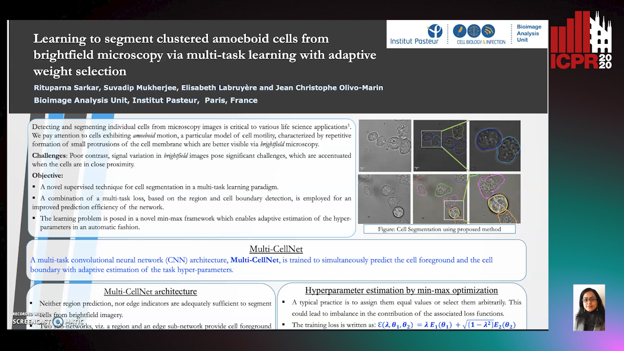
Auto-TLDR; Supervised Cell Segmentation from Microscopy Images using Multi-task Learning in a Multi-Task Learning Paradigm

Poster Similar

Detecting and segmenting individual cells from microscopy images is critical to various life science applications. Traditional cell segmentation tools are often ill-suited for applications in brightfield microscopy due to poor contrast and intensity heterogeneity, and only a small subset are applicable to segment cells in a cluster. In this regard, we introduce a novel supervised technique for cell segmentation in a multi-task learning paradigm. A combination of a multi-task loss, based on the region and cell boundary detection, is employed for an improved prediction efficiency of the network. The learning problem is posed in a novel min-max framework which enables adaptive estimation of the hyper-parameters in an automatic fashion. The region and cell boundary predictions are combined via morphological operations and active contour model to segment individual cells. The proposed methodology is particularly suited to segment touching cells from brightfield microscopy images without manual interventions. Quantitatively, we observe an overall Dice score of 0.93 on the validation set, which is an improvement of over 15.9% on a recent unsupervised method, and outperforms the popular supervised U-net algorithm by at least 5.8% on average.

BCAU-Net: A Novel Architecture with Binary Channel Attention Module for MRI Brain Segmentation

Yongpei Zhu, Zicong Zhou, Guojun Liao, Kehong Yuan

Responsive image

Auto-TLDR; BCAU-Net: Binary Channel Attention U-Net for MRI brain segmentation

Slides Poster Similar

Recently deep learning-based networks have achieved advanced performance in medical image segmentation. However, the development of deep learning is slow in magnetic resonance image (MRI) segmentation of normal brain tissues. In this paper, inspired by channel attention module, we propose a new architecture, Binary Channel Attention U-Net (BCAU-Net), by introducing a novel Binary Channel Attention Module (BCAM) into skip connection of U-Net, which can take full advantages of the channel information extracted from the encoding path and corresponding decoding path. To better aggregate multi-scale spatial information of the feature map, spatial pyramid pooling (SPP) modules with different pooling operations are used in BCAM instead of original average-pooling and max-pooling operations. We verify this model on two datasets including IBSR and MRBrainS18, and obtain better performance on MRI brain segmentation compared with other methods. We believe the proposed method can advance the performance in brain segmentation and clinical diagnosis.

MTGAN: Mask and Texture-Driven Generative Adversarial Network for Lung Nodule Segmentation

Wei Chen, Qiuli Wang, Kun Wang, Dan Yang, Xiaohong Zhang, Chen Liu, Yucong Li

Responsive image

Auto-TLDR; Mask and Texture-driven Generative Adversarial Network for Lung Nodule Segmentation

Slides Poster Similar

Accurate segmentation for lung nodules in lung computed tomography (CT) scans plays a key role in the early diagnosis of lung cancer. Many existing methods, especially UNet, have made significant progress in lung nodule segmentation. However, due to the complex shapes of lung nodules and the similarity of visual characteristics between nodules and lung tissues, an accurate segmentation with few false positives of lung nodules is still a challenging problem. Considering the fact that both boundary and texture information of lung nodules are important for obtaining an accurate segmentation result, we propose a novel Mask and Texture-driven Generative Adversarial Network (MTGAN) with a joint multi-scale L1 loss for lung nodule segmentation, which takes full advantages of U-Net and adversarial training. The proposed MTGAN leverages adversarial learning strategy guided by the boundary and texture information of lung nodules to generate more accurate segmentation results with lesser false positives. We validate our model with the LIDC–IDRI dataset, and experimental results show that our method achieves excellent segmentation results for a variety of lung nodules, especially for juxtapleural nodules and low-dense nodules. Without any bells and whistles, the proposed MTGAN achieves significant segmentation performance with the Dice similarity coefficient (DSC) of 85.24% on the LIDC–IDRI dataset.

A Lumen Segmentation Method in Ureteroscopy Images Based on a Deep Residual U-Net Architecture

Jorge Lazo, Marzullo Aldo, Sara Moccia, Michele Catellani, Benoit Rosa, Elena De Momi, Michel De Mathelin, Francesco Calimeri

Responsive image

Auto-TLDR; A Deep Neural Network for Ureteroscopy with Residual Units

Slides Poster Similar

Ureteroscopy is becoming the first surgical treatment option for the majority of urinary affections. This procedure is carried out using an endoscope which provides the surgeon with the visual and spatial information necessary to navigate inside the urinary tract. Having in mind the development of surgical assistance systems, that could enhance the performance of surgeon, the task of lumen segmentation is a fundamental part since this is the visual reference which marks the path that the endoscope should follow. This is something that has not been analyzed in ureteroscopy data before. However, this task presents several challenges given the image quality and the conditions itself of ureteroscopy procedures. In this paper, we study the implementation of a Deep Neural Network which exploits the advantage of residual units in an architecture based on U-Net. For the training of these networks, we analyze the use of two different color spaces: gray-scale and RGB data images. We found that training on gray-scale images gives the best results obtaining mean values of Dice Score, Precision, and Recall of 0.73, 0.58, and 0.92 respectively. The results obtained show that the use of residual U-Net could be a suitable model for further development for a computer-aided system for navigation and guidance through the urinary system.

Deep Learning-Based Type Identification of Volumetric MRI Sequences

Jean Pablo De Mello, Thiago Paixão, Rodrigo Berriel, Mauricio Reyes, Alberto F. De Souza, Claudine Badue, Thiago Oliveira-Santos

Responsive image

Auto-TLDR; Deep Learning for Brain MRI Sequences Identification Using Convolutional Neural Network

Slides Poster Similar

The analysis of Magnetic Resonance Imaging (MRI) sequences enables clinical professionals to monitor the progression of a brain tumor. As the interest for automatizing brain volume MRI analysis increases, it becomes convenient to have each sequence well identified. However, the unstandardized naming of MRI sequences make their identification difficult for automated systems, as well as make it difficult for researches to generate or use datasets for machine learning research. In face of that, we propose a system for identifying types of brain MRI sequences based on deep learning. By training a Convolutional Neural Network (CNN) based on 18-layer ResNet architecture, our system is able to classify a volumetric brain MRI as a T1, T1c, T2 or FLAIR sequence, or whether it does not belong to any of these classes. The network was trained with both pre-processed (BraTS dataset) and non-pre-processed (TCGA-GBM dataset) images with diverse acquisition protocols, requiring only a few layers of the volume for training. Our system is able to classify among sequence types with an accuracy of 96.27%.

FOANet: A Focus of Attention Network with Application to Myocardium Segmentation

Zhou Zhao, Elodie Puybareau, Nicolas Boutry, Thierry Geraud

Responsive image

Auto-TLDR; FOANet: A Hybrid Loss Function for Myocardium Segmentation of Cardiac Magnetic Resonance Images

Slides Poster Similar

In myocardium segmentation of cardiac magnetic resonance images, ambiguities often appear near the boundaries of the target domains due to tissue similarities. To address this issue, we propose a new architecture, called FOANet, which can be decomposed in three main steps: a localization step, a Gaussian-based contrast enhancement step, and a segmentation step. This architecture is supplied with a hybrid loss function that guides the FOANet to study the transformation relationship between the input image and the corresponding label in a threelevel hierarchy (pixel-, patch- and map-level), which is helpful to improve segmentation and recovery of the boundaries. We demonstrate the efficiency of our approach on two public datasets in terms of regional and boundary segmentations.

SA-UNet: Spatial Attention U-Net for Retinal Vessel Segmentation

Changlu Guo, Marton Szemenyei, Yugen Yi, Wenle Wang, Buer Chen, Changqi Fan

Responsive image

Auto-TLDR; Spatial Attention U-Net for Segmentation of Retinal Blood Vessels

Slides Poster Similar

The precise segmentation of retinal blood vessels is of great significance for early diagnosis of eye-related diseases such as diabetes and hypertension. In this work, we propose a lightweight network named Spatial Attention U-Net (SA-UNet) that does not require thousands of annotated training samples and can be utilized in a data augmentation manner to use the available annotated samples more efficiently. SA-UNet introduces a spatial attention module which infers the attention map along the spatial dimension, and multiplies the attention map by the input feature map for adaptive feature refinement. In addition, the proposed network employs structured dropout convolutional blocks instead of the original convolutional blocks of U-Net to prevent the network from overfitting. We evaluate SA-UNet based on two benchmark retinal datasets: the Vascular Extraction (DRIVE) dataset and the Child Heart and Health Study (CHASE_DB1) dataset. The results show that the proposed SA-UNet achieves state-of-the-art performance on both datasets.The implementation and the trained networks are available on Github1.

A Multi-Task Contextual Atrous Residual Network for Brain Tumor Detection & Segmentation

Ngan Le, Kashu Yamazaki, Quach Kha Gia, Thanh-Dat Truong, Marios Savvides

Responsive image

Auto-TLDR; Contextual Brain Tumor Segmentation Using 3D atrous Residual Networks and Cascaded Structures

Poster Similar

In recent years, deep neural networks have achieved state-of-the-art performance in a variety of recognition and segmentation tasks in medical imaging including brain tumor segmentation. We investigate that segmenting brain tumor is facing to the imbalanced data problem where the number of pixels belonging to background class (non tumor pixel) is much larger than the number of pixels belonging to foreground class (tumor pixel). To address this problem, we propose a multi-task network which is formed as a cascaded structure and designed to share the feature maps. Our model consists of two targets, i.e., (i) effectively differentiating brain tumor regions and (ii) estimating brain tumor masks. The first task is performed by our proposed contextual brain tumor detection network, which plays the role of an attention gate and focuses on the region around brain tumor only while ignore the background (non tumor area). Instead of processing every pixel, our contextual brain tumor detection network only processes contextual regions around ground-truth instances and this strategy helps to produce meaningful regions proposals. The second task is built upon a 3D atrous residual network and under an encode-decode network in order to effectively segment both large and small objects (brain tumor). Our 3D atrous residual network is designed with a skip connection to enables the gradient from the deep layers to be directly propagated to shallow layers, thus, features of different depths are preserved and used for refining each other. In order to incorporate larger contextual information in volume MRI data, our network is designed by 3D atrous convolution with various kernel sizes, which enlarges the receptive field of filters. Our proposed network has been evaluated on various datasets including BRATS2015, BRATS2017 and BRATS2018 datasets with both validation set and testing set. Our performance has been benchmarked by both region-based metrics and surface-based metrics. We also have conducted comparisons against state-of-the-art approaches.

Inception Based Deep Learning Architecture for Tuberculosis Screening of Chest X-Rays

Dipayan Das, K.C. Santosh, Umapada Pal

Responsive image

Auto-TLDR; End to End CNN-based Chest X-ray Screening for Tuberculosis positive patients in the severely resource constrained regions of the world

Slides Poster Similar

The motivation for this work is the primary need of screening Tuberculosis (TB) positive patients in the severely resource constrained regions of the world. Chest X-ray (CXR) is considered to be a promising indicator for the onset of TB, but the lack of skilled radiologists in such regions degrades the situation. Therefore, several computer aided diagnosis (CAD) systems have been proposed to solve the decision making problem, which includes hand engineered feature extraction methods to deep learning or Convolutional Neural Network (CNN) based methods. Feature extraction, being a time and resource intensive process, often delays the process of mass screening. Hence an end to end CNN architecture is proposed in this work to solve the problem. Two benchmark CXR datasets have been used in this work, collected from Shenzhen (China) and Montgomery County (USA), on which the proposed methodology achieved a maximum abnormality detection accuracy (ACC) of 91.7\% (0.96 AUC) and 87.47\% (0.92 AUC) respectively. To the greatest of our knowledge, the obtained results are marginally superior to the state of the art results that have solely used deep learning methodologies on the aforementioned datasets.

Early Wildfire Smoke Detection in Videos

Taanya Gupta, Hengyue Liu, Bir Bhanu

Responsive image

Auto-TLDR; Semi-supervised Spatio-Temporal Video Object Segmentation for Automatic Detection of Smoke in Videos during Forest Fire

Poster Similar

Recent advances in unmanned aerial vehicles and camera technology have proven useful for the detection of smoke that emerges above the trees during a forest fire. Automatic detection of smoke in videos is of great interest to Fire department. To date, in most parts of the world, the fire is not detected in its early stage and generally it turns catastrophic. This paper introduces a novel technique that integrates spatial and temporal features in a deep learning framework using semi-supervised spatio-temporal video object segmentation and dense optical flow. However, detecting this smoke in the presence of haze and without the labeled data is difficult. Considering the visibility of haze in the sky, a dark channel pre-processing method is used that reduces the amount of haze in video frames and consequently improves the detection results. Online training is performed on a video at the time of testing that reduces the need for ground-truth data. Tests using the publicly available video datasets show that the proposed algorithms outperform previous work and they are robust across different wildfire-threatened locations.

SAGE: Sequential Attribute Generator for Analyzing Glioblastomas Using Limited Dataset

Padmaja Jonnalagedda, Brent Weinberg, Jason Allen, Taejin Min, Shiv Bhanu, Bir Bhanu

Responsive image

Auto-TLDR; SAGE: Generative Adversarial Networks for Imaging Biomarker Detection and Prediction

Slides Poster Similar

While deep learning approaches have shown remarkable performance in many imaging tasks, most of these methods rely on availability of large quantities of data. Medical image data, however, is scarce and fragmented. Generative Adversarial Networks (GANs) have recently been very effective in handling such datasets by generating more data. If the datasets are very small, however, GANs cannot learn the data distribution properly, resulting in less diverse or low-quality results. One such limited dataset is that for the concurrent gain of 19/20 chromosomes (19/20 co-gain), a mutation with positive prognostic value in Glioblastomas (GBM). In this paper, we detect imaging biomarkers for the mutation to streamline the extensive and invasive prognosis pipeline. Since this mutation is relatively rare, i.e. small dataset, we propose a novel generative framework – the Sequential Attribute GEnerator (SAGE), that generates detailed tumor imaging features while learning from a limited dataset. Experiments show that not only does SAGE generate high quality tumors when compared to standard Deep Convolutional GAN (DC-GAN) and Wasserstein GAN with Gradient Penalty (WGAN-GP), it also captures the imaging biomarkers accurately.

Vesselness Filters: A Survey with Benchmarks Applied to Liver Imaging

Jonas Lamy, Odyssée Merveille, Bertrand Kerautret, Nicolas Passat, Antoine Vacavant

Responsive image

Auto-TLDR; Comparison of Vessel Enhancement Filters for Liver Vascular Network Segmentation

Slides Poster Similar

The accurate knowledge of vascular network geometry is crucial for many clinical applications such as cardiovascular disease diagnosis and surgery planning. Vessel enhancement algorithms are often a key step to improve the robustness of vessel segmentation. A wide variety of enhancement filters exists in the literature, but they are often difficult to compare as the applications and datasets differ from a paper to another and the code is rarely available. In this article, we compare seven vessel enhancement filters covering the last twenty years literature in a unique common framework. We focus our study on the liver vascular network which is under-represented in the literature. The evaluation is made from three points of view: in the whole liver, in the vessel neighborhood and near the bifurcations. The study is performed on two publicly available datasets: the Ircad dataset (CT images) and the VascuSynth dataset adapted for MRI simulation. We discuss the strengths and weaknesses of each method in the hepatic context. In addition, the benchmark framework including a C++ implementation of each compared method is provided. An online demonstration ensures the reproducibility of the results without requiring any additional software.

EM-Net: Deep Learning for Electron Microscopy Image Segmentation

Afshin Khadangi, Thomas Boudier, Vijay Rajagopal

Responsive image

Auto-TLDR; EM-net: Deep Convolutional Neural Network for Electron Microscopy Image Segmentation

Similar

Recent high-throughput electron microscopy techniques such as focused ion-beam scanning electron microscopy (FIB-SEM) provide thousands of serial sections which assist the biologists in studying sub-cellular structures at high resolution and large volume. Low contrast of such images hinder image segmentation and 3D visualisation of these datasets. With recent advances in computer vision and deep learning, such datasets can be segmented and reconstructed in 3D with greater ease and speed than with previous approaches. However, these methods still rely on thousands of ground-truth samples for training and electron microscopy datasets require significant amounts of time for carefully curated manual annotations. We address these bottlenecks with EM-net, a scalable deep convolutional neural network for EM image segmentation. We have evaluated EM-net using two datasets, one of which belongs to an ongoing competition on EM stack segmentation since 2012. We show that EM-net variants achieve better performances than current deep learning methods using small- and medium-sized ground-truth datasets. We also show that the ensemble of top EM-net base classifiers outperforms other methods across a wide variety of evaluation metrics.

End-To-End Multi-Task Learning for Lung Nodule Segmentation and Diagnosis

Wei Chen, Qiuli Wang, Dan Yang, Xiaohong Zhang, Chen Liu, Yucong Li

Responsive image

Auto-TLDR; A novel multi-task framework for lung nodule diagnosis based on deep learning and medical features

Slides Similar

Computer-Aided Diagnosis (CAD) systems for lung nodule diagnosis based on deep learning have attracted much attention in recent years. However, most existing methods ignore the relationships between the segmentation and classification tasks, which leads to unstable performances. To address this problem, we propose a novel multi-task framework, which can provide lung nodule segmentation mask, malignancy prediction, and medical features for interpretable diagnosis at the same time. Our framework mainly contains two sub-network: (1) Multi-Channel Segmentation Sub-network (MSN) for lung nodule segmentation, and (2) Joint Classification Sub-network (JCN) for interpretable lung nodule diagnosis. In the proposed framework, we use U-Net down-sampling processes for extracting low-level deep learning features, which are shared by two sub-networks. The JCN forces the down-sampling processes to learn better lowlevel deep features, which lead to a better construct of segmentation masks. Meanwhile, two additional channels constructed by OTSU and super-pixel (SLIC) methods, are utilized as the guideline of the feature extraction. The proposed framework takes advantages of deep learning methods and classical methods, which can significantly improve the performances of all tasks. We evaluate the proposed framework on public dataset LIDCIDRI. Our framework achieves a promising Dice score of 86.43% in segmentation, 87.07% in malignancy level prediction, and convincing results in interpretable medical feature predictions.

Multi-focus Image Fusion for Confocal Microscopy Using U-Net Regression Map

Md Maruf Hossain Shuvo, Yasmin M. Kassim, Filiz Bunyak, Olga V. Glinskii, Leike Xie, Vladislav V Glinsky, Virginia H. Huxley, Kannappan Palaniappan

Responsive image

Auto-TLDR; Independent Single Channel U-Net Fusion for Multi-focus Microscopy Images

Slides Poster Similar

Multi-focus image fusion plays an important role to better visualize the detailed information and anatomical structures of microscopy images. We propose a new approach to fuse all single-focus microscopy images in each Z-stack. As the structures are different in different channels, input images are separated into red and green channels. Red for blood vessels, and green for lymphatics like structures . Taking the maximum likelihood of U-Net regression likelihood map along Z, we obtain the focus selection map for each channel. We named this approach as Independent Single Channel U-Net (ISCU) fusion. We combined each channel fusion result to get the final dual channel composite RGB image. The dataset used is extremely challenging with complex microscopy images of mice dura mater attached to bone. We compared our results with one of the popular and widely used derivative based fusion method [7] using multiscale Hessian. We found that multiscale Hessian-based approach produces banding effects with nonhomogeneous background lacking detailed anatomical structures. So, we took the advantages of Convolutional Neural Network (CNN), and used the U-Net regression likelihood map to fuse the images. Perception based no-reference image quality assessment parameters like PIQUE, NIQE, and BRISQUE confirms the effectiveness of the proposed method.

Aerial Road Segmentation in the Presence of Topological Label Noise

Corentin Henry, Friedrich Fraundorfer, Eleonora Vig

Responsive image

Auto-TLDR; Improving Road Segmentation with Noise-Aware U-Nets for Fine-Grained Topology delineation

Slides Poster Similar

The availability of large-scale annotated datasets has enabled Fully-Convolutional Neural Networks to reach outstanding performance on road extraction in aerial images. However, high-quality pixel-level annotation is expensive to produce and even manually labeled data often contains topological errors. Trading off quality for quantity, many datasets rely on already available yet noisy labels, for example from OpenStreetMap. In this paper, we explore the training of custom U-Nets built with ResNet and DenseNet backbones using noise-aware losses that are robust towards label omission and registration noise. We perform an extensive evaluation of standard and noise-aware losses, including a novel Bootstrapped DICE-Coefficient loss, on two challenging road segmentation benchmarks. Our losses yield a consistent improvement in overall extraction quality and exhibit a strong capacity to cope with severe label noise. Our method generalizes well to two other fine-grained topology delineation tasks: surface crack detection for quality inspection and cell membrane extraction in electron microscopy imagery.

Deep Recurrent-Convolutional Model for AutomatedSegmentation of Craniomaxillofacial CT Scans

Francesca Murabito, Simone Palazzo, Federica Salanitri Proietto, Francesco Rundo, Ulas Bagci, Daniela Giordano, Rosalia Leonardi, Concetto Spampinato

Responsive image

Auto-TLDR; Automated Segmentation of Anatomical Structures in Craniomaxillofacial CT Scans using Fully Convolutional Deep Networks

Slides Poster Similar

In this paper we define a deep learning architecture for automated segmentation of anatomical structures in Craniomaxillofacial (CMF) CT scans that leverages the recent success of encoder-decoder models for semantic segmentation of natural images. In particular, we propose a fully convolutional deep network that combines the advantages of recent fully convolutional models, such as Tiramisu, with squeeze-and-excitation blocks for feature recalibration, integrated with convolutional LSTMs to model spatio-temporal correlations between consecutive slices. The proposed segmentation network shows superior performance and generalization capabilities (to different structures and imaging modalities) than state of the art methods on automated segmentation of CMF structures (e.g., mandibles and airways) in several standard benchmarks (e.g., MICCAI datasets) and on new datasets proposed herein, effectively facing shape variability.

A GAN-Based Blind Inpainting Method for Masonry Wall Images

Yahya Ibrahim, Balázs Nagy, Csaba Benedek

Responsive image

Auto-TLDR; An End-to-End Blind Inpainting Algorithm for Masonry Wall Images

Slides Poster Similar

In this paper we introduce a novel end-to-end blind inpainting algorithm for masonry wall images, performing the automatic detection and virtual completion of occluded or damaged wall regions. For this purpose, we propose a three-stage deep neural network that comprises a U-Net-based sub-network for wall segmentation into brick, mortar and occluded regions, which is followed by a two-stage adversarial inpainting model. The first adversarial network predicts the schematic mortar-brick pattern of the occluded areas based on the observed wall structure, providing in itself valuable structural information for archeological and architectural applications. Finally, the second adversarial network predicts the RGB pixel values yielding a realistic visual experience for the observer. While the three stages implement a sequential pipeline, they interact through dependencies of their loss functions admitting the consideration of hidden feature dependencies between the different network components. For training and testing the network a new dataset has been created, and an extensive qualitative and quantitative evaluation versus the state-of-the-art is given.

Weakly Supervised Geodesic Segmentation of Egyptian Mummy CT Scans

Avik Hati, Matteo Bustreo, Diego Sona, Vittorio Murino, Alessio Del Bue

Responsive image

Auto-TLDR; A Weakly Supervised and Efficient Interactive Segmentation of Ancient Egyptian Mummies CT Scans Using Geodesic Distance Measure and GrabCut

Slides Poster Similar

In this paper, we tackle the task of automatically analyzing 3D volumetric scans obtained from computed tomography (CT) devices. In particular, we address a particular task for which data is very limited: the segmentation of ancient Egyptian mummies CT scans. We aim at digitally unwrapping the mummy and identify different segments such as body, bandages and jewelry. The problem is complex because of the lack of annotated data for the different semantic regions to segment, thus discouraging the use of strongly supervised approaches. We, therefore, propose a weakly supervised and efficient interactive segmentation method to solve this challenging problem. After segmenting the wrapped mummy from its exterior region using histogram analysis and template matching, we first design a voxel distance measure to find an approximate solution for the body and bandage segments. Here, we use geodesic distances since voxel features as well as spatial relationship among voxels is incorporated in this measure. Next, we refine the solution using a GrabCut based segmentation together with a tracking method on the slices of the scan that assigns labels to different regions in the volume, using limited supervision in the form of scribbles drawn by the user. The efficiency of the proposed method is demonstrated using visualizations and validated through quantitative measures and qualitative unwrapping of the mummy.

Learning Defects in Old Movies from Manually Assisted Restoration

Arthur Renaudeau, Travis Seng, Axel Carlier, Jean-Denis Durou, Fabien Pierre, Francois Lauze, Jean-François Aujol

Responsive image

Auto-TLDR; U-Net: Detecting Defects in Old Movies by Inpainting Techniques

Slides Poster Similar

We propose to detect defects in old movies, as the first step of a larger framework of old movies restoration by inpainting techniques. The specificity of our work is to learn a film restorer's expertise from a pair of sequences, composed of a movie with defects, and the same movie which was semi-automatically restored with the help of a specialized software. In order to detect those defects with minimal human interaction and further reduce the time spent for a restoration, we feed a U-Net with consecutive defective frames as input to detect the unexpected variations of pixel intensity over space and time. Since the output of the network is a mask of defect location, we first have to create the dataset of mask frames on the basis of restored frames from the software used by the film restorer, instead of classical synthetic ground truth, which is not available. These masks are estimated by computing the absolute difference between restored frames and defectuous frames, combined with thresholding and morphological closing. Our network succeeds in automatically detecting real defects with more precision than the manual selection with an all-encompassing shape, including some the expert restorer could have missed for lack of time.

Learn to Segment Retinal Lesions and Beyond

Qijie Wei, Xirong Li, Weihong Yu, Xiao Zhang, Yongpeng Zhang, Bojie Hu, Bin Mo, Di Gong, Ning Chen, Dayong Ding, Youxin Chen

Responsive image

Auto-TLDR; Multi-task Lesion Segmentation and Disease Classification for Diabetic Retinopathy Grading

Poster Similar

Towards automated retinal screening, this paper makes an endeavor to simultaneously achieve pixel-level retinal lesion segmentation and image-level disease classification. Such a multi-task approach is crucial for accurate and clinically interpretable disease diagnosis. Prior art is insufficient due to three challenges, i.e., lesions lacking objective boundaries, clinical importance of lesions irrelevant to their size, and the lack of one-to-one correspondence between lesion and disease classes. This paper attacks the three challenges in the context of diabetic retinopathy (DR) grading. We propose Lesion-Net, a new variant of fully convolutional networks, with its expansive path re- designed to tackle the first challenge. A dual Dice loss that leverages both semantic segmentation and image classification losses is introduced to resolve the second challenge. Lastly, we build a multi-task network that employs Lesion-Net as a side- attention branch for both DR grading and result interpretation. A set of 12K fundus images is manually segmented by 45 ophthalmologists for 8 DR-related lesions, resulting in 290K manual segments in total. Extensive experiments on this large- scale dataset show that our proposed approach surpasses the prior art for multiple tasks including lesion segmentation, lesion classification and DR grading.

An Evaluation of DNN Architectures for Page Segmentation of Historical Newspapers

Manuel Burghardt, Bernhard Liebl

Responsive image

Auto-TLDR; Evaluation of Backbone Architectures for Optical Character Segmentation of Historical Documents

Slides Poster Similar

One important and particularly challenging step in the optical character recognition of historical documents with complex layouts, such as newspapers, is the separation of text from non-text content (e.g. page borders or illustrations). This step is commonly referred to as page segmentation. While various rule-based algorithms have been proposed, the applicability of Deep Neural Networks for this task recently has gained a lot of attention. In this paper, we perform a systematic evaluation of 11 different published backbone architectures and 9 different tiling and scaling configurations for separating text, tables or table column lines. We also show the influence of the number of labels and the number of training pages on the segmentation quality, which we measure using the Matthews Correlation Coefficient. Our results show that (depending on the task) Inception-ResNet-v2 and EfficientNet backbones work best, vertical tiling is generally preferable to other tiling approaches, and training data that comprises 30 to 40 pages will be sufficient most of the time.

End-To-End Training of a Two-Stage Neural Network for Defect Detection

Jakob Božič, Domen Tabernik, Danijel Skocaj

Responsive image

Auto-TLDR; End-to-End Training of Segmentation-based Neural Network for Surface Defect Detection

Slides Poster Similar

Segmentation-based, two-stage neural network has shown excellent results in the surface defect detection, enabling the network to learn from a relatively small number of samples. In this work, we introduce end-to-end training of the two-stage network together with several extensions to the training process, which reduce the amount of training time and improve results on surface defect detection tasks. To enable end-to-end training we carefully balance the contributions of both the segmentation and the classification loss throughout the learning. We adjust the gradient flow from the classification into the segmentation network in order to prevent the unstable features from corrupting the learning. As additional extension to the learning, we propose frequency-of-use sampling scheme of negative samples to address the issue of over- and under-sampling of images during the training, while we employ the distance transform algorithm on the region-based segmentation masks as weights for positive pixels, giving greater importance to areas with higher probability of presence of defect without requiring a detailed annotation. We demonstrate the performance of the end-to-end training scheme and the proposed extensions on three defect detection datasets---DAGM, KolektorSDD and Severstal Steel defect dataset--- where we show state-of-the-art results. On the DAGM and the KolektorSDD we demonstrate 100\% detection rate, therefore completely solving the datasets. Additional ablation study performed on all three datasets quantitatively demonstrates the contribution to the overall result improvements for each of the proposed extensions.

Street-Map Based Validation of Semantic Segmentation in Autonomous Driving

Laura Von Rueden, Tim Wirtz, Fabian Hueger, Jan David Schneider, Nico Piatkowski, Christian Bauckhage

Responsive image

Auto-TLDR; Semantic Segmentation Mask Validation Using A-priori Knowledge from Street Maps

Slides Poster Similar

Artificial intelligence for autonomous driving must meet strict requirements on safety and robustness, which motivates the thorough validation of learned models. However, current validation approaches mostly require ground truth data and are thus both cost-intensive and limited in their applicability. We propose to overcome these limitations by a model agnostic validation using a-priori knowledge from street maps. In particular, we show how to validate semantic segmentation masks and demonstrate the potential of our approach using OpenStreetMap. We introduce validation metrics that indicate false positive or negative road segments. Besides the validation approach, we present a method to correct the vehicle's GPS position so that a more accurate localization can be used for the street map based validation. Lastly, we present quantitative results on the Cityscapes dataset indicating that our validation approach can indeed uncover errors in semantic segmentation masks.

Fine-Tuning Convolutional Neural Networks: A Comprehensive Guide and Benchmark Analysis for Glaucoma Screening

Amed Mvoulana, Rostom Kachouri, Mohamed Akil

Responsive image

Auto-TLDR; Fine-tuning Convolutional Neural Networks for Glaucoma Screening

Slides Poster Similar

This work aimed at giving a comprehensive and in-detailed guide on the route to fine-tuning Convolutional Neural Networks (CNNs) for glaucoma screening. Transfer learning consists in a promising alternative to train CNNs from stratch, to avoid the huge data and resources requirements. After a thorough study of five state-of-the-art CNNs architectures, a complete and well-explained strategy for fine-tuning these networks is proposed, using hyperparameter grid-searching and two-phase training approach. Excellent performance is reached on model evaluation, with a 0.9772 AUROC validation rate, giving arise to reliable glaucoma diagosis-help systems. Also, a benchmark analysis is conducted across all fine-tuned models, studying them according to performance indices such as model complexity and size, AUROC density and inference time. This in-depth analysis allows a rigorous comparison between model characteristics, and is useful for giving practioners important trademarks for prospective applications and deployments.

BiLuNet: A Multi-Path Network for Semantic Segmentation on X-Ray Images

Van Luan Tran, Huei-Yung Lin, Rachel Liu, Chun-Han Tseng, Chun-Han Tseng

Responsive image

Auto-TLDR; BiLuNet: Multi-path Convolutional Neural Network for Semantic Segmentation of Lumbar vertebrae, sacrum,

Similar

Semantic segmentation and shape detection of lumbar vertebrae, sacrum, and femoral heads from clinical X-ray images are important and challenging tasks. In this paper, we propose a new multi-path convolutional neural network, BiLuNet, for semantic segmentation on X-ray images. The network is capable of medical image segmentation with very limited training data. With the shape fitting of the bones, we can identify the location of the target regions very accurately for lumbar vertebra inspection. We collected our dataset and annotated by doctors for model training and performance evaluation. Compared to the state-of-the-art methods, the proposed technique provides better mIoUs and higher success rates with the same training data. The experimental results have demonstrated the feasibility of our network to perform semantic segmentation for lumbar vertebrae, sacrum, and femoral heads.

Extended Depth of Field Preserving Color Fidelity for Automated Digital Cytology

Alexandre Bouyssoux, Riadh Fezzani, Jean-Christophe Olivo-Marin

Responsive image

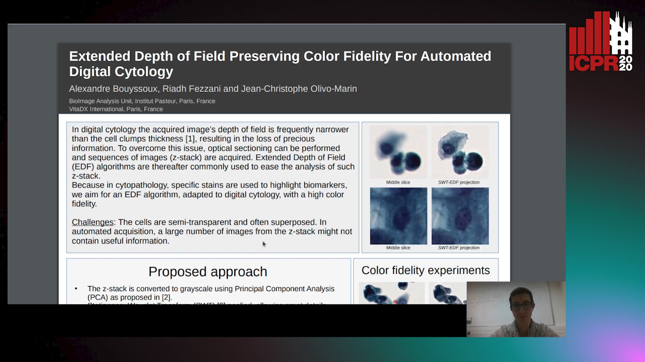
Auto-TLDR; Multi-Channel Extended Depth of Field for Digital cytology based on the stationary wavelet transform

Poster Similar

This paper presents a multi-channel Extended Depth of Field (EDF) method for digital cytology based on the stationary wavelet transform. With a coefficient selection rule adapted to a precise color recovery, a sharp image can be reconstructed even on images with transparent overlapping cells. The precision and the color fidelity of the proposed method is analyzed. Moreover, an experiment demonstrating the necessity of volume analysis in cytology to achieve precise segmentation on cell clumps is conducted, and the importance of color fidelity in this context is asserted. The proposed method was tested on pap-stained urothelial cells and gray-scale cervical cells with important overlapping.

Robust Localization of Retinal Lesions Via Weakly-Supervised Learning

Ruohan Zhao, Qin Li, Jane You

Responsive image

Auto-TLDR; Weakly Learning of Lesions in Fundus Images Using Multi-level Feature Maps and Classification Score

Slides Poster Similar

Retinal fundus images reveal the condition of retina, blood vessels and optic nerve. Retinal imaging is becoming widely adopted in clinical work because any subtle changes to the structures at the back of the eyes can affect the eyes and indicate the overall health. Machine learning, in particular deep learning by convolutional neural network (CNN), has been increasingly adopted for computer-aided detection (CAD) of retinal lesions. However, a significant barrier to the high performance of CNN based CAD approach is caused by the lack of sufficient labeled ground-truth image samples for training. Unlike the fully-supervised learning which relies on pixel-level annotation of pathology in fundus images, this paper presents a new approach to discriminate the location of various lesions based on image-level labels via weakly learning. More specifically, our proposed method leverages multi-level feature maps and classification score to cope with both bright and red lesions in fundus images. To enhance capability of learning less discriminative parts of objects (e.g. small blobs of microaneurysms opposed to bulk of exudates), the classifier is regularized by refining images with corresponding labels. The experimental results of the performance evaluation and benchmarking at both image-level and pixel-level on the public DIARETDB1 dataset demonstrate the feasibility and excellent potentials of our method in practice.

Motion and Region Aware Adversarial Learning for Fall Detection with Thermal Imaging

Vineet Mehta, Abhinav Dhall, Sujata Pal, Shehroz Khan

Responsive image

Auto-TLDR; Automatic Fall Detection with Adversarial Network using Thermal Imaging Camera

Slides Poster Similar

Automatic fall detection is a vital technology for ensuring health and safety of people. Home based camera systems for fall detection often put people's privacy at risk. Thermal cameras can partially/fully obfuscate facial features, thus preserving the privacy of a person. Another challenge is the less occurrence of falls in comparison to normal activities of daily living. As fall occurs rarely, it is non-trivial to learn algorithms due to class imbalance. To handle these problems, we formulate fall detection as an anomaly detection within an adversarial framework using thermal imaging camera. We present a novel adversarial network that comprise of two channel 3D convolutional auto encoders; one each handling video sequences and optical flow, which then reconstruct the thermal data and the optical flow input sequences. We introduce a differential constraint, a technique to track the region of interest and a joint discriminator to compute the reconstruction error. Larger reconstruction error indicates the occurrence of fall in a video sequence. The experiments on a publicly available thermal fall dataset show the superior results obtained in comparison to standard baseline.

Dual Stream Network with Selective Optimization for Skin Disease Recognition in Consumer Grade Images

Krishnam Gupta, Jaiprasad Rampure, Monu Krishnan, Ajit Narayanan, Nikhil Narayan

Responsive image

Auto-TLDR; A Deep Network Architecture for Skin Disease Localisation and Classification on Consumer Grade Images

Slides Poster Similar

Skin disease localisation and classification on consumer-grade images is more challenging compared to that on dermoscopic imaging. Consumer grade images refer to the images taken using commonly available imaging devices such as a mobile camera or a hand held digital camera. Such images, in addition to having the skin condition of interest in a very small area of the image, has other noisy non-clinical details introduced due to the lighting conditions and the distance of the hand held device from the anatomy at the time of acquisition. We propose a novel deep network architecture \& a new optimization strategy for classification with implicit localisation of skin diseases from clinical/consumer grade images. A weakly supervised segmentation algorithm is first employed to extract Region of Interests (RoI) from the image, the RoI and the original image form the two input streams of the proposed architecture. Each stream of the architecture learns high level and low level features from the original image and the RoI, respectively. The two streams are independently optimised until the loss stops decreasing after which both the streams are optimised collectively with the help of a third combiner sub-network. Such a strategy resulted in a 5% increase of accuracy over the current state-of-the-art methods on SD-198 dataset, which is publicly available. The proposed algorithm is also validated on a new dataset containing over 12,000 images across 75 different skin conditions. We intend to release this dataset as SD-75 to aid in the advancement of research on skin condition classification on consumer grade images.

OCT Image Segmentation Using NeuralArchitecture Search and SRGAN

Saba Heidari, Omid Dehzangi, Nasser M. Nasarabadi, Ali Rezai

Responsive image

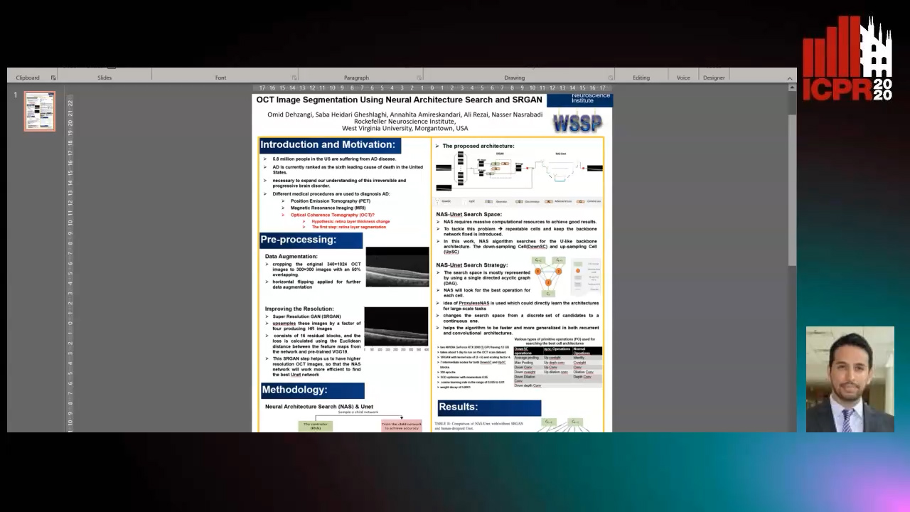
Auto-TLDR; Automatic Segmentation of Retinal Layers in Optical Coherence Tomography using Neural Architecture Search

Poster Similar

Alzheimer’s disease (AD) diagnosis is one of the major research areas in computational medicine. Optical coherence tomography (OCT) is a non-invasive, inexpensive, and timely efficient method that scans the human’s retina with depth. It has been hypothesized that the thickness of the retinal layers extracted from OCTs could be an efficient and effective biomarker for early diagnosis of AD. In this work, we aim to design a self-training model architecture for the task of segmenting the retinal layers in OCT scans. Neural architecture search (NAS) is a subfield of AutoML domain, which has a significant impact on improving the accuracy of machine vision tasks. We integrate the NAS algorithm with a Unet auto-encoder architecture as its backbone. Then, we employ our proposed model to segment the retinal nerve fiber layer in our preprocessed OCT images with the aim of AD diagnosis. In this work, we trained a super-resolution generative adversarial network on the raw OCT scans to improve the quality of the images before the modeling stage. In our architecture search strategy, different primitive operations suggested to find down- \& up-sampling Unet cell blocks and the binary gate method has been applied to make the search strategy more practical. Our architecture search method is empirically evaluated by training on the Unet and NAS-Unet from scratch. Specifically, the proposed NAS-Unet training significantly outperforms the baseline human-designed architecture by achieving 95.1\% in the mean Intersection over Union metric and 79.1\% in the Dice similarity coefficient.

Iterative Label Improvement: Robust Training by Confidence Based Filtering and Dataset Partitioning

Christian Haase-Schütz, Rainer Stal, Heinz Hertlein, Bernhard Sick

Responsive image

Auto-TLDR; Meta Training and Labelling for Unlabelled Data

Slides Poster Similar

State-of-the-art, high capacity deep neural networks not only require large amounts of labelled training data, they are also highly susceptible to labelling errors in this data, typically resulting in large efforts and costs and therefore limiting the applicability of deep learning. To alleviate this issue, we propose a novel meta training and labelling scheme that is able to use inexpensive unlabelled data by taking advantage of the generalization power of deep neural networks. We show experimentally that by solely relying on one network architecture and our proposed scheme of combining self-training with pseudolabels, both label quality and resulting model accuracy, can be improved significantly. Our method achieves state-of-the-art results, while being architecture agnostic and therefore broadly applicable. Compared to other methods dealing with erroneous labels, our approach does neither require another network to be trained, nor does it necessarily need an additional, highly accurate reference label set. Instead of removing samples from a labelled set, our technique uses additional sensor data without the need for manual labelling. Furthermore, our approach can be used for semi-supervised learning.

Automatic Tuberculosis Detection Using Chest X-Ray Analysis with Position Enhanced Structural Information

Hermann Jepdjio Nkouanga, Szilard Vajda

Responsive image

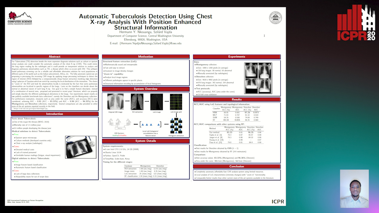
Auto-TLDR; Automatic Chest X-ray Screening for Tuberculosis in Rural Population using Localized Region on Interest

Slides Poster Similar

For Tuberculosis (TB) detection beside the more expensive diagnosis solutions such as culture or sputum smear analysis one could consider the automatic analysis of the chest X-ray (CXR). This could mimic the lung region reading by the radiologist and it could provide a cheap solution to analyze and diagnose pulmonary abnormalities such as TB which often co- occurs with HIV. This software based pulmonary screening can be a reliable and affordable solution for rural population in different parts of the world such as India, Africa, etc. Our fully automatic system is processing the incoming CXR image by applying image processing techniques to detect the region on interest (ROI) followed by a computationally cheap feature extraction involving edge detection using Laplacian of Gaussian which we enrich by counting the local distribution of the intensities. The choice to ”zoom in” the ROI and look for abnormalities locally is motivated by the fact that some pulmonary abnormalities are localized in specific regions of the lungs. Later on the classifiers can decide about the normal or abnormal nature of each lung X-ray. Our goal is to find a simple feature, instead of a combination of several ones, -proposed and promoted in recent years’ literature, which can properly describe the different pathological alterations in the lungs. Our experiments report results on two publicly available data collections1, namely the Shenzhen and the Montgomery collection. For performance evaluation, measures such as area under the curve (AUC), and accuracy (ACC) were considered, achieving AUC = 0.81 (ACC = 83.33%) and AUC = 0.96 (ACC = 96.35%) for the Montgomery and Schenzen collections, respectively. Several comparisons are also provided to other state- of-the-art systems reported recently in the field.

Segmenting Kidney on Multiple Phase CT Images Using ULBNet

Yanling Chi, Yuyu Xu, Gang Feng, Jiawei Mao, Sihua Wu, Guibin Xu, Weimin Huang

Responsive image

Auto-TLDR; A ULBNet network for kidney segmentation on multiple phase CT images

Poster Similar

Abstract—Segmentation of kidney on CT images is critical to computer-assisted surgical planning for kidney interventional therapy. Segmenting kidney manually is impractical in clinical, automatic segmentation is desirable. U-Net has been successful in medical image segmentation and is a promising candidate for the task. However, semantic gap still exists, especially when multiple phase images or multiple center images are involved. In this paper, we proposed an ULBNet to reduce the semantic gap and to improve segmentation performance. The proposed architecture includes new skip connections of local binary convolution (LBC). We also proposed a novel strategy of fast retraining a model for a new task without manually labelling required. We evaluated the network for kidney segmentation on multiple phase CT images. ULBNet resulted in an overall accuracy of 98.0% with comparison to Resunet 97.5%. Specifically, on the plain phase CT images, 98.1% resulted from ULBNet and 97.6% from Resunet; on the corticomedullay phase images, 97.8% from ULBNet and 97.2% from Resunet; on the nephrographic phase images, 97.6% from ULBNet and 97.4% from Resunet; on the excretory phase images, 98.1% from ULBNet and 97.4% from Resunet. The proposed network architecture performs better than Resunet on generalizing to multiple phase images.

Detection and Correspondence Matching of Corneal Reflections for Eye Tracking Using Deep Learning

Soumil Chugh, Braiden Brousseau, Jonathan Rose, Moshe Eizenman

Responsive image

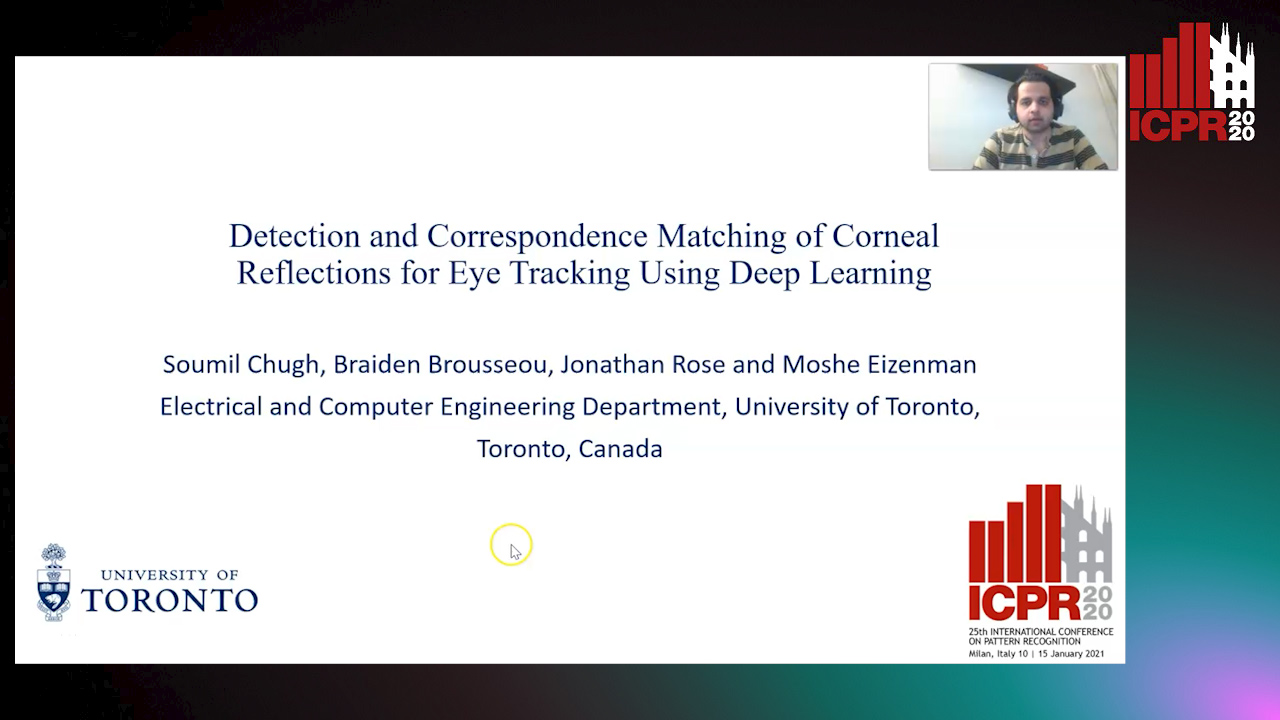
Auto-TLDR; A Fully Convolutional Neural Network for Corneal Reflection Detection and Matching in Extended Reality Eye Tracking Systems

Slides Poster Similar

Eye tracking systems that estimate the point-of-gaze are essential in extended reality (XR) systems as they enable new interaction paradigms and technological improvements. It is important for these systems to maintain accuracy when the headset moves relative to the head (known as device slippage) due to head movements or user adjustment. One of the most accurate eye tracking techniques, which is also insensitive to shifts of the system relative to the head, uses two or more infrared (IR) light emitting diodes to illuminate the eye and an IR camera to capture images of the eye. An essential step in estimating the point-of-gaze in these systems is the precise determination of the location of two or more corneal reflections (virtual images of the IR-LEDs that illuminate the eye) in images of the eye. Eye trackers tend to have multiple light sources to ensure at least one pair of reflections for each gaze position. The use of multiple light sources introduces a difficult problem: the need to match the corneal reflections with the corresponding light source over the range of expected eye movements. Corneal reflection detection and matching often fail in XR systems due to the proximity of camera and steep illumination angles of light sources with respect to the eye. The failures are caused by corneal reflections having varying shape and intensity levels or disappearance due to rotation of the eye, or the presence of spurious reflections. We have developed a fully convolutional neural network, based on the UNET architecture, that solves the detection and matching problem in the presence of spurious and missing reflections. Eye images of 25 people were collected in a virtual reality headset using a binocular eye tracking module consisting of five infrared light sources per eye. A set of 4,000 eye images were manually labelled for each of the corneal reflections, and data augmentation was used to generate a dataset of 40,000 images. The network is able to correctly identify and match 91% of corneal reflections present in the test set. This is comparable to a state-of-the-art deep learning system, but our approach requires 33 times less memory and executes 10 times faster. The proposed algorithm, when used in an eye tracker in a VR system, achieved an average mean absolute gaze error of 1°. This is a significant improvement over the state-of-the-art learning-based XR eye tracking systems that have reported gaze errors of 2-3°.

Prediction of Obstructive Coronary Artery Disease from Myocardial Perfusion Scintigraphy using Deep Neural Networks

Ida Arvidsson, Niels Christian Overgaard, Miguel Ochoa Figueroa, Jeronimo Rose, Anette Davidsson, Kalle Åström, Anders Heyden

Responsive image

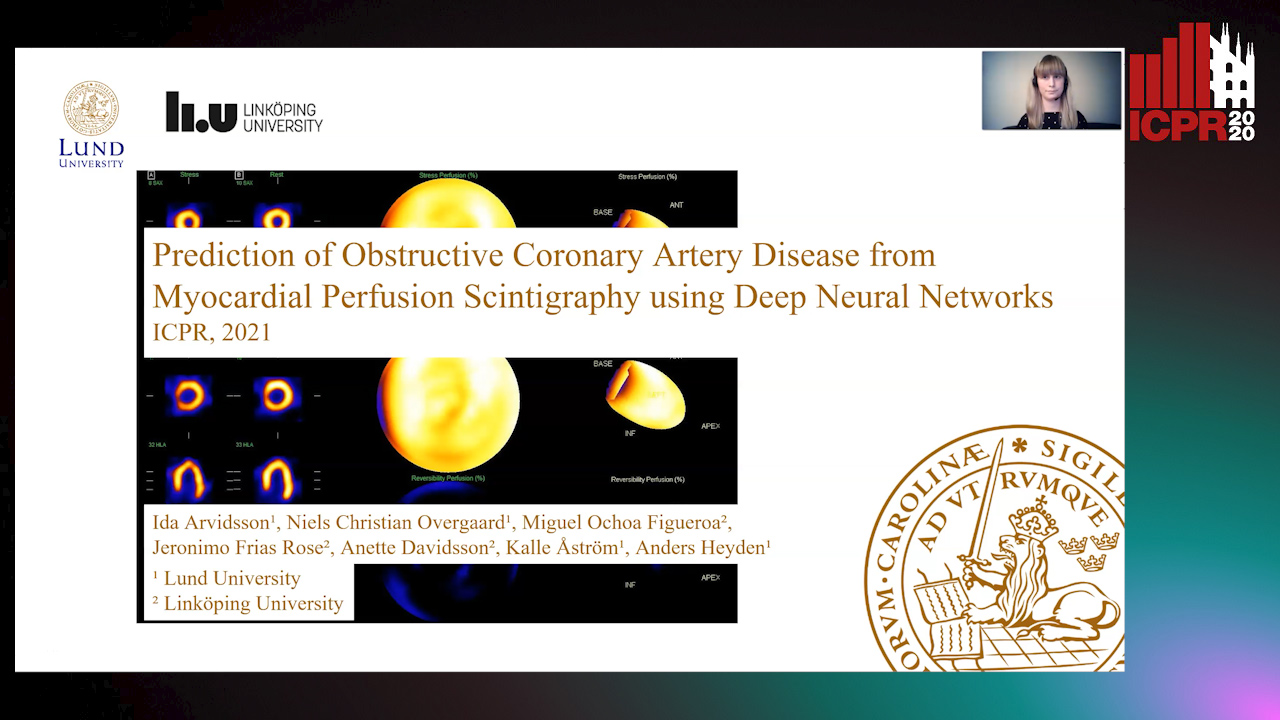
Auto-TLDR; A Deep Learning Algorithm for Multi-label Classification of Myocardial Perfusion Scintigraphy for Stable Ischemic Heart Disease

Slides Poster Similar

For diagnosis and risk assessment in patients with stable ischemic heart disease, myocardial perfusion scintigraphy is one of the most common cardiological examinations performed today. There are however many motivations for why an artificial intelligence algorithm would provide useful input to this task. For example to reduce the subjectiveness and save time for the nuclear medicine physicians working with this time consuming task. In this work we have developed a deep learning algorithm for multi-label classification based on a modified convolutional neural network to estimate probability of obstructive coronary artery disease in the left anterior artery, left circumflex artery and right coronary artery. The prediction is based on data from myocardial perfusion scintigraphy studies conducted in a dedicated Cadmium-Zinc-Telluride cardio camera (D-SPECT Spectrum Dynamics). Data from 588 patients was available, with stress images in both upright and supine position, as well as a number of auxiliary parameters such as angina symptoms and BMI. The data was used to train and evaluate the algorithm using 5-fold cross-validation. We achieve state-of-the-art results for this task with an area under the receiver operating characteristics curve of 0.89 as average on per-vessel level and 0.94 on per-patient level.

Automatically Gather Address Specific Dwelling Images Using Google Street View

Salman Khan, Carl Salvaggio

Responsive image

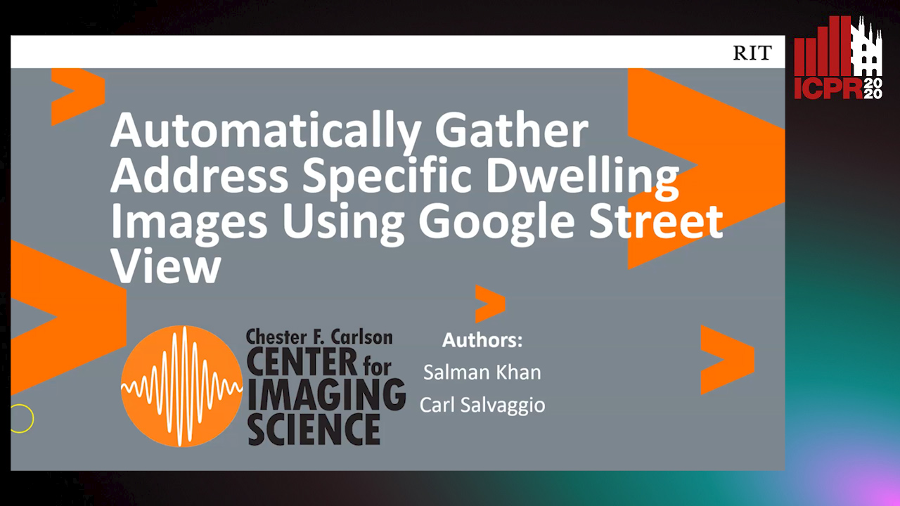
Auto-TLDR; Automatic Address Specific Dwelling Image Collection Using Google Street View Data

Slides Poster Similar

Exciting research is being conducted using Google’s street view imagery. Researchers can have access to training data that allows CNN training for topics ranging from assessing neighborhood environments to estimating the age of a building. However, due to the uncontrolled nature of imagery available via Google’s Street View API, data collection can be lengthy and tedious. In an effort to help researchers gather address specific dwelling images efficiently, we developed an innovative and novel way of automatically performing this task. It was accomplished by exploiting Google’s publicly available platform with a combination of 3 separate network types and postprocessing techniques. Our uniquely developed NMS technique helped achieve 99.4%, valid, address specific dwelling images.

Detecting Marine Species in Echograms Via Traditional, Hybrid, and Deep Learning Frameworks

Porto Marques Tunai, Alireza Rezvanifar, Melissa Cote, Alexandra Branzan Albu, Kaan Ersahin, Todd Mudge, Stephane Gauthier

Responsive image

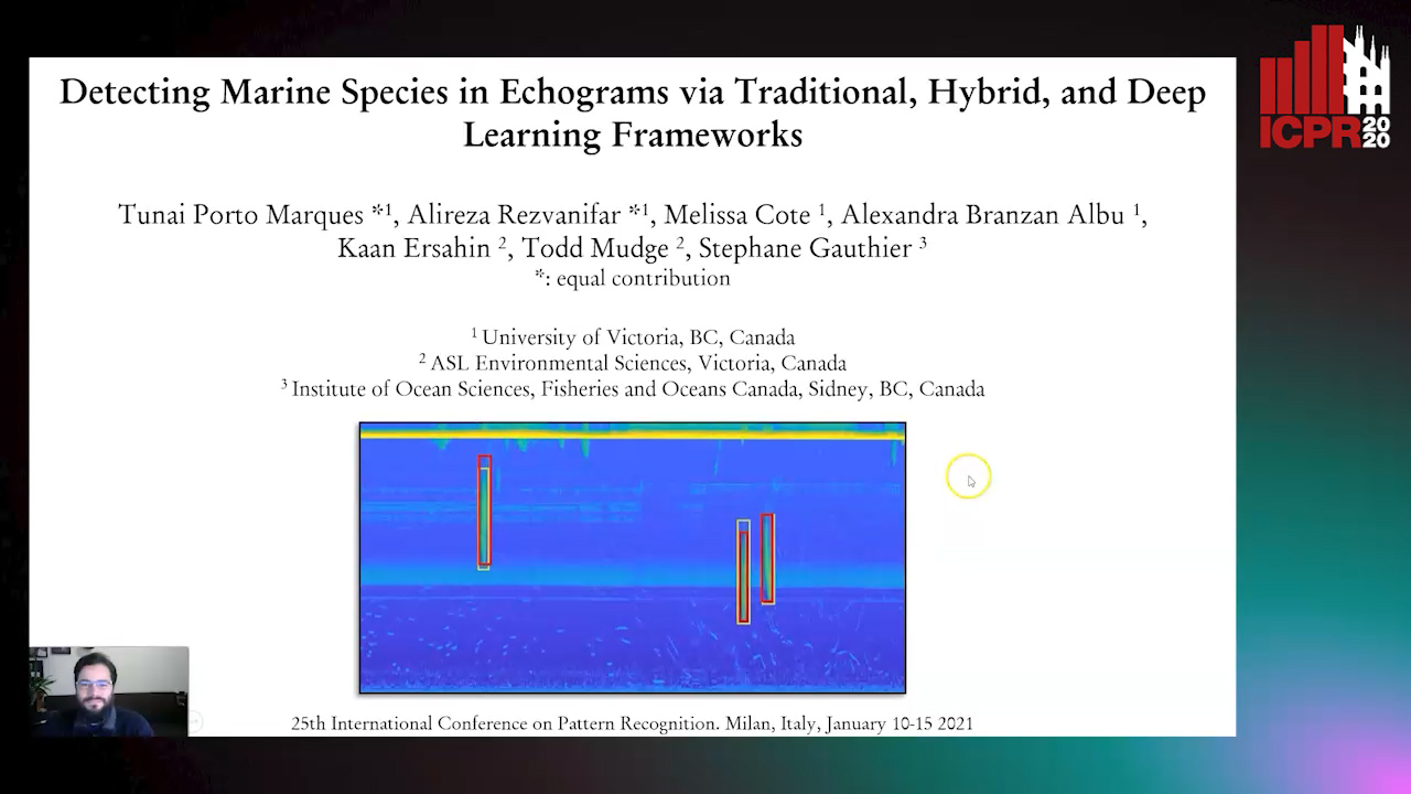
Auto-TLDR; End-to-End Deep Learning for Echogram Interpretation of Marine Species in Echograms

Slides Poster Similar

This paper provides a comprehensive comparative study of traditional, hybrid, and deep learning (DL) methods for detecting marine species in echograms. Acoustic backscatter data obtained from multi-frequency echosounders is visualized as echograms and typically interpreted by marine biologists via manual or semi-automatic methods, which are time-consuming. Challenges related to automatic echogram interpretation are the variable size and acoustic properties of the biological targets (marine life), along with significant inter-class similarities. Our study explores and compares three types of approaches that cover the entire range of machine learning methods. Based on our experimental results, we conclude that an end-to-end DL-based framework, that can be readily scaled to accommodate new species, is overall preferable to other learning approaches for echogram interpretation, even when only a limited number of annotated training samples is available.

DE-Net: Dilated Encoder Network for Automated Tongue Segmentation

Hui Tang, Bin Wang, Jun Zhou, Yongsheng Gao

Responsive image

Auto-TLDR; Automated Tongue Image Segmentation using De-Net

Slides Poster Similar

Automated tongue recognition is a growing research field due to global demand for personal health care. Using mobile devices to take tongue pictures is convenient and of low cost for tongue recognition. It is particularly suitable for self-health evaluation of the public. However, images taken by mobile devices are easily affected by various imaging environment, which makes fine segmentation a more challenging task compared with those taken by specialized acquisition devices. Deep learning approaches are promising for tongue image segmentation because they have powerful feature learning and representation capability. However, the successive pooling operations in these methods lead to loss of information on image details, making them fail when segmenting low-quality images captured by mobile devices. To address this issue, we propose a dilated encoder network (DE-Net) to capture more high-level features and get high-resolution output for automated tongue image segmentation. In addition, we construct two tongue image datasets which contain images taken by specialized devices and mobile devices, respectively, to verify the effectiveness of the proposed method. Experimental results on both datasets demonstrate that the proposed method outperforms the state-of-the-art methods in tongue image segmentation.