Fine-Tuning Convolutional Neural Networks: A Comprehensive Guide and Benchmark Analysis for Glaucoma Screening

Amed Mvoulana, Rostom Kachouri, Mohamed Akil

Responsive image

Auto-TLDR; Fine-tuning Convolutional Neural Networks for Glaucoma Screening

Slides Poster

This work aimed at giving a comprehensive and in-detailed guide on the route to fine-tuning Convolutional Neural Networks (CNNs) for glaucoma screening. Transfer learning consists in a promising alternative to train CNNs from stratch, to avoid the huge data and resources requirements. After a thorough study of five state-of-the-art CNNs architectures, a complete and well-explained strategy for fine-tuning these networks is proposed, using hyperparameter grid-searching and two-phase training approach. Excellent performance is reached on model evaluation, with a 0.9772 AUROC validation rate, giving arise to reliable glaucoma diagosis-help systems. Also, a benchmark analysis is conducted across all fine-tuned models, studying them according to performance indices such as model complexity and size, AUROC density and inference time. This in-depth analysis allows a rigorous comparison between model characteristics, and is useful for giving practioners important trademarks for prospective applications and deployments.

Similar papers

A Systematic Investigation on Deep Architectures for Automatic Skin Lesions Classification

Pierluigi Carcagni, Marco Leo, Andrea Cuna, Giuseppe Celeste, Cosimo Distante

Responsive image

Auto-TLDR; RegNet: Deep Investigation of Convolutional Neural Networks for Automatic Classification of Skin Lesions

Slides Poster Similar

Computer vision-based techniques are more and more employed in healthcare and medical fields nowadays in order, principally, to be as a support to the experienced medical staff to help them to make a quick and correct diagnosis. One of the hot topics in this arena concerns the automatic classification of skin lesions. Several promising works exist about it, mainly leveraging Convolutional Neural Networks (CNN), but proposed pipeline mainly rely on complex data preprocessing and there is no systematic investigation about how available deep models can actually reach the accuracy needed for real applications. In order to overcome these drawbacks, in this work, an end-to-end pipeline is introduced and some of the most recent Convolutional Neural Networks (CNNs) architectures are included in it and compared on the largest common benchmark dataset recently introduced. To this aim, for the first time in this application context, a new network design paradigm, namely RegNet, has been exploited to get the best models among a population of configurations. The paper introduces a threefold level of contribution and novelty with respect the previous literature: the deep investigation of several CNN architectures driving to a consistent improvement of the lesions recognition accuracy, the exploitation of a new network design paradigm able to study the behavior of populations of models and a deep discussion about pro and cons of each analyzed method paving the path towards new research lines.

Investigating and Exploiting Image Resolution for Transfer Learning-Based Skin Lesion Classification

Amirreza Mahbod, Gerald Schaefer, Chunliang Wang, Rupert Ecker, Georg Dorffner, Isabella Ellinger

Responsive image

Auto-TLDR; Fine-tuned Neural Networks for Skin Lesion Classification Using Dermoscopic Images

Slides Poster Similar

Skin cancer is among the most common cancer types. Dermoscopic image analysis improves the diagnostic accuracy for detection of malignant melanoma and other pigmented skin lesions when compared to unaided visual inspection. Hence, computer-based methods to support medical experts in the diagnostic procedure are of great interest. Fine-tuning pre-trained convolutional neural networks (CNNs) has been shown to work well for skin lesion classification. Pre-trained CNNs are usually trained with natural images of a fixed image size which is typically significantly smaller than captured skin lesion images and consequently dermoscopic images are downsampled for fine-tuning. However, useful medical information may be lost during this transformation. In this paper, we explore the effect of input image size on skin lesion classification performance of fine-tuned CNNs. For this, we resize dermoscopic images to different resolutions, ranging from 64x64 to 768x768 pixels and investigate the resulting classification performance of three well-established CNNs, namely DenseNet-121, ResNet-18, and ResNet-50. Our results show that using very small images (of size 64x64 pixels) degrades the classification performance, while images of size 128x128 pixels and above support good performance with larger image sizes leading to slightly improved classification. We further propose a novel fusion approach based on a three-level ensemble strategy that exploits multiple fine-tuned networks trained with dermoscopic images at various sizes. When applied on the ISIC 2017 skin lesion classification challenge, our fusion approach yields an area under the receiver operating characteristic curve of 89.2% and 96.6% for melanoma classification and seborrheic keratosis classification, respectively, outperforming state-of-the-art algorithms.

Bridging the Gap between Natural and Medical Images through Deep Colorization

Lia Morra, Luca Piano, Fabrizio Lamberti, Tatiana Tommasi

Responsive image

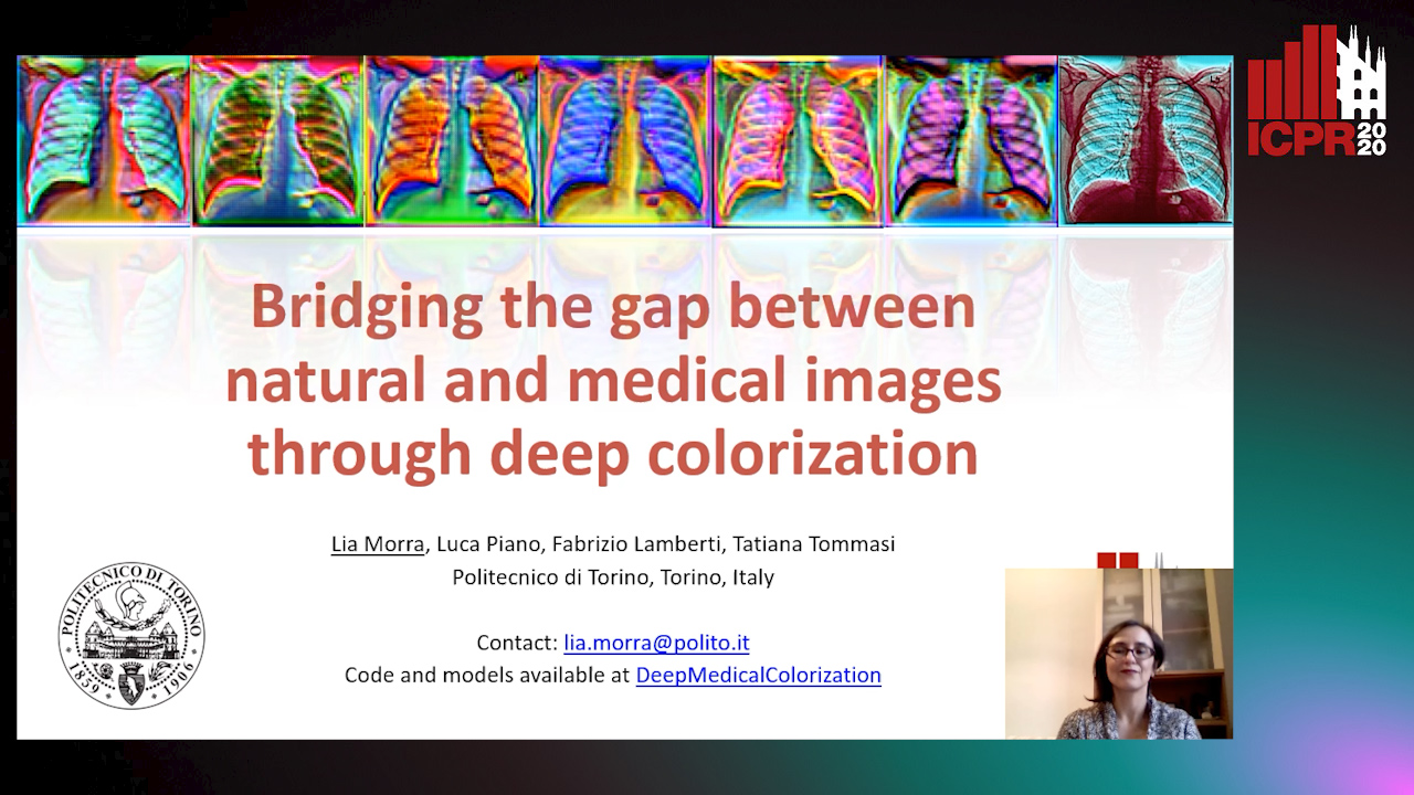
Auto-TLDR; Transfer Learning for Diagnosis on X-ray Images Using Color Adaptation

Slides Poster Similar

Deep learning has thrived by training on large-scale datasets. However, in many applications, as for medical image diagnosis, getting massive amount of data is still prohibitive due to privacy, lack of acquisition homogeneity and annotation cost. In this scenario transfer learning from natural image collections is a standard practice that attempts to tackle shape, texture and color discrepancy all at once through pretrained model fine-tuning. In this work we propose to disentangle those challenges and design a dedicated network module that focuses on color adaptation. We combine learning from scratch of the color module with transfer learning of different classification backbones obtaining an end-to-end, easy-to-train architecture for diagnostic image recognition on X-ray images. Extensive experiments show how our approach is particularly efficient in case of data scarcity and provides a new path for further transferring the learned color information across multiple medical datasets.

Inception Based Deep Learning Architecture for Tuberculosis Screening of Chest X-Rays

Dipayan Das, K.C. Santosh, Umapada Pal

Responsive image

Auto-TLDR; End to End CNN-based Chest X-ray Screening for Tuberculosis positive patients in the severely resource constrained regions of the world

Slides Poster Similar

The motivation for this work is the primary need of screening Tuberculosis (TB) positive patients in the severely resource constrained regions of the world. Chest X-ray (CXR) is considered to be a promising indicator for the onset of TB, but the lack of skilled radiologists in such regions degrades the situation. Therefore, several computer aided diagnosis (CAD) systems have been proposed to solve the decision making problem, which includes hand engineered feature extraction methods to deep learning or Convolutional Neural Network (CNN) based methods. Feature extraction, being a time and resource intensive process, often delays the process of mass screening. Hence an end to end CNN architecture is proposed in this work to solve the problem. Two benchmark CXR datasets have been used in this work, collected from Shenzhen (China) and Montgomery County (USA), on which the proposed methodology achieved a maximum abnormality detection accuracy (ACC) of 91.7\% (0.96 AUC) and 87.47\% (0.92 AUC) respectively. To the greatest of our knowledge, the obtained results are marginally superior to the state of the art results that have solely used deep learning methodologies on the aforementioned datasets.

Transfer Learning through Weighted Loss Function and Group Normalization for Vessel Segmentation from Retinal Images

Abdullah Sarhan, Jon Rokne, Reda Alhajj, Andrew Crichton

Responsive image

Auto-TLDR; Deep Learning for Segmentation of Blood Vessels in Retinal Images

Slides Poster Similar

The vascular structure of blood vessels is important in diagnosing retinal conditions such as glaucoma and diabetic retinopathy. Accurate segmentation of these vessels can help in detecting retinal objects such as the optic disc and optic cup and hence determine if there are damages to these areas. Moreover, the structure of the vessels can help in diagnosing glaucoma. The rapid development of digital imaging and computer-vision techniques has increased the potential for developing approaches for segmenting retinal vessels. In this paper, we propose an approach for segmenting retinal vessels that uses deep learning along with transfer learning. We adapted the U-Net structure to use a customized InceptionV3 as the encoder and used multiple skip connections to form the decoder. Moreover, we used a weighted loss function to handle the issue of class imbalance in retinal images. Furthermore, we contributed a new dataset to this field. We tested our approach on six publicly available datasets and a newly created dataset. We achieved an average accuracy of 95.60\% and a Dice coefficient of 80.98\%. The results obtained from comprehensive experiments demonstrate the robustness of our approach to the segmentation of blood vessels in retinal images obtained from different sources. Our approach results in greater segmentation accuracy than other approaches.

Robust Localization of Retinal Lesions Via Weakly-Supervised Learning

Ruohan Zhao, Qin Li, Jane You

Responsive image

Auto-TLDR; Weakly Learning of Lesions in Fundus Images Using Multi-level Feature Maps and Classification Score

Slides Poster Similar

Retinal fundus images reveal the condition of retina, blood vessels and optic nerve. Retinal imaging is becoming widely adopted in clinical work because any subtle changes to the structures at the back of the eyes can affect the eyes and indicate the overall health. Machine learning, in particular deep learning by convolutional neural network (CNN), has been increasingly adopted for computer-aided detection (CAD) of retinal lesions. However, a significant barrier to the high performance of CNN based CAD approach is caused by the lack of sufficient labeled ground-truth image samples for training. Unlike the fully-supervised learning which relies on pixel-level annotation of pathology in fundus images, this paper presents a new approach to discriminate the location of various lesions based on image-level labels via weakly learning. More specifically, our proposed method leverages multi-level feature maps and classification score to cope with both bright and red lesions in fundus images. To enhance capability of learning less discriminative parts of objects (e.g. small blobs of microaneurysms opposed to bulk of exudates), the classifier is regularized by refining images with corresponding labels. The experimental results of the performance evaluation and benchmarking at both image-level and pixel-level on the public DIARETDB1 dataset demonstrate the feasibility and excellent potentials of our method in practice.

Semi-Supervised Generative Adversarial Networks with a Pair of Complementary Generators for Retinopathy Screening

Yingpeng Xie, Qiwei Wan, Hai Xie, En-Leng Tan, Yanwu Xu, Baiying Lei

Responsive image

Auto-TLDR; Generative Adversarial Networks for Retinopathy Diagnosis via Fundus Images

Slides Poster Similar

Several typical types of retinopathy are major causes of blindness. However, early detection of retinopathy is quite not easy since few symptoms are observable in the early stage, attributing to the development of non-mydriatic retinal camera. These camera produces high-resolution retinal fundus images provide the possibility of Computer-Aided-Diagnosis (CAD) via deep learning to assist diagnosing retinopathy. Deep learning algorithms usually rely on a great number of labelled images which are expensive and time-consuming to obtain in the medical imaging area. Moreover, the random distribution of various lesions which often vary greatly in size also brings significant challenges to learn discriminative information from high-resolution fundus image. In this paper, we present generative adversarial networks simultaneously equipped with "good" generator and "bad" generator (GBGANs) to make up for the incomplete data distribution provided by limited fundus images. To improve the generative feasibility of generator, we introduce into pre-trained feature extractor to acquire condensed feature for each fundus image in advance. Experimental results on integrated three public iChallenge datasets show that the proposed GBGANs could fully utilize the available fundus images to identify retinopathy with little label cost.

Improving Model Accuracy for Imbalanced Image Classification Tasks by Adding a Final Batch Normalization Layer: An Empirical Study

Veysel Kocaman, Ofer M. Shir, Thomas Baeck

Responsive image

Auto-TLDR; Exploiting Batch Normalization before the Output Layer in Deep Learning for Minority Class Detection in Imbalanced Data Sets

Slides Poster Similar

Some real-world domains, such as Agriculture and Healthcare, comprise early-stage disease indications whose recording constitutes a rare event, and yet, whose precise detection at that stage is critical. In this type of highly imbalanced classification problems, which encompass complex features, deep learning (DL) is much needed because of its strong detection capabilities. At the same time, DL is observed in practice to favor majority over minority classes and consequently suffer from inaccurate detection of the targeted early-stage indications. To simulate such scenarios, we artificially generate skewness (99% vs. 1%) for certain plant types out of the PlantVillage dataset as a basis for classification of scarce visual cues through transfer learning. By randomly and unevenly picking healthy and unhealthy samples from certain plant types to form a training set, we consider a base experiment as fine-tuning ResNet34 and VGG19 architectures and then testing the model performance on a balanced dataset of healthy and unhealthy images. We empirically observe that the initial F1 test score jumps from 0.29 to 0.95 for the minority class upon adding a final Batch Normalization (BN) layer just before the output layer in VGG19. We demonstrate that utilizing an additional BN layer before the output layer in modern CNN architectures has a considerable impact in terms of minimizing the training time and testing error for minority classes in highly imbalanced data sets. Moreover, when the final BN is employed, trying to minimize validation and training losses may not be an optimal way for getting a high F1 test score for minority classes in anomaly detection problems. That is, the network might perform better even if it is not ‘confident’ enough while making a prediction; leading to another discussion about why softmax output is not a good uncertainty measure for DL models.

A Comparison of Neural Network Approaches for Melanoma Classification

Maria Frasca, Michele Nappi, Michele Risi, Genoveffa Tortora, Alessia Auriemma Citarella

Responsive image

Auto-TLDR; Classification of Melanoma Using Deep Neural Network Methodologies

Slides Poster Similar

Melanoma is the deadliest form of skin cancer and it is diagnosed mainly visually, starting from initial clinical screening and followed by dermoscopic analysis, biopsy and histopathological examination. A dermatologist’s recognition of melanoma may be subject to errors and may take some time to diagnose it. In this regard, deep learning can be useful in the study and classification of skin cancer. In particular, by classifying images with Deep Neural Network methodologies, it is possible to obtain comparable or even superior results compared to those of dermatologists. In this paper, we propose a methodology for the classification of melanoma by adopting different deep learning techniques applied to a common dataset, composed of images from the ISIC dataset and consisting of different types of skin diseases, including melanoma on which we applied a specific pre-processing phase. In particular, a comparison of the results is performed in order to select the best effective neural network to be applied to the problem of recognition and classification of melanoma. Moreover, we also evaluate the impact of the pre- processing phase on the final classification. Different metrics such as accuracy, sensitivity, and specificity have been selected to assess the goodness of the adopted neural networks and compare them also with the manual classification of dermatologists.

Learn to Segment Retinal Lesions and Beyond

Qijie Wei, Xirong Li, Weihong Yu, Xiao Zhang, Yongpeng Zhang, Bojie Hu, Bin Mo, Di Gong, Ning Chen, Dayong Ding, Youxin Chen

Responsive image

Auto-TLDR; Multi-task Lesion Segmentation and Disease Classification for Diabetic Retinopathy Grading

Poster Similar

Towards automated retinal screening, this paper makes an endeavor to simultaneously achieve pixel-level retinal lesion segmentation and image-level disease classification. Such a multi-task approach is crucial for accurate and clinically interpretable disease diagnosis. Prior art is insufficient due to three challenges, i.e., lesions lacking objective boundaries, clinical importance of lesions irrelevant to their size, and the lack of one-to-one correspondence between lesion and disease classes. This paper attacks the three challenges in the context of diabetic retinopathy (DR) grading. We propose Lesion-Net, a new variant of fully convolutional networks, with its expansive path re- designed to tackle the first challenge. A dual Dice loss that leverages both semantic segmentation and image classification losses is introduced to resolve the second challenge. Lastly, we build a multi-task network that employs Lesion-Net as a side- attention branch for both DR grading and result interpretation. A set of 12K fundus images is manually segmented by 45 ophthalmologists for 8 DR-related lesions, resulting in 290K manual segments in total. Extensive experiments on this large- scale dataset show that our proposed approach surpasses the prior art for multiple tasks including lesion segmentation, lesion classification and DR grading.

Automatic Semantic Segmentation of Structural Elements related to the Spinal Cord in the Lumbar Region by Using Convolutional Neural Networks

Jhon Jairo Sáenz Gamboa, Maria De La Iglesia-Vaya, Jon Ander Gómez

Responsive image

Auto-TLDR; Semantic Segmentation of Lumbar Spine Using Convolutional Neural Networks

Slides Poster Similar

This work addresses the problem of automatically segmenting the MR images corresponding to the lumbar spine. The purpose is to detect and delimit the different structural elements like vertebrae, intervertebral discs, nerves, blood vessels, etc. This task is known as semantic segmentation. The approach proposed in this work is based on convolutional neural networks whose output is a mask where each pixel from the input image is classified into one of the possible classes. Classes were defined by radiologists and correspond to structural elements and tissues. The proposed network architectures are variants of the U-Net. Several complementary blocks were used to define the variants: spatial attention models, deep supervision and multi-kernels at input, this last block type is based on the idea of inception. Those architectures which got the best results are described in this paper, and their results are discussed. Two of the proposed architectures outperform the standard U-Net used as baseline.

Planar 3D Transfer Learning for End to End Unimodal MRI Unbalanced Data Segmentation

Martin Kolarik, Radim Burget, Carlos M. Travieso-Gonzalez, Jan Kocica

Responsive image

Auto-TLDR; Planar 3D Res-U-Net Network for Unbalanced 3D Image Segmentation using Fluid Attenuation Inversion Recover

Slides Similar

We present a novel approach of 2D to 3D transfer learning based on mapping pre-trained 2D convolutional neural network weights into planar 3D kernels. The method is validated by proposed planar 3D res-u-net network with encoder transferred from the 2D VGG-16 which is applied for a single-stage unbalanced 3D image data segmentation. In particular, we evaluate the method on the MICCAI 2016 MS lesion segmentation challenge dataset utilizing solely Fluid Attenuation Inversion Recover (FLAIR) sequence without brain extraction for training and inference to simulate real medical praxis. The planar 3D res-u-net network performed the best both in sensitivity and Dice score amongst end to end methods processing raw MRI scans and achieved comparable Dice score to a state-of-the-art unimodal not end to end approach. Complete source code was released under the open-source license and this paper is in compliance with the Machine learning Reproducibility Checklist. By implementing practical transfer learning for 3D data representation we were able to successfully segment heavily unbalanced data without selective sampling and achieved more reliable results using less training data in single modality. From medical perspective, the unimodal approach gives an advantage in real praxis as it does not require co-registration nor additional scanning time during examination. Although modern medical imaging methods capture high resolution 3D anatomy scans suitable for computer aided detection system processing, deployment of automatic systems for interpretation of radiology imaging is still rather theoretical in many medical areas. Our work aims to bridge the gap offering solution for partial research questions.

Confidence Calibration for Deep Renal Biopsy Immunofluorescence Image Classification

Federico Pollastri, Juan Maroñas, Federico Bolelli, Giulia Ligabue, Roberto Paredes, Riccardo Magistroni, Costantino Grana

Responsive image

Auto-TLDR; A Probabilistic Convolutional Neural Network for Immunofluorescence Classification in Renal Biopsy

Slides Poster Similar

With this work we tackle immunofluorescence classification in renal biopsy, employing state-of-the-art Convolutional Neural Networks. In this setting, the aim of the probabilistic model is to assist an expert practitioner towards identifying the location pattern of antibody deposits within a glomerulus. Since modern neural networks often provide overconfident outputs, we stress the importance of having a reliable prediction, demonstrating that Temperature Scaling, a recently introduced re-calibration technique, can be successfully applied to immunofluorescence classification in renal biopsy. Experimental results demonstrate that the designed model yields good accuracy on the specific task, and that Temperature Scaling is able to provide reliable probabilities, which are highly valuable for such a task given the low inter-rater agreement.

Which are the factors affecting the performance of audio surveillance systems?

Antonio Greco, Antonio Roberto, Alessia Saggese, Mario Vento

Responsive image

Auto-TLDR; Sound Event Recognition Using Convolutional Neural Networks and Visual Representations on MIVIA Audio Events

Slides Similar

Sound event recognition systems are rapidly becoming part of our life, since they can be profitably used in several vertical markets, ranging from audio security applications to scene classification and multi-modal analysis in social robotics. In the last years, a not negligible part of the scientific community started to apply Convolutional Neural Networks (CNNs) to image-based representations of the audio stream, due to their successful adoption in almost all the computer vision tasks. In this paper, we carry out a detailed benchmark of various widely used CNN architectures and visual representations on a popular dataset, namely the MIVIA Audio Events database. Our analysis is aimed at understanding how these factors affect the sound event recognition performance with a particular focus on the false positive rate, very relevant in audio surveillance solutions. In fact, although most of the proposed solutions achieve a high recognition rate, the capability of distinguishing the events-of-interest from the background is often not yet sufficient for real systems, and prevent its usage in real applications. Our comprehensive experimental analysis investigates this aspect and allows to identify useful design guidelines for increasing the specificity of sound event recognition systems.

Deep Transfer Learning for Alzheimer’s Disease Detection

Nicole Cilia, Claudio De Stefano, Francesco Fontanella, Claudio Marrocco, Mario Molinara, Alessandra Scotto Di Freca

Responsive image

Auto-TLDR; Automatic Detection of Handwriting Alterations for Alzheimer's Disease Diagnosis using Dynamic Features

Slides Poster Similar

Early detection of Alzheimer’s Disease (AD) is essential in order to initiate therapies that can reduce the effects of such a disease, improving both life quality and life expectancy of patients. Among all the activities carried out in our daily life, handwriting seems one of the first to be influenced by the arise of neurodegenerative diseases. For this reason, the analysis of handwriting and the study of its alterations has become of great interest in this research field in order to make a diagnosis as early as possible. In recent years, many studies have tried to use classification algorithms applied to handwritings to implement decision support systems for AD diagnosis. A key issue for the use of these techniques is the detection of effective features, that allow the system to distinguish the natural handwriting alterations due to age, from those caused by neurodegenerative disorders. In this context, many interesting results have been published in the literature in which the features have been typically selected by hand, generally considering the dynamics of the handwriting process in order to detect motor disorders closely related to AD. Features directly derived from handwriting generation models can be also very helpful for AD diagnosis. It should be remarked, however, that the above features do not consider changes in the shape of handwritten traces, which may occur as a consequence of neurodegenerative diseases, as well as the correlation among shape alterations and changes in the dynamics of the handwriting process. Moving from these considerations, the aim of this study is to verify if the combined use of both shape and dynamic features allows a decision support system to improve performance for AD diagnosis. To this purpose, starting from a database of on-line handwriting samples, we generated for each of them a synthetic off-line colour image, where the colour of each elementary trait encodes, in the three RGB channels, the dynamic information associated to that trait. Finally, we exploited the capability of Deep Neural Networks (DNN) to automatically extract features from raw images. The experimental comparison of the results obtained by using standard features and features extracted according the above procedure, confirmed the effectiveness of our approach.

Supporting Skin Lesion Diagnosis with Content-Based Image Retrieval

Stefano Allegretti, Federico Bolelli, Federico Pollastri, Sabrina Longhitano, Giovanni Pellacani, Costantino Grana

Responsive image

Auto-TLDR; Skin Images Retrieval Using Convolutional Neural Networks for Skin Lesion Classification and Segmentation

Slides Poster Similar

Given the relevance of skin cancer, many attempts have been dedicated to the creation of automated devices that could assist both expert and beginner dermatologists towards fast and early diagnosis of skin lesions. In recent years, tasks such as skin lesion classification and segmentation have been extensively addressed with deep learning algorithms, which in some cases reach a diagnostic accuracy comparable to that of expert physicians. However, the general lack of interpretability and reliability severely hinders the ability of those approaches to actually support dermatologists in the diagnosis process. In this paper a novel skin images retrieval system is presented, which exploits features extracted by Convolutional Neural Networks to gather similar images from a publicly available dataset, in order to assist the diagnosis process of both expert and novice practitioners. In the proposed framework, Resnet-50 is initially trained for the classification of dermoscopic images; then, the feature extraction part is isolated, and an embedding network is build on top of it. The embedding learns an alternative representation, which allows to check image similarity by means of a distance measure. Experimental results reveal that the proposed method is able to select meaningful images, which can effectively boost the classification accuracy of human dermatologists.

PCANet: Pyramid Context-Aware Network for Retinal Vessel Segmentation

Yi Zhang, Yixuan Chen, Kai Zhang

Responsive image

Auto-TLDR; PCANet: Adaptive Context-Aware Network for Automated Retinal Vessel Segmentation

Slides Poster Similar

Automated retinal vessel segmentation plays an important role in the diagnosis of some diseases such as diabetes, arteriosclerosis and hypertension. Recent works attempt to improve segmentation performance by exploring either global or local contexts. However, the context demands are varying from regions in each image and different levels of network. To address these problems, we propose Pyramid Context-aware Network (PCANet), which can adaptively capture multi-scale context representations. Specifically, PCANet is composed of multiple Adaptive Context-aware (ACA) blocks arranged in parallel, each of which can adaptively obtain the context-aware features by estimating affinity coefficients at a specific scale under the guidance of global contextual dependencies. Meanwhile, we import ACA blocks with specific scales in different levels of the network to obtain a coarse-to-fine result. Furthermore, an integrated test-time augmentation method is developed to further boost the performance of PCANet. Finally, extensive experiments demonstrate the effectiveness of the proposed PCANet, and state-of-the-art performances are achieved with AUCs of 0.9866, 0.9886 and F1 Scores of 0.8274, 0.8371 on two public datasets, DRIVE and STARE, respectively.

Weight Estimation from an RGB-D Camera in Top-View Configuration

Marco Mameli, Marina Paolanti, Nicola Conci, Filippo Tessaro, Emanuele Frontoni, Primo Zingaretti

Responsive image

Auto-TLDR; Top-View Weight Estimation using Deep Neural Networks

Slides Poster Similar

The development of so-called soft-biometrics aims at providing information related to the physical and behavioural characteristics of a person. This paper focuses on bodyweight estimation based on the observation from a top-view RGB-D camera. In fact, the capability to estimate the weight of a person can be of help in many different applications, from health-related scenarios to business intelligence and retail analytics. To deal with this issue, a TVWE (Top-View Weight Estimation) framework is proposed with the aim of predicting the weight. The approach relies on the adoption of Deep Neural Networks (DNNs) that have been trained on depth data. Each network has also been modified in its top section to replace classification with prediction inference. The performance of five state-of-art DNNs has been compared, namely VGG16, ResNet, Inception, DenseNet and Efficient-Net. In addition, a convolutional auto-encoder has also been included for completeness. Considering the limited literature in this domain, the TVWE framework has been evaluated on a new publicly available dataset: “VRAI Weight estimation Dataset”, which also collects, for each subject, labels related to weight, gender, and height. The experimental results have demonstrated that the proposed methods are suitable for this task, bringing different and significant insights for the application of the solution in different domains.

Trainable Spectrally Initializable Matrix Transformations in Convolutional Neural Networks

Michele Alberti, Angela Botros, Schuetz Narayan, Rolf Ingold, Marcus Liwicki, Mathias Seuret

Responsive image

Auto-TLDR; Trainable and Spectrally Initializable Matrix Transformations for Neural Networks

Slides Poster Similar

In this work, we introduce a new architectural component to Neural Networks (NN), i.e., trainable and spectrally initializable matrix transformations on feature maps. While previous literature has already demonstrated the possibility of adding static spectral transformations as feature processors, our focus is on more general trainable transforms. We study the transforms in various architectural configurations on four datasets of different nature: from medical (ColorectalHist, HAM10000) and natural (Flowers) images to historical documents (CB55). With rigorous experiments that control for the number of parameters and randomness, we show that networks utilizing the introduced matrix transformations outperform vanilla neural networks. The observed accuracy increases appreciably across all datasets. In addition, we show that the benefit of spectral initialization leads to significantly faster convergence, as opposed to randomly initialized matrix transformations. The transformations are implemented as auto-differentiable PyTorch modules that can be incorporated into any neural network architecture. The entire code base is open-source.

Enhancing Semantic Segmentation of Aerial Images with Inhibitory Neurons

Ihsan Ullah, Sean Reilly, Michael Madden

Responsive image

Auto-TLDR; Lateral Inhibition in Deep Neural Networks for Object Recognition and Semantic Segmentation

Slides Poster Similar

In a Convolutional Neural Network, each neuron in the output feature map takes input from the neurons in its receptive field. This receptive field concept plays a vital role in today's deep neural networks. However, inspired by neuro-biological research, it has been proposed to add inhibitory neurons outside the receptive field, which may enhance the performance of neural network models. In this paper, we begin with deep network architectures such as VGG and ResNet, and propose an approach to add lateral inhibition in each output neuron to reduce its impact on its neighbours, both in fine-tuning pre-trained models and training from scratch. Our experiments show that notable improvements upon prior baseline deep models can be achieved. A key feature of our approach is that it is easy to add to baseline models; it can be adopted in any model containing convolution layers, and we demonstrate its value in applications including object recognition and semantic segmentation of aerial images, where we show state-of-the-art result on the Aeroscape dataset. On semantic segmentation tasks, our enhancement shows 17.43% higher mIoU than a single baseline model on a single source (the Aeroscape dataset), 13.43% higher performance than an ensemble model on the same single source, and 7.03% higher than an ensemble model on multiple sources (segmentation datasets). Our experiments illustrate the potential impact of using inhibitory neurons in deep learning models, and they also show better results than the baseline models that have standard convolutional layer.

BAT Optimized CNN Model Identifies Water Stress in Chickpea Plant Shoot Images

Shiva Azimi, Taranjit Kaur, Tapan Gandhi

Responsive image

Auto-TLDR; BAT Optimized ResNet-18 for Stress Classification of chickpea shoot images under water deficiency

Slides Poster Similar

Stress due to water deficiency in plants can significantly lower the agricultural yield. It can affect many visible plant traits such as size and surface area, the number of leaves and their color, etc. In recent years, computer vision-based plant phenomics has emerged as a promising tool for plant research and management. Such techniques have the advantage of being non-destructive, non-evasive, fast, and offer high levels of automation. Pulses like chickpeas play an important role in ensuring food security in poor countries owing to their high protein and nutrition content. In the present work, we have built a dataset comprising of two varieties of chickpea plant shoot images under different moisture stress conditions. Specifically, we propose a BAT optimized ResNet-18 model for classifying stress induced by water deficiency using chickpea shoot images. BAT algorithm identifies the optimal value of the mini-batch size to be used for training rather than employing the traditional manual approach of trial and error. Experimentation on two crop varieties (JG and Pusa) reveals that BAT optimized approach achieves an accuracy of 96% and 91% for JG and Pusa varieties that is better than the traditional method by 4%. The experimental results are also compared with state of the art CNN models like Alexnet, GoogleNet, and ResNet-50. The comparison results demonstrate that the proposed BAT optimized ResNet-18 model achieves higher performance than the comparison counterparts.

Dual Stream Network with Selective Optimization for Skin Disease Recognition in Consumer Grade Images

Krishnam Gupta, Jaiprasad Rampure, Monu Krishnan, Ajit Narayanan, Nikhil Narayan

Responsive image

Auto-TLDR; A Deep Network Architecture for Skin Disease Localisation and Classification on Consumer Grade Images

Slides Poster Similar

Skin disease localisation and classification on consumer-grade images is more challenging compared to that on dermoscopic imaging. Consumer grade images refer to the images taken using commonly available imaging devices such as a mobile camera or a hand held digital camera. Such images, in addition to having the skin condition of interest in a very small area of the image, has other noisy non-clinical details introduced due to the lighting conditions and the distance of the hand held device from the anatomy at the time of acquisition. We propose a novel deep network architecture \& a new optimization strategy for classification with implicit localisation of skin diseases from clinical/consumer grade images. A weakly supervised segmentation algorithm is first employed to extract Region of Interests (RoI) from the image, the RoI and the original image form the two input streams of the proposed architecture. Each stream of the architecture learns high level and low level features from the original image and the RoI, respectively. The two streams are independently optimised until the loss stops decreasing after which both the streams are optimised collectively with the help of a third combiner sub-network. Such a strategy resulted in a 5% increase of accuracy over the current state-of-the-art methods on SD-198 dataset, which is publicly available. The proposed algorithm is also validated on a new dataset containing over 12,000 images across 75 different skin conditions. We intend to release this dataset as SD-75 to aid in the advancement of research on skin condition classification on consumer grade images.

Rethinking of Deep Models Parameters with Respect to Data Distribution

Shitala Prasad, Dongyun Lin, Yiqun Li, Sheng Dong, Zaw Min Oo

Responsive image

Auto-TLDR; A progressive stepwise training strategy for deep neural networks

Slides Poster Similar

The performance of deep learning models are driven by various parameters but to tune all of them every time, for every dataset, is a heuristic practice. In this paper, unlike the common practice of decaying the learning rate, we propose a step-wise training strategy where the learning rate and the batch size are tuned based on the dataset size. Here, the given dataset size is progressively increased during the training to boost the network performance without saturating the learning curve, after certain epochs. We conducted extensive experiments on multiple networks and datasets to validate the proposed training strategy. The experimental results proves our hypothesis that the learning rate, the batch size and the data size are interrelated and can improve the network accuracy if an optimal progressive stepwise training strategy is applied. The proposed strategy also the overall training computational cost is reduced.

Conditional Multi-Task Learning for Plant Disease Identification

Sue Han Lee, Herve Goëau, Pierre Bonnet, Alexis Joly

Responsive image

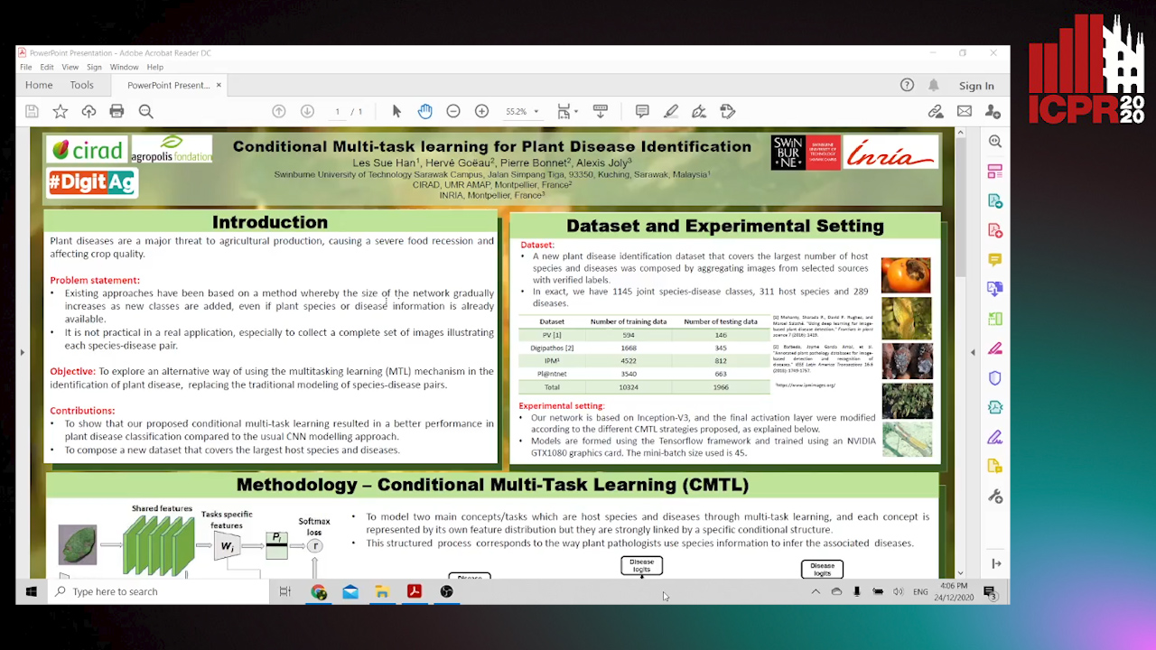
Auto-TLDR; A conditional multi-task learning approach for plant disease identification

Slides Poster Similar

Several recent studies have proposed an automatic plant disease identification system based on deep learning. Although successful, these approaches are generally based on learned classification models with target classes of joint host species-disease pairs that may not allow optimal use of the available information. This is due to the fact that they require distinguishing between similar host species or diseases. In fact, these approaches have limited scalability because the size of a network gradually increases as new classes are added, even if information on host species or diseases is already available. This constraint is all the more important as it can be difficult to collect/establish a specific list of all diseases for each host plant species in an actual application. In this paper, we address the problems by proposing a new conditional multi-task learning (CMTL) approach which allows the distribution of host species and disease characteristics learned simultaneously with a conditional link between them. This conditioning is formed in such a way that the knowledge to infer the prediction of one concept (the diseases) depends on the other concept (the host species), which corresponds to the way plant pathologists used to infer the diseases of the host species. We show that our approach can improve the performance of plant disease identification compared to the usual species-disease pair modeling in the previous studies. Meanwhile, we also compose a new dataset on plant disease identification that could serve as an important benchmark in this field.

A Deep Learning Approach for the Segmentation of Myocardial Diseases

Khawala Brahim, Abdull Qayyum, Alain Lalande, Arnaud Boucher, Anis Sakly, Fabrice Meriaudeau

Responsive image

Auto-TLDR; Segmentation of Myocardium Infarction Using Late GADEMRI and SegU-Net

Slides Poster Similar

Cardiac left ventricular (LV) segmentation is of paramount essential step for both diagnosis and treatment of cardiac pathologies such as ischemia, myocardial infarction, arrhythmia and myocarditis. However, this segmentation is challenging due to high variability across patients and the potential lack of contrast between structures. In this work, we propose and evaluate a (2.5D) SegU-Net model based on the fusion of two deep learning techniques (U-Net and Seg-Net) for automated LGEMRI (Late gadolinium enhanced magnetic resonance imaging) myocardial disease (infarct core and no reflow region) quantification in a new multifield expert annotated dataset. Given that the scar tissue represents a small part of the whole MRI slices, we focused on myocardium area. Segmentation results show that this preprocessing step facilitate the learning procedure. In order to solve the class imbalance problem, we propose to apply the Jaccard loss and the Focal Loss as optimization loss function and to integrate a class weights strategy into the objective function. Late combination has been used to merge the output of the best trained models on a different set of hyperparameters. The final network segmentation performances will be useful for future comparison of new method to the current related work for this task. A total number of 2237 of slices (320 cases) were used for training/validation and 210 slices (35 cases) were used for testing. Experiments over our proposed dataset, using several evaluation metrics such Jaccard distance (IOU), Accuracy and Dice similarity coefficient (DSC), demonstrate efficiency performance in quantifying different zones of myocardium infarction across various patients. As compared to the second intra-observer study, our testing results showed that the SegUNet prediction model leads to these average dice coefficients over all segmented tissue classes, respectively : 'Background': 0.99999, 'Myocardium': 0.99434, 'Infarctus': 0.95587, 'Noreflow': 0.78187.

Deep Multiple Instance Learning with Spatial Attention for ROP Case Classification, Instance Selection and Abnormality Localization

Xirong Li, Wencui Wan, Yang Zhou, Jianchun Zhao, Qijie Wei, Junbo Rong, Pengyi Zhou, Limin Xu, Lijuan Lang, Yuying Liu, Chengzhi Niu, Dayong Ding, Xuemin Jin

Responsive image

Auto-TLDR; MIL-SA: Deep Multiple Instance Learning for Automated Screening of Retinopathy of Prematurity

Similar

This paper tackles automated screening of Retinopathy of Prematurity (ROP), one of the most common causes of visual loss in childhood. Clinically, ROP screening per case requires multiple color fundus images capturing different zones of the premature retina. A desirable model shall not only make a decision at the case level, but also pinpoint which instances and what part of the instances are responsible for the decision. This paper makes the first attempt to accomplish three tasks, i.e, ROP case classification, instance selection and abnormality localization in a unified framework. To that end, we propose a new model that effectively combines instance-attention based deep multiple instance learning (MIL) and spatial attention (SA). The propose model, which we term MIL-SA, identifies positive instances in light of their contributions to case-level decision. Meanwhile, abnormal regions in the identified instances are automatically localized by the SA mechanism. Moreover, MIL-SA is learned from case-level binary labels exclusively, and in an end-to-end manner. Experiments on a large clinical dataset of 2,186 cases with 11,053 fundus images show the viability of the proposed model for all the three tasks.

Multimodal Side-Tuning for Document Classification

Stefano Zingaro, Giuseppe Lisanti, Maurizio Gabbrielli

Responsive image

Auto-TLDR; Side-tuning for Multimodal Document Classification

Slides Poster Similar

In this paper, we propose to exploit the side-tuning framework for multimodal document classification. Side-tuning is a methodology for network adaptation recently introduced to solve some of the problems related to previous approaches. Thanks to this technique it is actually possible to overcome model rigidity and catastrophic forgetting of transfer learning by fine-tuning. The proposed solution uses off-the-shelf deep learning architectures leveraging the side-tuning framework to combine a base model with a tandem of two side networks. We show that side-tuning can be successfully employed also when different data sources are considered, e.g. text and images in document classification. The experimental results show that this approach pushes further the limit for document classification accuracy with respect to the state of the art.

Dealing with Scarce Labelled Data: Semi-Supervised Deep Learning with Mix Match for Covid-19 Detection Using Chest X-Ray Images

Saúl Calderón Ramirez, Raghvendra Giri, Shengxiang Yang, Armaghan Moemeni, Mario Umaña, David Elizondo, Jordina Torrents-Barrena, Miguel A. Molina-Cabello

Responsive image

Auto-TLDR; Semi-supervised Deep Learning for Covid-19 Detection using Chest X-rays

Slides Poster Similar

Coronavirus (Covid-19) is spreading fast, infecting people through contact in various forms including droplets from sneezing and coughing. Therefore, the detection of infected subjects in an early, quick and cheap manner is urgent. Currently available tests are scarce and limited to people in danger of serious illness. The application of deep learning to chest X-ray images for Covid-19 detection is an attractive approach. However, this technology usually relies on the availability of large labelled datasets, a requirement hard to meet in the context of a virus outbreak. To overcome this challenge, a semi-supervised deep learning model using both labelled and unlabelled data is proposed. We developed and tested a semi-supervised deep learning framework based on the Mix Match architecture to classify chest X-rays into Covid-19, pneumonia and healthy cases. The presented approach was calibrated using two publicly available datasets. The results show an accuracy increase of around $15\%$ under low labelled / unlabelled data ratio. This indicates that our semi-supervised framework can help improve performance levels towards Covid-19 detection when the amount of high-quality labelled data is scarce. Also, we introduce a semi-supervised deep learning boost coefficient which is meant to ease the scalability of our approach and performance comparison.

Prediction of Obstructive Coronary Artery Disease from Myocardial Perfusion Scintigraphy using Deep Neural Networks

Ida Arvidsson, Niels Christian Overgaard, Miguel Ochoa Figueroa, Jeronimo Rose, Anette Davidsson, Kalle Åström, Anders Heyden

Responsive image

Auto-TLDR; A Deep Learning Algorithm for Multi-label Classification of Myocardial Perfusion Scintigraphy for Stable Ischemic Heart Disease

Slides Poster Similar

For diagnosis and risk assessment in patients with stable ischemic heart disease, myocardial perfusion scintigraphy is one of the most common cardiological examinations performed today. There are however many motivations for why an artificial intelligence algorithm would provide useful input to this task. For example to reduce the subjectiveness and save time for the nuclear medicine physicians working with this time consuming task. In this work we have developed a deep learning algorithm for multi-label classification based on a modified convolutional neural network to estimate probability of obstructive coronary artery disease in the left anterior artery, left circumflex artery and right coronary artery. The prediction is based on data from myocardial perfusion scintigraphy studies conducted in a dedicated Cadmium-Zinc-Telluride cardio camera (D-SPECT Spectrum Dynamics). Data from 588 patients was available, with stress images in both upright and supine position, as well as a number of auxiliary parameters such as angina symptoms and BMI. The data was used to train and evaluate the algorithm using 5-fold cross-validation. We achieve state-of-the-art results for this task with an area under the receiver operating characteristics curve of 0.89 as average on per-vessel level and 0.94 on per-patient level.

The Color Out of Space: Learning Self-Supervised Representations for Earth Observation Imagery

Stefano Vincenzi, Angelo Porrello, Pietro Buzzega, Marco Cipriano, Pietro Fronte, Roberto Cuccu, Carla Ippoliti, Annamaria Conte, Simone Calderara

Responsive image

Auto-TLDR; Satellite Image Representation Learning for Remote Sensing

Slides Poster Similar

The recent growth in the number of satellite images fosters the development of effective deep-learning techniques for Remote Sensing (RS). However, their full potential is untapped due to the lack of large annotated datasets. Such a problem is usually countered by fine-tuning a feature extractor that is previously trained on the ImageNet dataset. Unfortunately, the domain of natural images differs from the RS one, which hinders the final performance. In this work, we propose to learn meaningful representations from satellite imagery, leveraging its high-dimensionality spectral bands to reconstruct the visible colors. We conduct experiments on land cover classification (BigEarthNet) and West Nile Virus detection, showing that colorization is a solid pretext task for training a feature extractor. Furthermore, we qualitatively observe that guesses based on natural images and colorization rely on different parts of the input. This paves the way to an ensemble model that eventually outperforms both the above-mentioned techniques.

Self-Supervised Learning for Astronomical Image Classification

Ana Martinazzo, Mateus Espadoto, Nina S. T. Hirata

Responsive image

Auto-TLDR; Unlabeled Astronomical Images for Deep Neural Network Pre-training

Slides Poster Similar

In Astronomy, a huge amount of image data is generated daily by photometric surveys, which scan the sky to collect data from stars, galaxies and other celestial objects. In this paper, we propose a technique to leverage unlabeled astronomical images to pre-train deep convolutional neural networks, in order to learn a domain-specific feature extractor which improves the results of machine learning techniques in setups with small amounts of labeled data available. We show that our technique produces results which are in many cases better than using ImageNet pre-training.

Deep Learning-Based Type Identification of Volumetric MRI Sequences

Jean Pablo De Mello, Thiago Paixão, Rodrigo Berriel, Mauricio Reyes, Alberto F. De Souza, Claudine Badue, Thiago Oliveira-Santos

Responsive image

Auto-TLDR; Deep Learning for Brain MRI Sequences Identification Using Convolutional Neural Network

Slides Poster Similar

The analysis of Magnetic Resonance Imaging (MRI) sequences enables clinical professionals to monitor the progression of a brain tumor. As the interest for automatizing brain volume MRI analysis increases, it becomes convenient to have each sequence well identified. However, the unstandardized naming of MRI sequences make their identification difficult for automated systems, as well as make it difficult for researches to generate or use datasets for machine learning research. In face of that, we propose a system for identifying types of brain MRI sequences based on deep learning. By training a Convolutional Neural Network (CNN) based on 18-layer ResNet architecture, our system is able to classify a volumetric brain MRI as a T1, T1c, T2 or FLAIR sequence, or whether it does not belong to any of these classes. The network was trained with both pre-processed (BraTS dataset) and non-pre-processed (TCGA-GBM dataset) images with diverse acquisition protocols, requiring only a few layers of the volume for training. Our system is able to classify among sequence types with an accuracy of 96.27%.

Automatic Tuberculosis Detection Using Chest X-Ray Analysis with Position Enhanced Structural Information

Hermann Jepdjio Nkouanga, Szilard Vajda

Responsive image

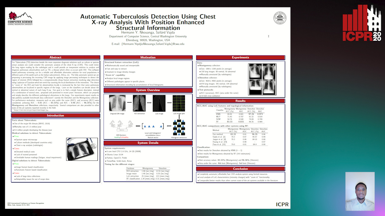
Auto-TLDR; Automatic Chest X-ray Screening for Tuberculosis in Rural Population using Localized Region on Interest

Slides Poster Similar

For Tuberculosis (TB) detection beside the more expensive diagnosis solutions such as culture or sputum smear analysis one could consider the automatic analysis of the chest X-ray (CXR). This could mimic the lung region reading by the radiologist and it could provide a cheap solution to analyze and diagnose pulmonary abnormalities such as TB which often co- occurs with HIV. This software based pulmonary screening can be a reliable and affordable solution for rural population in different parts of the world such as India, Africa, etc. Our fully automatic system is processing the incoming CXR image by applying image processing techniques to detect the region on interest (ROI) followed by a computationally cheap feature extraction involving edge detection using Laplacian of Gaussian which we enrich by counting the local distribution of the intensities. The choice to ”zoom in” the ROI and look for abnormalities locally is motivated by the fact that some pulmonary abnormalities are localized in specific regions of the lungs. Later on the classifiers can decide about the normal or abnormal nature of each lung X-ray. Our goal is to find a simple feature, instead of a combination of several ones, -proposed and promoted in recent years’ literature, which can properly describe the different pathological alterations in the lungs. Our experiments report results on two publicly available data collections1, namely the Shenzhen and the Montgomery collection. For performance evaluation, measures such as area under the curve (AUC), and accuracy (ACC) were considered, achieving AUC = 0.81 (ACC = 83.33%) and AUC = 0.96 (ACC = 96.35%) for the Montgomery and Schenzen collections, respectively. Several comparisons are also provided to other state- of-the-art systems reported recently in the field.

OCT Image Segmentation Using NeuralArchitecture Search and SRGAN

Saba Heidari, Omid Dehzangi, Nasser M. Nasarabadi, Ali Rezai

Responsive image

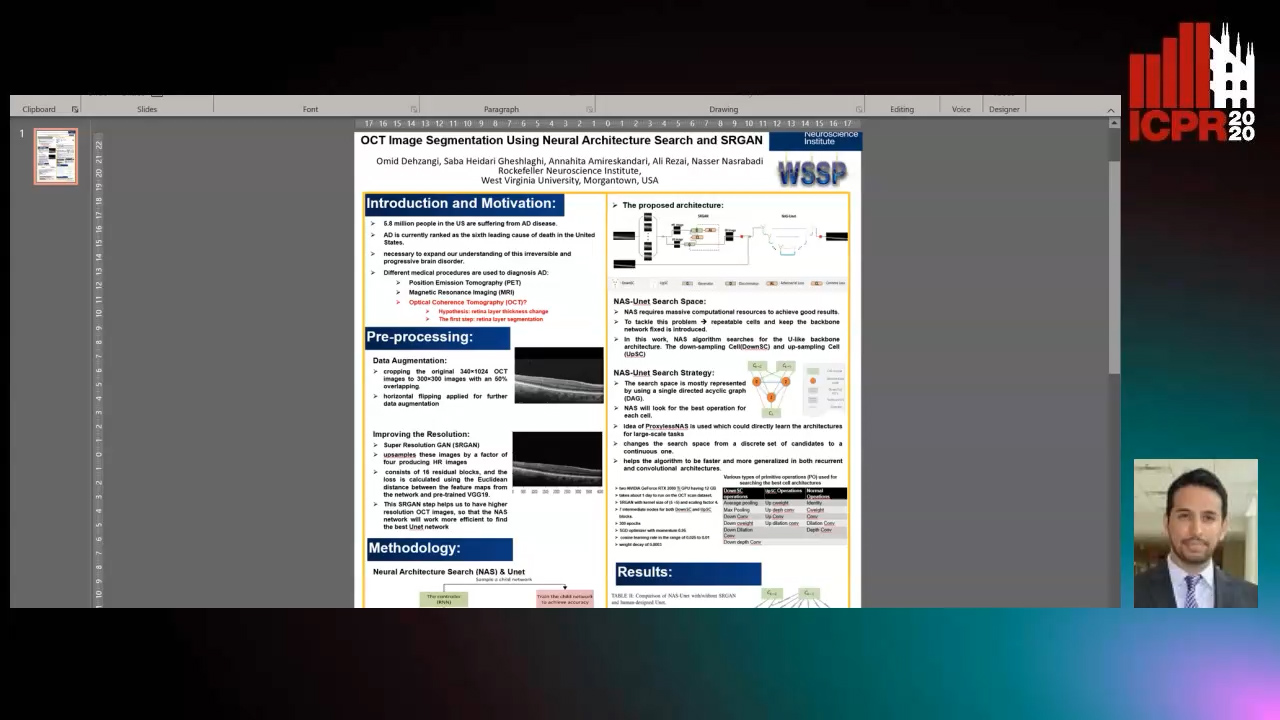
Auto-TLDR; Automatic Segmentation of Retinal Layers in Optical Coherence Tomography using Neural Architecture Search

Poster Similar

Alzheimer’s disease (AD) diagnosis is one of the major research areas in computational medicine. Optical coherence tomography (OCT) is a non-invasive, inexpensive, and timely efficient method that scans the human’s retina with depth. It has been hypothesized that the thickness of the retinal layers extracted from OCTs could be an efficient and effective biomarker for early diagnosis of AD. In this work, we aim to design a self-training model architecture for the task of segmenting the retinal layers in OCT scans. Neural architecture search (NAS) is a subfield of AutoML domain, which has a significant impact on improving the accuracy of machine vision tasks. We integrate the NAS algorithm with a Unet auto-encoder architecture as its backbone. Then, we employ our proposed model to segment the retinal nerve fiber layer in our preprocessed OCT images with the aim of AD diagnosis. In this work, we trained a super-resolution generative adversarial network on the raw OCT scans to improve the quality of the images before the modeling stage. In our architecture search strategy, different primitive operations suggested to find down- \& up-sampling Unet cell blocks and the binary gate method has been applied to make the search strategy more practical. Our architecture search method is empirically evaluated by training on the Unet and NAS-Unet from scratch. Specifically, the proposed NAS-Unet training significantly outperforms the baseline human-designed architecture by achieving 95.1\% in the mean Intersection over Union metric and 79.1\% in the Dice similarity coefficient.

Neuron-Based Network Pruning Based on Majority Voting

Ali Alqahtani, Xianghua Xie, Ehab Essa, Mark W. Jones

Responsive image

Auto-TLDR; Large-Scale Neural Network Pruning using Majority Voting

Slides Poster Similar

The achievement of neural networks in a variety of applications is accompanied by a dramatic increase in computational costs and memory requirements. In this paper, we propose an efficient method to simultaneously identify the critical neurons and prune the model during training without involving any pre-training or fine-tuning procedures. Unlike existing methods, which accomplish this task in a greedy fashion, we propose a majority voting technique to compare the activation values among neurons and assign a voting score to quantitatively evaluate their importance.This mechanism helps to effectively reduce model complexity by eliminating the less influential neurons and aims to determine a subset of the whole model that can represent the reference model with much fewer parameters within the training process. Experimental results show that majority voting efficiently compresses the network with no drop in model accuracy, pruning more than 79\% of the original model parameters on CIFAR10 and more than 91\% of the original parameters on MNIST. Moreover, we show that with our proposed method, sparse models can be further pruned into even smaller models by removing more than 60\% of the parameters, whilst preserving the reference model accuracy.

A Systematic Investigation on End-To-End Deep Recognition of Grocery Products in the Wild

Marco Leo, Pierluigi Carcagni, Cosimo Distante

Responsive image

Auto-TLDR; Automatic Recognition of Products on grocery shelf images using Convolutional Neural Networks

Slides Poster Similar

Automatic recognition of products on grocery shelf images is a new and attractive topic in computer vision and machine learning since, it can be exploited in different application areas. This paper introduces a complete end-to-end pipeline (without preliminary radiometric and spatial transformations usually involved while dealing with the considered issue) and it provides a systematic investigation of recent machine learning models based on convolutional neural networks for addressing the product recognition task by exploiting the proposed pipeline on a recent challenging grocery product dataset. The investigated models were never been used in this context: they derive from the successful and more generic object recognition task and have been properly tuned to address this specific issue. Besides, also ensembles of nets built by most advanced theoretical fundaments have been taken into account. Gathered classification results were very encouraging since the recognition accuracy has been improved up to 15\% with respect to the leading approaches in the state of art on the same dataset. A discussion about the pros and cons of the investigated solutions are discussed by paving the path towards new research lines.

A Lumen Segmentation Method in Ureteroscopy Images Based on a Deep Residual U-Net Architecture

Jorge Lazo, Marzullo Aldo, Sara Moccia, Michele Catellani, Benoit Rosa, Elena De Momi, Michel De Mathelin, Francesco Calimeri

Responsive image

Auto-TLDR; A Deep Neural Network for Ureteroscopy with Residual Units

Slides Poster Similar

Ureteroscopy is becoming the first surgical treatment option for the majority of urinary affections. This procedure is carried out using an endoscope which provides the surgeon with the visual and spatial information necessary to navigate inside the urinary tract. Having in mind the development of surgical assistance systems, that could enhance the performance of surgeon, the task of lumen segmentation is a fundamental part since this is the visual reference which marks the path that the endoscope should follow. This is something that has not been analyzed in ureteroscopy data before. However, this task presents several challenges given the image quality and the conditions itself of ureteroscopy procedures. In this paper, we study the implementation of a Deep Neural Network which exploits the advantage of residual units in an architecture based on U-Net. For the training of these networks, we analyze the use of two different color spaces: gray-scale and RGB data images. We found that training on gray-scale images gives the best results obtaining mean values of Dice Score, Precision, and Recall of 0.73, 0.58, and 0.92 respectively. The results obtained show that the use of residual U-Net could be a suitable model for further development for a computer-aided system for navigation and guidance through the urinary system.

Deep Recurrent-Convolutional Model for AutomatedSegmentation of Craniomaxillofacial CT Scans

Francesca Murabito, Simone Palazzo, Federica Salanitri Proietto, Francesco Rundo, Ulas Bagci, Daniela Giordano, Rosalia Leonardi, Concetto Spampinato

Responsive image

Auto-TLDR; Automated Segmentation of Anatomical Structures in Craniomaxillofacial CT Scans using Fully Convolutional Deep Networks

Slides Poster Similar

In this paper we define a deep learning architecture for automated segmentation of anatomical structures in Craniomaxillofacial (CMF) CT scans that leverages the recent success of encoder-decoder models for semantic segmentation of natural images. In particular, we propose a fully convolutional deep network that combines the advantages of recent fully convolutional models, such as Tiramisu, with squeeze-and-excitation blocks for feature recalibration, integrated with convolutional LSTMs to model spatio-temporal correlations between consecutive slices. The proposed segmentation network shows superior performance and generalization capabilities (to different structures and imaging modalities) than state of the art methods on automated segmentation of CMF structures (e.g., mandibles and airways) in several standard benchmarks (e.g., MICCAI datasets) and on new datasets proposed herein, effectively facing shape variability.

Facial Expression Recognition Using Residual Masking Network

Luan Pham, Vu Huynh, Tuan Anh Tran

Responsive image

Auto-TLDR; Deep Residual Masking for Automatic Facial Expression Recognition

Slides Poster Similar

Automatic facial expression recognition (FER) has gained much attention due to its applications in human-computer interaction. Among the approaches to improve FER tasks, this paper focuses on deep architecture with the attention mechanism. We propose a novel Masking idea to boost the performance of CNN in facial expression task. It uses a segmentation network to refine feature maps, enabling the network to focus on relevant information to make correct decisions. In experiments, we combine the ubiquitous Deep Residual Network and Unet-like architecture to produce a Residual Masking Network. The proposed method holds state-of-the-art (SOTA) accuracy on the well-known FER2013 and private VEMO datasets. Our works are available on Github.

Merged 1D-2D Deep Convolutional Neural Networks for Nerve Detection in Ultrasound Images

Mohammad Alkhatib, Adel Hafiane, Pierre Vieyres

Responsive image

Auto-TLDR; A Deep Neural Network for Deep Neural Networks to Detect Median Nerve in Ultrasound-Guided Regional Anesthesia

Slides Poster Similar

Ultrasound-Guided Regional Anesthesia (UGRA) becomes a standard procedure in surgical operations and contributes to pain management. It offers the advantages of the targeted nerve detection and provides the visualization of regions of interest such as anatomical structures. However, nerve detection is one of the most challenging tasks that anesthetists can encounter in the UGRA procedure. A computer-aided system that can detect automatically the nerve region would facilitate the anesthetist's daily routine and allow them to concentrate more on the anesthetic delivery. In this paper, we propose a new method based on merging deep learning models from different data to detect the median nerve. The merged architecture consists of two branches, one being one dimensional (1D) convolutional neural networks (CNN) branch and another 2D CNN branch. The merged architecture aims to learn the high-level features from 1D handcrafted noise-robust features and 2D ultrasound images. The obtained results show the validity and high accuracy of the proposed approach and its robustness.

Deep Learning in the Ultrasound Evaluation of Neonatal Respiratory Status

Michela Gravina, Diego Gragnaniello, Giovanni Poggi, Luisa Verdoliva, Carlo Sansone, Iuri Corsini, Carlo Dani, Fabio Meneghin, Gianluca Lista, Salvatore Aversa, Migliaro Migliaro, Raimondi Francesco

Responsive image

Auto-TLDR; Lung Ultrasound Imaging with Deep Learning Networks and Training Strategies: An Analysis and Adaptation

Slides Poster Similar

Lung ultrasound imaging is reaching growing interest from the scientific community. On one side, thanks to its harmlessness and high descriptive power, this kind of diagnostic imaging became largely adopted in sensitive applications, like the diagnosis and follow-up of preterm newborns in neonatal intensive care units. At the same time, novel image analysis and pattern recognition approaches can fully exploit the rich information contained in this data, making them attractive for the research community. In this work, we present a thorough analysis of recent deep learning networks and training strategies conducted on a vast and challenging multicenter dataset comprising 87 patients with different diseases and gestational ages. These approaches are firstly discussed in the context of lung respiratory status assessing through ultrasound imaging and then evaluated against a reference marker. The conducted analysis shed some light on this problem, by relating the criticisms that can mislead the training procedure and proposing some adaptations to the specific problem. The achieved results sensibly outperform that obtained by previous work, based on textural features, and narrow the gap with the visual score predicted by the human experts.

VPU Specific CNNs through Neural Architecture Search

Ciarán Donegan, Hamza Yous, Saksham Sinha, Jonathan Byrne

Responsive image

Auto-TLDR; Efficient Convolutional Neural Networks for Edge Devices using Neural Architecture Search

Slides Poster Similar

The success of deep learning at computer vision tasks has led to an ever-increasing number of applications on edge devices. Often with the use of edge AI hardware accelerators like the Intel Movidius Vision Processing Unit (VPU). Performing computer vision tasks on edge devices is challenging. Many Convolutional Neural Networks (CNNs) are too complex to run on edge devices with limited computing power. This has created large interest in designing efficient CNNs and one promising way of doing this is through Neural Architecture Search (NAS). NAS aims to automate the design of neural networks. NAS can also optimize multiple different objectives together, like accuracy and efficiency, which is difficult for humans. In this paper, we use a differentiable NAS method to find efficient CNNs for VPU that achieves state-of-the-art classification accuracy on ImageNet. Our NAS designed model outperforms MobileNetV2, having almost 1\% higher top-1 accuracy while being 13\% faster on MyriadX VPU. To the best of our knowledge, this is the first time a VPU specific CNN has been designed using a NAS algorithm. Our results also reiterate the fact that efficient networks must be designed for each specific hardware. We show that efficient networks targeted at different devices do not perform as well on the VPU.

Documents Counterfeit Detection through a Deep Learning Approach

Darwin Danilo Saire Pilco, Salvatore Tabbone

Responsive image

Auto-TLDR; End-to-End Learning for Counterfeit Documents Detection using Deep Neural Network

Slides Poster Similar

The main topic of this work is on the detection of counterfeit documents and especially banknotes. We propose an end-to-end learning model using a deep learning approach based on Adapnet++ which manages feature extraction at multiple scale levels using several residual units. Unlike previous models based on regions of interest (ROI) and high-resolution documents, our network is feed with simple input images (i.e., a single patch) and we do not need high resolution images. Besides, discriminative regions can be visualized at different scales. Our network learns by itself which regions of interest predict the better results. Experimental results show that we are competitive compared with the state-of-the-art and our deep neural network has good ability to generalize and can be applied to other kind of documents like identity or administrative one.

SA-UNet: Spatial Attention U-Net for Retinal Vessel Segmentation

Changlu Guo, Marton Szemenyei, Yugen Yi, Wenle Wang, Buer Chen, Changqi Fan

Responsive image

Auto-TLDR; Spatial Attention U-Net for Segmentation of Retinal Blood Vessels

Slides Poster Similar

The precise segmentation of retinal blood vessels is of great significance for early diagnosis of eye-related diseases such as diabetes and hypertension. In this work, we propose a lightweight network named Spatial Attention U-Net (SA-UNet) that does not require thousands of annotated training samples and can be utilized in a data augmentation manner to use the available annotated samples more efficiently. SA-UNet introduces a spatial attention module which infers the attention map along the spatial dimension, and multiplies the attention map by the input feature map for adaptive feature refinement. In addition, the proposed network employs structured dropout convolutional blocks instead of the original convolutional blocks of U-Net to prevent the network from overfitting. We evaluate SA-UNet based on two benchmark retinal datasets: the Vascular Extraction (DRIVE) dataset and the Child Heart and Health Study (CHASE_DB1) dataset. The results show that the proposed SA-UNet achieves state-of-the-art performance on both datasets.The implementation and the trained networks are available on Github1.

A Versatile Crack Inspection Portable System Based on Classifier Ensemble and Controlled Illumination

Milind Gajanan Padalkar, Carlos Beltran-Gonzalez, Matteo Bustreo, Alessio Del Bue, Vittorio Murino

Responsive image

Auto-TLDR; Lighting Conditions for Crack Detection in Ceramic Tile

Slides Poster Similar

This paper presents a novel setup for automatic visual inspection of cracks in ceramic tile as well as studies the effect of various classifiers and height-varying illumination conditions for this task. The intuition behind this setup is that cracks can be better visualized under specific lighting conditions than others. Our setup, which is designed for field work with constraints in its maximum dimensions, can acquire images for crack detection with multiple lighting conditions using the illumination sources placed at multiple heights. Crack detection is then performed by classifying patches extracted from the acquired images in a sliding window fashion. We study the effect of lights placed at various heights by training classifiers both on customized as well as state-of-the-art architectures and evaluate their performance both at patch-level and image-level, demonstrating the effectiveness of our setup. More importantly, ours is the first study that demonstrates how height-varying illumination conditions can affect crack detection with the use of existing state-of-the-art classifiers. We provide an insight about the illumination conditions that can help in improving crack detection in a challenging real-world industrial environment.

LiNet: A Lightweight Network for Image Super Resolution

Armin Mehri, Parichehr Behjati Ardakani, Angel D. Sappa

Responsive image

Auto-TLDR; LiNet: A Compact Dense Network for Lightweight Super Resolution

Slides Poster Similar

This paper proposes a new lightweight network, LiNet, that enhancing technical efficiency in lightweight super resolution and operating approximately like very large and costly networks in terms of number of network parameters and operations. The proposed architecture allows the network to learn more abstract properties by avoiding low-level information via multiple links. LiNet introduces a Compact Dense Module, which contains set of inner and outer blocks, to efficiently extract meaningful information, to better leverage multi-level representations before upsampling stage, and to allow an efficient information and gradient flow within the network. Experiments on benchmark datasets show that the proposed LiNet achieves favorable performance against lightweight state-of-the-art methods.

Stage-Wise Neural Architecture Search

Artur Jordão, Fernando Akio Yamada, Maiko Lie, William Schwartz

Responsive image

Auto-TLDR; Efficient Neural Architecture Search for Deep Convolutional Networks

Slides Poster Similar

Modern convolutional networks such as ResNet and NASNet have achieved state-of-the-art results in many computer vision applications. These architectures consist of stages, which are sets of layers that operate on representations in the same resolution. It has been demonstrated that increasing the number of layers in each stage improves the prediction ability of the network. However, the resulting architecture becomes computationally expensive in terms of floating point operations, memory requirements and inference time. Thus, significant human effort is necessary to evaluate different trade-offs between depth and performance. To handle this problem, recent works have proposed to automatically design high-performance architectures, mainly by means of neural architecture search (NAS). Current NAS strategies analyze a large set of possible candidate architectures and, hence, require vast computational resources and take many GPUs days. Motivated by this, we propose a NAS approach to efficiently design accurate and low-cost convolutional architectures and demonstrate that an efficient strategy for designing these architectures is to learn the depth stage-by-stage. For this purpose, our approach increases depth incrementally in each stage taking into account its importance, such that stages with low importance are kept shallow while stages with high importance become deeper. We conduct experiments on the CIFAR and different versions of ImageNet datasets, where we show that architectures discovered by our approach achieve better accuracy and efficiency than human-designed architectures. Additionally, we show that architectures discovered on CIFAR-10 can be successfully transferred to large datasets. Compared to previous NAS approaches, our method is substantially more efficient, as it evaluates one order of magnitude fewer models and yields architectures on par with the state-of-the-art.

Contextual Classification Using Self-Supervised Auxiliary Models for Deep Neural Networks

Sebastian Palacio, Philipp Engler, Jörn Hees, Andreas Dengel

Responsive image

Auto-TLDR; Self-Supervised Autogenous Learning for Deep Neural Networks

Slides Poster Similar

Classification problems solved with deep neural networks (DNNs) typically rely on a closed world paradigm, and optimize over a single objective (e.g., minimization of the cross- entropy loss). This setup dismisses all kinds of supporting signals that can be used to reinforce the existence or absence of particular patterns. The increasing need for models that are interpretable by design makes the inclusion of said contextual signals a crucial necessity. To this end, we introduce the notion of Self-Supervised Autogenous Learning (SSAL). A SSAL objective is realized through one or more additional targets that are derived from the original supervised classification task, following architectural principles found in multi-task learning. SSAL branches impose low-level priors into the optimization process (e.g., grouping). The ability of using SSAL branches during inference, allow models to converge faster, focusing on a richer set of class-relevant features. We equip state-of-the-art DNNs with SSAL objectives and report consistent improvements for all of them on CIFAR100 and Imagenet. We show that SSAL models outperform similar state-of-the-art methods focused on contextual loss functions, auxiliary branches and hierarchical priors.