Robust Localization of Retinal Lesions Via Weakly-Supervised Learning

Ruohan Zhao, Qin Li, Jane You

Responsive image

Auto-TLDR; Weakly Learning of Lesions in Fundus Images Using Multi-level Feature Maps and Classification Score

Slides Poster

Retinal fundus images reveal the condition of retina, blood vessels and optic nerve. Retinal imaging is becoming widely adopted in clinical work because any subtle changes to the structures at the back of the eyes can affect the eyes and indicate the overall health. Machine learning, in particular deep learning by convolutional neural network (CNN), has been increasingly adopted for computer-aided detection (CAD) of retinal lesions. However, a significant barrier to the high performance of CNN based CAD approach is caused by the lack of sufficient labeled ground-truth image samples for training. Unlike the fully-supervised learning which relies on pixel-level annotation of pathology in fundus images, this paper presents a new approach to discriminate the location of various lesions based on image-level labels via weakly learning. More specifically, our proposed method leverages multi-level feature maps and classification score to cope with both bright and red lesions in fundus images. To enhance capability of learning less discriminative parts of objects (e.g. small blobs of microaneurysms opposed to bulk of exudates), the classifier is regularized by refining images with corresponding labels. The experimental results of the performance evaluation and benchmarking at both image-level and pixel-level on the public DIARETDB1 dataset demonstrate the feasibility and excellent potentials of our method in practice.

Similar papers

Learn to Segment Retinal Lesions and Beyond

Qijie Wei, Xirong Li, Weihong Yu, Xiao Zhang, Yongpeng Zhang, Bojie Hu, Bin Mo, Di Gong, Ning Chen, Dayong Ding, Youxin Chen

Responsive image

Auto-TLDR; Multi-task Lesion Segmentation and Disease Classification for Diabetic Retinopathy Grading

Poster Similar

Towards automated retinal screening, this paper makes an endeavor to simultaneously achieve pixel-level retinal lesion segmentation and image-level disease classification. Such a multi-task approach is crucial for accurate and clinically interpretable disease diagnosis. Prior art is insufficient due to three challenges, i.e., lesions lacking objective boundaries, clinical importance of lesions irrelevant to their size, and the lack of one-to-one correspondence between lesion and disease classes. This paper attacks the three challenges in the context of diabetic retinopathy (DR) grading. We propose Lesion-Net, a new variant of fully convolutional networks, with its expansive path re- designed to tackle the first challenge. A dual Dice loss that leverages both semantic segmentation and image classification losses is introduced to resolve the second challenge. Lastly, we build a multi-task network that employs Lesion-Net as a side- attention branch for both DR grading and result interpretation. A set of 12K fundus images is manually segmented by 45 ophthalmologists for 8 DR-related lesions, resulting in 290K manual segments in total. Extensive experiments on this large- scale dataset show that our proposed approach surpasses the prior art for multiple tasks including lesion segmentation, lesion classification and DR grading.

Deep Multiple Instance Learning with Spatial Attention for ROP Case Classification, Instance Selection and Abnormality Localization

Xirong Li, Wencui Wan, Yang Zhou, Jianchun Zhao, Qijie Wei, Junbo Rong, Pengyi Zhou, Limin Xu, Lijuan Lang, Yuying Liu, Chengzhi Niu, Dayong Ding, Xuemin Jin

Responsive image

Auto-TLDR; MIL-SA: Deep Multiple Instance Learning for Automated Screening of Retinopathy of Prematurity

Similar

This paper tackles automated screening of Retinopathy of Prematurity (ROP), one of the most common causes of visual loss in childhood. Clinically, ROP screening per case requires multiple color fundus images capturing different zones of the premature retina. A desirable model shall not only make a decision at the case level, but also pinpoint which instances and what part of the instances are responsible for the decision. This paper makes the first attempt to accomplish three tasks, i.e, ROP case classification, instance selection and abnormality localization in a unified framework. To that end, we propose a new model that effectively combines instance-attention based deep multiple instance learning (MIL) and spatial attention (SA). The propose model, which we term MIL-SA, identifies positive instances in light of their contributions to case-level decision. Meanwhile, abnormal regions in the identified instances are automatically localized by the SA mechanism. Moreover, MIL-SA is learned from case-level binary labels exclusively, and in an end-to-end manner. Experiments on a large clinical dataset of 2,186 cases with 11,053 fundus images show the viability of the proposed model for all the three tasks.

Dual Stream Network with Selective Optimization for Skin Disease Recognition in Consumer Grade Images

Krishnam Gupta, Jaiprasad Rampure, Monu Krishnan, Ajit Narayanan, Nikhil Narayan

Responsive image

Auto-TLDR; A Deep Network Architecture for Skin Disease Localisation and Classification on Consumer Grade Images

Slides Poster Similar

Skin disease localisation and classification on consumer-grade images is more challenging compared to that on dermoscopic imaging. Consumer grade images refer to the images taken using commonly available imaging devices such as a mobile camera or a hand held digital camera. Such images, in addition to having the skin condition of interest in a very small area of the image, has other noisy non-clinical details introduced due to the lighting conditions and the distance of the hand held device from the anatomy at the time of acquisition. We propose a novel deep network architecture \& a new optimization strategy for classification with implicit localisation of skin diseases from clinical/consumer grade images. A weakly supervised segmentation algorithm is first employed to extract Region of Interests (RoI) from the image, the RoI and the original image form the two input streams of the proposed architecture. Each stream of the architecture learns high level and low level features from the original image and the RoI, respectively. The two streams are independently optimised until the loss stops decreasing after which both the streams are optimised collectively with the help of a third combiner sub-network. Such a strategy resulted in a 5% increase of accuracy over the current state-of-the-art methods on SD-198 dataset, which is publicly available. The proposed algorithm is also validated on a new dataset containing over 12,000 images across 75 different skin conditions. We intend to release this dataset as SD-75 to aid in the advancement of research on skin condition classification on consumer grade images.

PCANet: Pyramid Context-Aware Network for Retinal Vessel Segmentation

Yi Zhang, Yixuan Chen, Kai Zhang

Responsive image

Auto-TLDR; PCANet: Adaptive Context-Aware Network for Automated Retinal Vessel Segmentation

Slides Poster Similar

Automated retinal vessel segmentation plays an important role in the diagnosis of some diseases such as diabetes, arteriosclerosis and hypertension. Recent works attempt to improve segmentation performance by exploring either global or local contexts. However, the context demands are varying from regions in each image and different levels of network. To address these problems, we propose Pyramid Context-aware Network (PCANet), which can adaptively capture multi-scale context representations. Specifically, PCANet is composed of multiple Adaptive Context-aware (ACA) blocks arranged in parallel, each of which can adaptively obtain the context-aware features by estimating affinity coefficients at a specific scale under the guidance of global contextual dependencies. Meanwhile, we import ACA blocks with specific scales in different levels of the network to obtain a coarse-to-fine result. Furthermore, an integrated test-time augmentation method is developed to further boost the performance of PCANet. Finally, extensive experiments demonstrate the effectiveness of the proposed PCANet, and state-of-the-art performances are achieved with AUCs of 0.9866, 0.9886 and F1 Scores of 0.8274, 0.8371 on two public datasets, DRIVE and STARE, respectively.

Fine-Tuning Convolutional Neural Networks: A Comprehensive Guide and Benchmark Analysis for Glaucoma Screening

Amed Mvoulana, Rostom Kachouri, Mohamed Akil

Responsive image

Auto-TLDR; Fine-tuning Convolutional Neural Networks for Glaucoma Screening

Slides Poster Similar

This work aimed at giving a comprehensive and in-detailed guide on the route to fine-tuning Convolutional Neural Networks (CNNs) for glaucoma screening. Transfer learning consists in a promising alternative to train CNNs from stratch, to avoid the huge data and resources requirements. After a thorough study of five state-of-the-art CNNs architectures, a complete and well-explained strategy for fine-tuning these networks is proposed, using hyperparameter grid-searching and two-phase training approach. Excellent performance is reached on model evaluation, with a 0.9772 AUROC validation rate, giving arise to reliable glaucoma diagosis-help systems. Also, a benchmark analysis is conducted across all fine-tuned models, studying them according to performance indices such as model complexity and size, AUROC density and inference time. This in-depth analysis allows a rigorous comparison between model characteristics, and is useful for giving practioners important trademarks for prospective applications and deployments.

Zoom-CAM: Generating Fine-Grained Pixel Annotations from Image Labels

Xiangwei Shi, Seyran Khademi, Yunqiang Li, Jan Van Gemert

Responsive image

Auto-TLDR; Zoom-CAM for Weakly Supervised Object Localization and Segmentation

Slides Poster Similar

Current weakly supervised object localization and segmentation rely on class-discriminative visualization techniques for convolutional neural networks (CNN) to generate pseudo-labels for pixel-level training. However, visualization methods, including CAM and Grad-CAM, focus on most discriminative object parts summarized in the last convolutional layer, missing the complete pixel mapping in intermediate layers. We propose Zoom-CAM: going beyond the last lowest resolution layer by integrating the importance maps over all activations in intermediate layers. Zoom-CAM captures fine-grained small-scale objects for various discriminative class instances, which are commonly missed by the baseline visualization methods. We focus on generating pixel-level pseudo-labels from class labels. The quality of our pseudo-labels evaluated on the ImageNet localization task exhibits more than 2.8% improvement on top-1 error. For weakly supervised semantic segmentation our generated pseudo-labels improve a state of the art model by 1.1%.

Transfer Learning through Weighted Loss Function and Group Normalization for Vessel Segmentation from Retinal Images

Abdullah Sarhan, Jon Rokne, Reda Alhajj, Andrew Crichton

Responsive image

Auto-TLDR; Deep Learning for Segmentation of Blood Vessels in Retinal Images

Slides Poster Similar

The vascular structure of blood vessels is important in diagnosing retinal conditions such as glaucoma and diabetic retinopathy. Accurate segmentation of these vessels can help in detecting retinal objects such as the optic disc and optic cup and hence determine if there are damages to these areas. Moreover, the structure of the vessels can help in diagnosing glaucoma. The rapid development of digital imaging and computer-vision techniques has increased the potential for developing approaches for segmenting retinal vessels. In this paper, we propose an approach for segmenting retinal vessels that uses deep learning along with transfer learning. We adapted the U-Net structure to use a customized InceptionV3 as the encoder and used multiple skip connections to form the decoder. Moreover, we used a weighted loss function to handle the issue of class imbalance in retinal images. Furthermore, we contributed a new dataset to this field. We tested our approach on six publicly available datasets and a newly created dataset. We achieved an average accuracy of 95.60\% and a Dice coefficient of 80.98\%. The results obtained from comprehensive experiments demonstrate the robustness of our approach to the segmentation of blood vessels in retinal images obtained from different sources. Our approach results in greater segmentation accuracy than other approaches.

Skin Lesion Classification Using Weakly-Supervised Fine-Grained Method

Xi Xue, Sei-Ichiro Kamata, Daming Luo

Responsive image

Auto-TLDR; Different Region proposal module for skin lesion classification

Slides Poster Similar

In recent years, skin cancer has become one of the most common cancers. Among all types of skin cancers, melanoma is the most fatal one and many people die of this disease every year. Early detection can greatly reduce the death rate and save more lives. Skin lesions are one of the early symptoms of melanoma and other types of skin cancer. So accurately recognizing various skin lesions in early stage are of great significance. There have been lots of existing works based on convolutional neural networks (CNN) to solve skin lesion classification but seldom do them involve the similarity among different lesions. For example, we find that some lesions of melanoma and nevi look similar in appearance which is hard for neural network to distinguish categories of skin lesions. Inspired by fine-grained image classification, we propose a novel network to distinguish each category accurately. In our paper, we design an effective module, distinct region proposal module (DRPM), to extract the distinct regions from each image. Spatial attention and channel-wise attention are both utilized to enrich feature maps and guide the network to focus on the highlighted areas in a weakly-supervised way. In addition, two preprocessing steps are added to ensure the network to get better results. We demonstrate the potential of the proposed method on ISIC 2017 dataset. Experiments show that our approach is effective and efficient.

Semi-Supervised Generative Adversarial Networks with a Pair of Complementary Generators for Retinopathy Screening

Yingpeng Xie, Qiwei Wan, Hai Xie, En-Leng Tan, Yanwu Xu, Baiying Lei

Responsive image

Auto-TLDR; Generative Adversarial Networks for Retinopathy Diagnosis via Fundus Images

Slides Poster Similar

Several typical types of retinopathy are major causes of blindness. However, early detection of retinopathy is quite not easy since few symptoms are observable in the early stage, attributing to the development of non-mydriatic retinal camera. These camera produces high-resolution retinal fundus images provide the possibility of Computer-Aided-Diagnosis (CAD) via deep learning to assist diagnosing retinopathy. Deep learning algorithms usually rely on a great number of labelled images which are expensive and time-consuming to obtain in the medical imaging area. Moreover, the random distribution of various lesions which often vary greatly in size also brings significant challenges to learn discriminative information from high-resolution fundus image. In this paper, we present generative adversarial networks simultaneously equipped with "good" generator and "bad" generator (GBGANs) to make up for the incomplete data distribution provided by limited fundus images. To improve the generative feasibility of generator, we introduce into pre-trained feature extractor to acquire condensed feature for each fundus image in advance. Experimental results on integrated three public iChallenge datasets show that the proposed GBGANs could fully utilize the available fundus images to identify retinopathy with little label cost.

A Systematic Investigation on Deep Architectures for Automatic Skin Lesions Classification

Pierluigi Carcagni, Marco Leo, Andrea Cuna, Giuseppe Celeste, Cosimo Distante

Responsive image

Auto-TLDR; RegNet: Deep Investigation of Convolutional Neural Networks for Automatic Classification of Skin Lesions

Slides Poster Similar

Computer vision-based techniques are more and more employed in healthcare and medical fields nowadays in order, principally, to be as a support to the experienced medical staff to help them to make a quick and correct diagnosis. One of the hot topics in this arena concerns the automatic classification of skin lesions. Several promising works exist about it, mainly leveraging Convolutional Neural Networks (CNN), but proposed pipeline mainly rely on complex data preprocessing and there is no systematic investigation about how available deep models can actually reach the accuracy needed for real applications. In order to overcome these drawbacks, in this work, an end-to-end pipeline is introduced and some of the most recent Convolutional Neural Networks (CNNs) architectures are included in it and compared on the largest common benchmark dataset recently introduced. To this aim, for the first time in this application context, a new network design paradigm, namely RegNet, has been exploited to get the best models among a population of configurations. The paper introduces a threefold level of contribution and novelty with respect the previous literature: the deep investigation of several CNN architectures driving to a consistent improvement of the lesions recognition accuracy, the exploitation of a new network design paradigm able to study the behavior of populations of models and a deep discussion about pro and cons of each analyzed method paving the path towards new research lines.

End-To-End Training of a Two-Stage Neural Network for Defect Detection

Jakob Božič, Domen Tabernik, Danijel Skocaj

Responsive image

Auto-TLDR; End-to-End Training of Segmentation-based Neural Network for Surface Defect Detection

Slides Poster Similar

Segmentation-based, two-stage neural network has shown excellent results in the surface defect detection, enabling the network to learn from a relatively small number of samples. In this work, we introduce end-to-end training of the two-stage network together with several extensions to the training process, which reduce the amount of training time and improve results on surface defect detection tasks. To enable end-to-end training we carefully balance the contributions of both the segmentation and the classification loss throughout the learning. We adjust the gradient flow from the classification into the segmentation network in order to prevent the unstable features from corrupting the learning. As additional extension to the learning, we propose frequency-of-use sampling scheme of negative samples to address the issue of over- and under-sampling of images during the training, while we employ the distance transform algorithm on the region-based segmentation masks as weights for positive pixels, giving greater importance to areas with higher probability of presence of defect without requiring a detailed annotation. We demonstrate the performance of the end-to-end training scheme and the proposed extensions on three defect detection datasets---DAGM, KolektorSDD and Severstal Steel defect dataset--- where we show state-of-the-art results. On the DAGM and the KolektorSDD we demonstrate 100\% detection rate, therefore completely solving the datasets. Additional ablation study performed on all three datasets quantitatively demonstrates the contribution to the overall result improvements for each of the proposed extensions.

A Benchmark Dataset for Segmenting Liver, Vasculature and Lesions from Large-Scale Computed Tomography Data

Bo Wang, Zhengqing Xu, Wei Xu, Qingsen Yan, Liang Zhang, Zheng You

Responsive image

Auto-TLDR; The Biggest Treatment-Oriented Liver Cancer Dataset for Segmentation

Slides Poster Similar

How to build a high-performance liver-related computer assisted diagnosis system is an open question of great interest. However, the performance of the state-of-art algorithm is always limited by the amount of data and quality of the label. To address this problem, we propose the biggest treatment-oriented liver cancer dataset for liver surgery and treatment planning. This dataset provides 216 cases (totally about 268K frames) scanned images in contrast-enhanced computed tomography (CT). We labeled all the CT images with the liver, liver vasculature and liver tumor segmentation ground truth for train and tune segmentation algorithms in advance. Based on that, we evaluate several recent and state-of-the-art segmentation algorithms, including 7 deep learning methods, on CT sequences. All results are compared to reference segmentations five error metrics that highlight different aspects of segmentation accuracy. In general, compared with previous datasets, our dataset is really a challenging dataset. To our knowledge, the proposed dataset and benchmark allow for the first time systematic exploration of such issues, and will be made available to allow for further research in this field.

Documents Counterfeit Detection through a Deep Learning Approach

Darwin Danilo Saire Pilco, Salvatore Tabbone

Responsive image

Auto-TLDR; End-to-End Learning for Counterfeit Documents Detection using Deep Neural Network

Slides Poster Similar

The main topic of this work is on the detection of counterfeit documents and especially banknotes. We propose an end-to-end learning model using a deep learning approach based on Adapnet++ which manages feature extraction at multiple scale levels using several residual units. Unlike previous models based on regions of interest (ROI) and high-resolution documents, our network is feed with simple input images (i.e., a single patch) and we do not need high resolution images. Besides, discriminative regions can be visualized at different scales. Our network learns by itself which regions of interest predict the better results. Experimental results show that we are competitive compared with the state-of-the-art and our deep neural network has good ability to generalize and can be applied to other kind of documents like identity or administrative one.

Investigating and Exploiting Image Resolution for Transfer Learning-Based Skin Lesion Classification

Amirreza Mahbod, Gerald Schaefer, Chunliang Wang, Rupert Ecker, Georg Dorffner, Isabella Ellinger

Responsive image

Auto-TLDR; Fine-tuned Neural Networks for Skin Lesion Classification Using Dermoscopic Images

Slides Poster Similar

Skin cancer is among the most common cancer types. Dermoscopic image analysis improves the diagnostic accuracy for detection of malignant melanoma and other pigmented skin lesions when compared to unaided visual inspection. Hence, computer-based methods to support medical experts in the diagnostic procedure are of great interest. Fine-tuning pre-trained convolutional neural networks (CNNs) has been shown to work well for skin lesion classification. Pre-trained CNNs are usually trained with natural images of a fixed image size which is typically significantly smaller than captured skin lesion images and consequently dermoscopic images are downsampled for fine-tuning. However, useful medical information may be lost during this transformation. In this paper, we explore the effect of input image size on skin lesion classification performance of fine-tuned CNNs. For this, we resize dermoscopic images to different resolutions, ranging from 64x64 to 768x768 pixels and investigate the resulting classification performance of three well-established CNNs, namely DenseNet-121, ResNet-18, and ResNet-50. Our results show that using very small images (of size 64x64 pixels) degrades the classification performance, while images of size 128x128 pixels and above support good performance with larger image sizes leading to slightly improved classification. We further propose a novel fusion approach based on a three-level ensemble strategy that exploits multiple fine-tuned networks trained with dermoscopic images at various sizes. When applied on the ISIC 2017 skin lesion classification challenge, our fusion approach yields an area under the receiver operating characteristic curve of 89.2% and 96.6% for melanoma classification and seborrheic keratosis classification, respectively, outperforming state-of-the-art algorithms.

Coarse to Fine: Progressive and Multi-Task Learning for Salient Object Detection

Dong-Goo Kang, Sangwoo Park, Joonki Paik

Responsive image

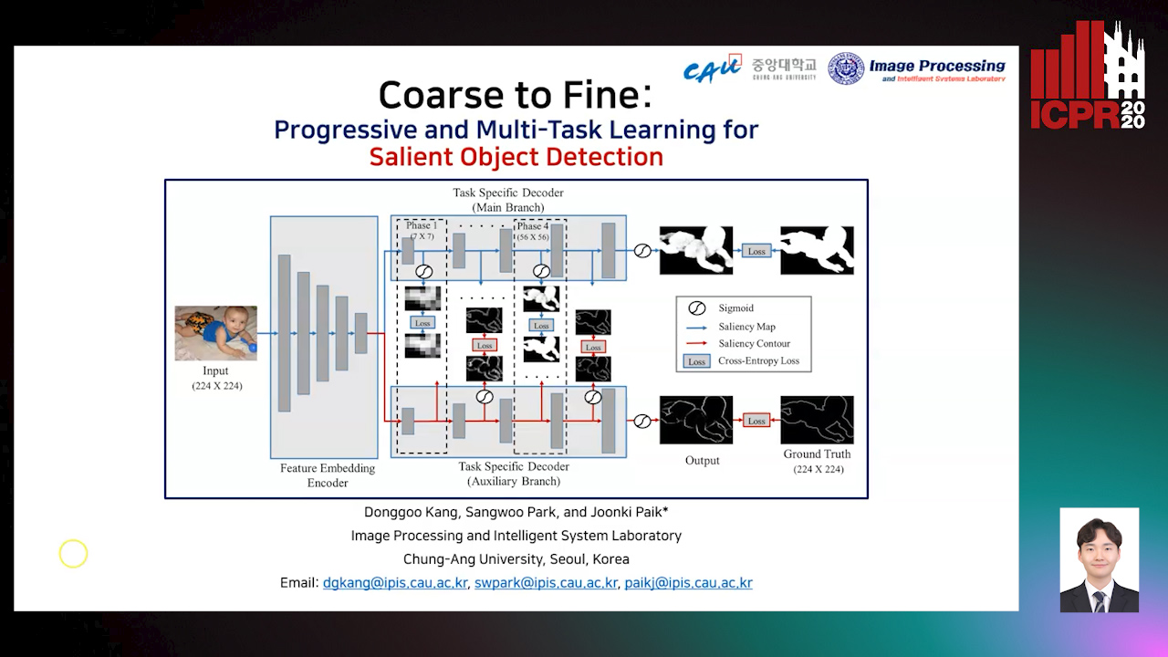
Auto-TLDR; Progressive and mutl-task learning scheme for salient object detection

Slides Poster Similar

Most deep learning-based salient object detection (SOD) methods tried to manipulate the convolution block to effectively capture the context of object. In this paper, we propose a novel method, called progressive and mutl-task learning scheme, to extract the context of object by only manipulating the learning scheme without changing the network architecture. The progressive learning scheme is a method to grow the decoder progressively in the train phase. In other words, starting from easier low-resolution layers, it gradually adds high-resolution layers. Although the progressive learning successfullyl captures the context of object, its output boundary tends to be rough. To solve this problem, we also propose a multi-task learning (MTL) scheme that processes the object saliency map and contour in a single network jointly. The proposed MTL scheme trains the network in an edge-preserved direction through an auxiliary branch that learns contours. The proposed a learning scheme can be combined with other convolution block manipulation methods. Extensive experiments on five datasets show that the proposed method performs best compared with state-of-the-art methods in most cases.

Supporting Skin Lesion Diagnosis with Content-Based Image Retrieval

Stefano Allegretti, Federico Bolelli, Federico Pollastri, Sabrina Longhitano, Giovanni Pellacani, Costantino Grana

Responsive image

Auto-TLDR; Skin Images Retrieval Using Convolutional Neural Networks for Skin Lesion Classification and Segmentation

Slides Poster Similar

Given the relevance of skin cancer, many attempts have been dedicated to the creation of automated devices that could assist both expert and beginner dermatologists towards fast and early diagnosis of skin lesions. In recent years, tasks such as skin lesion classification and segmentation have been extensively addressed with deep learning algorithms, which in some cases reach a diagnostic accuracy comparable to that of expert physicians. However, the general lack of interpretability and reliability severely hinders the ability of those approaches to actually support dermatologists in the diagnosis process. In this paper a novel skin images retrieval system is presented, which exploits features extracted by Convolutional Neural Networks to gather similar images from a publicly available dataset, in order to assist the diagnosis process of both expert and novice practitioners. In the proposed framework, Resnet-50 is initially trained for the classification of dermoscopic images; then, the feature extraction part is isolated, and an embedding network is build on top of it. The embedding learns an alternative representation, which allows to check image similarity by means of a distance measure. Experimental results reveal that the proposed method is able to select meaningful images, which can effectively boost the classification accuracy of human dermatologists.

Classify Breast Histopathology Images with Ductal Instance-Oriented Pipeline

Beibin Li, Ezgi Mercan, Sachin Mehta, Stevan Knezevich, Corey Arnold, Donald Weaver, Joann Elmore, Linda Shapiro

Responsive image

Auto-TLDR; DIOP: Ductal Instance-Oriented Pipeline for Diagnostic Classification

Slides Poster Similar

In this study, we propose the Ductal Instance-Oriented Pipeline (DIOP) that contains a duct-level instance segmentation model, a tissue-level semantic segmentation model, and three-levels of features for diagnostic classification. Based on recent advancements in instance segmentation and the Mask R-CNN model, our duct-level segmenter tries to identify each ductal individual inside a microscopic image; then, it extracts tissue-level information from the identified ductal instances. Leveraging three levels of information obtained from these ductal instances and also the histopathology image, the proposed DIOP outperforms previous approaches (both feature-based and CNN-based) in all diagnostic tasks; for the four-way classification task, the DIOP achieves comparable performance to general pathologists in this unique dataset. The proposed DIOP only takes a few seconds to run in the inference time, which could be used interactively on most modern computers. More clinical explorations are needed to study the robustness and generalizability of this system in the future.

Local Attention and Global Representation Collaborating for Fine-Grained Classification

He Zhang, Yunming Bai, Hui Zhang, Jing Liu, Xingguang Li, Zhaofeng He

Responsive image

Auto-TLDR; Weighted Region Network for Cosmetic Contact Lenses Detection

Slides Poster Similar

The cosmetic contact lenses over an iris may change its original textural pattern that is the foundation for iris recognition, making the cosmetic lenses a possible and easy-to-use iris presentation attack means. Aiming at cosmetic contact lenses detection of practical application system, some approaches have been proposed but still facing unsolved problems, such as low quality iris images and inaccurate localized iris boundaries. In this paper, we propose a novel framework called Weighted Region Network (WRN) for the cosmetic contact lenses detection. The WRN includes both the local attention Weight Network and the global classification Region Network. With the inherent attention mechanism, the proposed network is able to find the most discriminative regions, which reduces the requirement for target detection and improves the ability of classification based on some specific areas and patterns. The Weight Network can be trained by using Rank loss and MSE loss without manual discriminative region annotations. Experiments are conducted on several databases and a new collected low-quality iris image database. The proposed method outperforms state-of-the-art fake iris detection algorithms, and is also effective for the fine-grained image classification task.

Cross-View Relation Networks for Mammogram Mass Detection

Ma Jiechao, Xiang Li, Hongwei Li, Ruixuan Wang, Bjoern Menze, Wei-Shi Zheng

Responsive image

Auto-TLDR; Multi-view Modeling for Mass Detection in Mammogram

Slides Poster Similar

In medical image analysis, multi-view modeling is crucial for pathology detection when the target lesion is presented in different views, e.g. mass lesions in breast. Currently mammogram is the most effective imaging modality for mass lesion detection of breast cancer at the early stage. The pathological information from the two paired views (i.e., medio-lateral oblique and cranio-caudal) are highly relational and complementary, which is crucial for diagnosis in clinical practice. Existing mass detection methods do not consider learning synergistic features from the two relational views. For the first time, we propose a novel mass detection framework to capture the latent relation information from the two paired views of a same mass in mammogram. We evaluate our model on a public mammogram dataset and a large-scale private dataset, demonstrating that the proposed method outperforms existing feature fusion approaches and state-of-the-art mass detection methods. We further analyze the performance gains from the relation modeling. Our quantitative and qualitative results suggest that jointly learning cross-view features boosts the detection performance of existing models, which is a promising avenue for mass detection task in mammogram.

MTGAN: Mask and Texture-Driven Generative Adversarial Network for Lung Nodule Segmentation

Wei Chen, Qiuli Wang, Kun Wang, Dan Yang, Xiaohong Zhang, Chen Liu, Yucong Li

Responsive image

Auto-TLDR; Mask and Texture-driven Generative Adversarial Network for Lung Nodule Segmentation

Slides Poster Similar

Accurate segmentation for lung nodules in lung computed tomography (CT) scans plays a key role in the early diagnosis of lung cancer. Many existing methods, especially UNet, have made significant progress in lung nodule segmentation. However, due to the complex shapes of lung nodules and the similarity of visual characteristics between nodules and lung tissues, an accurate segmentation with few false positives of lung nodules is still a challenging problem. Considering the fact that both boundary and texture information of lung nodules are important for obtaining an accurate segmentation result, we propose a novel Mask and Texture-driven Generative Adversarial Network (MTGAN) with a joint multi-scale L1 loss for lung nodule segmentation, which takes full advantages of U-Net and adversarial training. The proposed MTGAN leverages adversarial learning strategy guided by the boundary and texture information of lung nodules to generate more accurate segmentation results with lesser false positives. We validate our model with the LIDC–IDRI dataset, and experimental results show that our method achieves excellent segmentation results for a variety of lung nodules, especially for juxtapleural nodules and low-dense nodules. Without any bells and whistles, the proposed MTGAN achieves significant segmentation performance with the Dice similarity coefficient (DSC) of 85.24% on the LIDC–IDRI dataset.

End-To-End Multi-Task Learning for Lung Nodule Segmentation and Diagnosis

Wei Chen, Qiuli Wang, Dan Yang, Xiaohong Zhang, Chen Liu, Yucong Li

Responsive image

Auto-TLDR; A novel multi-task framework for lung nodule diagnosis based on deep learning and medical features

Slides Similar

Computer-Aided Diagnosis (CAD) systems for lung nodule diagnosis based on deep learning have attracted much attention in recent years. However, most existing methods ignore the relationships between the segmentation and classification tasks, which leads to unstable performances. To address this problem, we propose a novel multi-task framework, which can provide lung nodule segmentation mask, malignancy prediction, and medical features for interpretable diagnosis at the same time. Our framework mainly contains two sub-network: (1) Multi-Channel Segmentation Sub-network (MSN) for lung nodule segmentation, and (2) Joint Classification Sub-network (JCN) for interpretable lung nodule diagnosis. In the proposed framework, we use U-Net down-sampling processes for extracting low-level deep learning features, which are shared by two sub-networks. The JCN forces the down-sampling processes to learn better lowlevel deep features, which lead to a better construct of segmentation masks. Meanwhile, two additional channels constructed by OTSU and super-pixel (SLIC) methods, are utilized as the guideline of the feature extraction. The proposed framework takes advantages of deep learning methods and classical methods, which can significantly improve the performances of all tasks. We evaluate the proposed framework on public dataset LIDCIDRI. Our framework achieves a promising Dice score of 86.43% in segmentation, 87.07% in malignancy level prediction, and convincing results in interpretable medical feature predictions.

Convolutional STN for Weakly Supervised Object Localization

Akhil Meethal, Marco Pedersoli, Soufiane Belharbi, Eric Granger

Responsive image

Auto-TLDR; Spatial Localization for Weakly Supervised Object Localization

Slides Similar

Weakly-supervised object localization is a challenging task in which the object of interest should be localized while learning its appearance. State-of-the-art methods recycle the architecture of a standard CNN by using the activation maps of the last layer for localizing the object. While this approach is simple and works relatively well, object localization relies on different features than classification, thus, a specialized localization mechanism is required during training to improve performance. In this paper, we propose a convolutional, multi-scale spatial localization network that provides accurate localization for the object of interest. Experimental results on CUB-200-2011 and ImageNet datasets show competitive performance of our proposed approach on Weakly supervised localization.

Point In: Counting Trees with Weakly Supervised Segmentation Network

Pinmo Tong, Shuhui Bu, Pengcheng Han

Responsive image

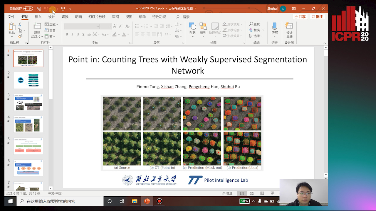
Auto-TLDR; Weakly Tree counting using Deep Segmentation Network with Localization and Mask Prediction

Slides Poster Similar

For tree counting tasks, since traditional image processing methods require expensive feature engineering and are not end-to-end frameworks, this will cause additional noise and cannot be optimized overall, so this method has not been widely used in recent trends of tree counting application. Recently, many deep learning based approaches are designed for this task because of the powerful feature extracting ability. The representative way is bounding box based supervised method, but time-consuming annotations are indispensable for them. Moreover, these methods are difficult to overcome the occlusion or overlap. To solve this problem, we propose a weakly tree counting network (WTCNet) based on deep segmentation network with only point supervision. It can simultaneously complete tree counting with localization and output mask of each tree at the same time. We first adopt a novel feature extractor network (FENet) to get features of input images, and then an effective strategy is introduced to deal with different mask predictions. In the end, we propose a basic localization guidance accompany with rectification guidance to train the network. We create two different datasets and select an existing challenging plant dataset to evaluate our method on three different tasks. Experimental results show the good performance improvement of our method compared with other existing methods. Further study shows that our method has great potential to reduce human labor and provide effective ground-truth masks and the results show the superiority of our method over the advanced methods.

Automatic Semantic Segmentation of Structural Elements related to the Spinal Cord in the Lumbar Region by Using Convolutional Neural Networks

Jhon Jairo Sáenz Gamboa, Maria De La Iglesia-Vaya, Jon Ander Gómez

Responsive image

Auto-TLDR; Semantic Segmentation of Lumbar Spine Using Convolutional Neural Networks

Slides Poster Similar

This work addresses the problem of automatically segmenting the MR images corresponding to the lumbar spine. The purpose is to detect and delimit the different structural elements like vertebrae, intervertebral discs, nerves, blood vessels, etc. This task is known as semantic segmentation. The approach proposed in this work is based on convolutional neural networks whose output is a mask where each pixel from the input image is classified into one of the possible classes. Classes were defined by radiologists and correspond to structural elements and tissues. The proposed network architectures are variants of the U-Net. Several complementary blocks were used to define the variants: spatial attention models, deep supervision and multi-kernels at input, this last block type is based on the idea of inception. Those architectures which got the best results are described in this paper, and their results are discussed. Two of the proposed architectures outperform the standard U-Net used as baseline.

Inception Based Deep Learning Architecture for Tuberculosis Screening of Chest X-Rays

Dipayan Das, K.C. Santosh, Umapada Pal

Responsive image

Auto-TLDR; End to End CNN-based Chest X-ray Screening for Tuberculosis positive patients in the severely resource constrained regions of the world

Slides Poster Similar

The motivation for this work is the primary need of screening Tuberculosis (TB) positive patients in the severely resource constrained regions of the world. Chest X-ray (CXR) is considered to be a promising indicator for the onset of TB, but the lack of skilled radiologists in such regions degrades the situation. Therefore, several computer aided diagnosis (CAD) systems have been proposed to solve the decision making problem, which includes hand engineered feature extraction methods to deep learning or Convolutional Neural Network (CNN) based methods. Feature extraction, being a time and resource intensive process, often delays the process of mass screening. Hence an end to end CNN architecture is proposed in this work to solve the problem. Two benchmark CXR datasets have been used in this work, collected from Shenzhen (China) and Montgomery County (USA), on which the proposed methodology achieved a maximum abnormality detection accuracy (ACC) of 91.7\% (0.96 AUC) and 87.47\% (0.92 AUC) respectively. To the greatest of our knowledge, the obtained results are marginally superior to the state of the art results that have solely used deep learning methodologies on the aforementioned datasets.

SA-UNet: Spatial Attention U-Net for Retinal Vessel Segmentation

Changlu Guo, Marton Szemenyei, Yugen Yi, Wenle Wang, Buer Chen, Changqi Fan

Responsive image

Auto-TLDR; Spatial Attention U-Net for Segmentation of Retinal Blood Vessels

Slides Poster Similar

The precise segmentation of retinal blood vessels is of great significance for early diagnosis of eye-related diseases such as diabetes and hypertension. In this work, we propose a lightweight network named Spatial Attention U-Net (SA-UNet) that does not require thousands of annotated training samples and can be utilized in a data augmentation manner to use the available annotated samples more efficiently. SA-UNet introduces a spatial attention module which infers the attention map along the spatial dimension, and multiplies the attention map by the input feature map for adaptive feature refinement. In addition, the proposed network employs structured dropout convolutional blocks instead of the original convolutional blocks of U-Net to prevent the network from overfitting. We evaluate SA-UNet based on two benchmark retinal datasets: the Vascular Extraction (DRIVE) dataset and the Child Heart and Health Study (CHASE_DB1) dataset. The results show that the proposed SA-UNet achieves state-of-the-art performance on both datasets.The implementation and the trained networks are available on Github1.

Attention-Based Selection Strategy for Weakly Supervised Object Localization

Zhenfei Zhang

Responsive image

Auto-TLDR; An Attention-based Selection Strategy for Weakly Supervised Object Localization

Slides Poster Similar

Weakly Supervised Object Localization (WSOL) task aims to recognize the object position by using only image-level labels. Some previous techniques remove the most discriminative parts for all input images or random images to capture the entire object location. However, these methods can not perform the correct operation on different images such as hiding the data or feature maps that should not be hidden. In this case, both classification and localization accuracy will be affected. Meanwhile, just erasing the most important regions tends to make the model learn the less discriminative parts from outside of the objects. To address these limitations, we propose an Attention-based Selection Strategy (ASS) method to choose images that do need to be erased. Moreover, we use different threshold self-attention maps to reduce the impact of unhelpful information in one of the branches of our selection strategy. Based on our experiments, the proposed method is simple but effective to improve the performance of WSOL. In particular, ASS achieves new state-of-the-art accuracy on CUB-200-2011 dataset and works very well on ILSVRC 2016 dataset.

A Comparison of Neural Network Approaches for Melanoma Classification

Maria Frasca, Michele Nappi, Michele Risi, Genoveffa Tortora, Alessia Auriemma Citarella

Responsive image

Auto-TLDR; Classification of Melanoma Using Deep Neural Network Methodologies

Slides Poster Similar

Melanoma is the deadliest form of skin cancer and it is diagnosed mainly visually, starting from initial clinical screening and followed by dermoscopic analysis, biopsy and histopathological examination. A dermatologist’s recognition of melanoma may be subject to errors and may take some time to diagnose it. In this regard, deep learning can be useful in the study and classification of skin cancer. In particular, by classifying images with Deep Neural Network methodologies, it is possible to obtain comparable or even superior results compared to those of dermatologists. In this paper, we propose a methodology for the classification of melanoma by adopting different deep learning techniques applied to a common dataset, composed of images from the ISIC dataset and consisting of different types of skin diseases, including melanoma on which we applied a specific pre-processing phase. In particular, a comparison of the results is performed in order to select the best effective neural network to be applied to the problem of recognition and classification of melanoma. Moreover, we also evaluate the impact of the pre- processing phase on the final classification. Different metrics such as accuracy, sensitivity, and specificity have been selected to assess the goodness of the adopted neural networks and compare them also with the manual classification of dermatologists.

Dealing with Scarce Labelled Data: Semi-Supervised Deep Learning with Mix Match for Covid-19 Detection Using Chest X-Ray Images

Saúl Calderón Ramirez, Raghvendra Giri, Shengxiang Yang, Armaghan Moemeni, Mario Umaña, David Elizondo, Jordina Torrents-Barrena, Miguel A. Molina-Cabello

Responsive image

Auto-TLDR; Semi-supervised Deep Learning for Covid-19 Detection using Chest X-rays

Slides Poster Similar

Coronavirus (Covid-19) is spreading fast, infecting people through contact in various forms including droplets from sneezing and coughing. Therefore, the detection of infected subjects in an early, quick and cheap manner is urgent. Currently available tests are scarce and limited to people in danger of serious illness. The application of deep learning to chest X-ray images for Covid-19 detection is an attractive approach. However, this technology usually relies on the availability of large labelled datasets, a requirement hard to meet in the context of a virus outbreak. To overcome this challenge, a semi-supervised deep learning model using both labelled and unlabelled data is proposed. We developed and tested a semi-supervised deep learning framework based on the Mix Match architecture to classify chest X-rays into Covid-19, pneumonia and healthy cases. The presented approach was calibrated using two publicly available datasets. The results show an accuracy increase of around $15\%$ under low labelled / unlabelled data ratio. This indicates that our semi-supervised framework can help improve performance levels towards Covid-19 detection when the amount of high-quality labelled data is scarce. Also, we introduce a semi-supervised deep learning boost coefficient which is meant to ease the scalability of our approach and performance comparison.

Planar 3D Transfer Learning for End to End Unimodal MRI Unbalanced Data Segmentation

Martin Kolarik, Radim Burget, Carlos M. Travieso-Gonzalez, Jan Kocica

Responsive image

Auto-TLDR; Planar 3D Res-U-Net Network for Unbalanced 3D Image Segmentation using Fluid Attenuation Inversion Recover

Slides Similar

We present a novel approach of 2D to 3D transfer learning based on mapping pre-trained 2D convolutional neural network weights into planar 3D kernels. The method is validated by proposed planar 3D res-u-net network with encoder transferred from the 2D VGG-16 which is applied for a single-stage unbalanced 3D image data segmentation. In particular, we evaluate the method on the MICCAI 2016 MS lesion segmentation challenge dataset utilizing solely Fluid Attenuation Inversion Recover (FLAIR) sequence without brain extraction for training and inference to simulate real medical praxis. The planar 3D res-u-net network performed the best both in sensitivity and Dice score amongst end to end methods processing raw MRI scans and achieved comparable Dice score to a state-of-the-art unimodal not end to end approach. Complete source code was released under the open-source license and this paper is in compliance with the Machine learning Reproducibility Checklist. By implementing practical transfer learning for 3D data representation we were able to successfully segment heavily unbalanced data without selective sampling and achieved more reliable results using less training data in single modality. From medical perspective, the unimodal approach gives an advantage in real praxis as it does not require co-registration nor additional scanning time during examination. Although modern medical imaging methods capture high resolution 3D anatomy scans suitable for computer aided detection system processing, deployment of automatic systems for interpretation of radiology imaging is still rather theoretical in many medical areas. Our work aims to bridge the gap offering solution for partial research questions.

Progressive Adversarial Semantic Segmentation

Abdullah-Al-Zubaer Imran, Demetri Terzopoulos

Responsive image

Auto-TLDR; Progressive Adversarial Semantic Segmentation for End-to-End Medical Image Segmenting

Slides Poster Similar

Medical image computing has advanced rapidly with the advent of deep learning techniques such as convolutional neural networks. Deep convolutional neural networks can perform exceedingly well given full supervision. However, the success of such fully-supervised models for various image analysis tasks (e.g., anatomy or lesion segmentation from medical images) is limited to the availability of massive amounts of labeled data. Given small sample sizes, such models are prohibitively data biased with large domain shift. To tackle this problem, we propose a novel end-to-end medical image segmentation model, namely Progressive Adversarial Semantic Segmentation (PASS), which can make improved segmentation predictions without requiring any domain-specific data during training time. Our extensive experimentation with 8 public diabetic retinopathy and chest X-ray datasets, confirms the effectiveness of PASS for accurate vascular and pulmonary segmentation, both for in-domain and cross-domain evaluations.

DA-RefineNet: Dual-Inputs Attention RefineNet for Whole Slide Image Segmentation

Ziqiang Li, Rentuo Tao, Qianrun Wu, Bin Li

Responsive image

Auto-TLDR; DA-RefineNet: A dual-inputs attention network for whole slide image segmentation

Slides Poster Similar

Automatic medical image segmentation techniques have wide applications for disease diagnosing, however, its much more challenging than natural optical image segmentation tasks due to the high-resolution of medical images and the corresponding huge computation cost. Sliding window was a commonly used technique for whole slide image (WSI) segmentation, however, for these methods that based on sliding window, the main drawback was lacking of global contextual information for supervision. In this paper, we proposed a dual-inputs attention network (denoted as DA-RefineNet) for WSI segmentation, where both local fine-grained information and global coarse information can be efficiently utilized. Sufficient comparative experiments were conducted to evaluate the effectiveness of the proposed method, the results proved that the proposed method can achieve better performance on WSI segmentation tasks compared to methods rely on single-input.

Unsupervised Detection of Pulmonary Opacities for Computer-Aided Diagnosis of COVID-19 on CT Images

Rui Xu, Xiao Cao, Yufeng Wang, Yen-Wei Chen, Xinchen Ye, Lin Lin, Wenchao Zhu, Chao Chen, Fangyi Xu, Yong Zhou, Hongjie Hu, Shoji Kido, Noriyuki Tomiyama

Responsive image

Auto-TLDR; A computer-aided diagnosis of COVID-19 from CT images using unsupervised pulmonary opacity detection

Slides Poster Similar

COVID-19 emerged towards the end of 2019 which was identified as a global pandemic by the world heath organization (WHO). With the rapid spread of COVID-19, the number of infected and suspected patients has increased dramatically. Chest computed tomography (CT) has been recognized as an efficient tool for the diagnosis of COVID-19. However, the huge CT data make it difficult for radiologist to fully exploit them on the diagnosis. In this paper, we propose a computer-aided diagnosis system that can automatically analyze CT images to distinguish the COVID-19 against to community-acquired pneumonia (CAP). The proposed system is based on an unsupervised pulmonary opacity detection method that locates opacity regions by a detector unsupervisedly trained from CT images with normal lung tissues. Radiomics based features are extracted insides the opacity regions, and fed into classifiers for classification. We evaluate the proposed CAD system by using 200 CT images collected from different patients in several hospitals. The accuracy, precision, recall, f1-score and AUC achieved are 95.5%, 100%, 91%, 95.1% and 95.9% respectively, exhibiting the promising capacity on the differential diagnosis of COVID-19 from CT images.

Dual-Attention Guided Dropblock Module for Weakly Supervised Object Localization

Junhui Yin, Siqing Zhang, Dongliang Chang, Zhanyu Ma, Jun Guo

Responsive image

Auto-TLDR; Dual-Attention Guided Dropblock for Weakly Supervised Object Localization

Slides Poster Similar

Attention mechanisms is frequently used to learn the discriminative features for better feature representations. In this paper, we extend the attention mechanism to the task of weakly supervised object localization (WSOL) and propose the dual-attention guided dropblock module (DGDM), which aims at learning the informative and complementary visual patterns for WSOL. This module contains two key components, the channel attention guided dropout (CAGD) and the spatial attention guided dropblock (SAGD). To model channel interdependencies, the CAGD ranks the channel attentions and treats the top-k attentions with the largest magnitudes as the important ones. It also keeps some low-valued elements to increase their value if they become important during training. The SAGD can efficiently remove the most discriminative information by erasing the contiguous regions of feature maps rather than individual pixels. This guides the model to capture the less discriminative parts for classification. Furthermore, it can also distinguish the foreground objects from the background regions to alleviate the attention misdirection. Experimental results demonstrate that the proposed method achieves new state-of-the-art localization performance.

MFPP: Morphological Fragmental Perturbation Pyramid for Black-Box Model Explanations

Qing Yang, Xia Zhu, Jong-Kae Fwu, Yun Ye, Ganmei You, Yuan Zhu

Responsive image

Auto-TLDR; Morphological Fragmental Perturbation Pyramid for Explainable Deep Neural Network

Slides Poster Similar

Deep neural networks (DNNs) have recently been applied and used in many advanced and diverse tasks, such as medical diagnosis, automatic driving, etc. Due to the lack of transparency of the deep models, DNNs are often criticized for their prediction that cannot be explainable by human. In this paper, we propose a novel Morphological Fragmental Perturbation Pyramid (MFPP) method to solve the Explainable AI problem. In particular, we focus on the black-box scheme, which can identify the input area responsible for the output of the DNN without having to understand the internal architecture of the DNN. In the MFPP method, we divide the input image into multi-scale fragments and randomly mask out fragments as perturbation to generate a saliency map, which indicates the significance of each pixel for the prediction result of the black box model. Compared with the existing input sampling perturbation method, the pyramid structure fragment has proved to be more effective. It can better explore the morphological information of the input image to match its semantic information, and does not need any value inside the DNN. We qualitatively and quantitatively prove that MFPP meets and exceeds the performance of state-of-the-art (SOTA) black-box interpretation method on multiple DNN models and datasets.

FOANet: A Focus of Attention Network with Application to Myocardium Segmentation

Zhou Zhao, Elodie Puybareau, Nicolas Boutry, Thierry Geraud

Responsive image

Auto-TLDR; FOANet: A Hybrid Loss Function for Myocardium Segmentation of Cardiac Magnetic Resonance Images

Slides Poster Similar

In myocardium segmentation of cardiac magnetic resonance images, ambiguities often appear near the boundaries of the target domains due to tissue similarities. To address this issue, we propose a new architecture, called FOANet, which can be decomposed in three main steps: a localization step, a Gaussian-based contrast enhancement step, and a segmentation step. This architecture is supplied with a hybrid loss function that guides the FOANet to study the transformation relationship between the input image and the corresponding label in a threelevel hierarchy (pixel-, patch- and map-level), which is helpful to improve segmentation and recovery of the boundaries. We demonstrate the efficiency of our approach on two public datasets in terms of regional and boundary segmentations.

Triplet-Path Dilated Network for Detection and Segmentation of General Pathological Images

Jiaqi Luo, Zhicheng Zhao, Fei Su, Limei Guo

Responsive image

Auto-TLDR; Triplet-path Network for One-Stage Object Detection and Segmentation in Pathological Images

Slides Similar

Deep learning has been widely applied in the field of medical image processing. However, compared with flourishing visual tasks in natural images, the progress achieved in pathological images is not remarkable, and detection and segmentation, which are among basic tasks of computer vision, are regarded as two independent tasks. In this paper, we make full use of existing datasets and construct a triplet-path network using dilated convolutions to cooperatively accomplish one-stage object detection and nuclei segmentation for general pathological images. First, in order to meet the requirement of detection and segmentation, a novel structure called triplet feature generation (TFG) is designed to extract high-resolution and multiscale features, where features from different layers can be properly integrated. Second, considering that pathological datasets are usually small, a location-aware and partially truncated loss function is proposed to improve the classification accuracy of datasets with few images and widely varying targets. We compare the performance of both object detection and instance segmentation with state-of-the-art methods. Experimental results demonstrate the effectiveness and efficiency of the proposed network on two datasets collected from multiple organs.

Attention Based Multi-Instance Thyroid Cytopathological Diagnosis with Multi-Scale Feature Fusion

Shuhao Qiu, Yao Guo, Chuang Zhu, Wenli Zhou, Huang Chen

Responsive image

Auto-TLDR; A weakly supervised multi-instance learning framework based on attention mechanism with multi-scale feature fusion for thyroid cytopathological diagnosis

Slides Poster Similar

In recent years, deep learning has been popular in combining with cytopathology diagnosis. Using the whole slide images (WSI) scanned by electronic scanners at clinics, researchers have developed many algorithms to classify the slide (benign or malignant). However, the key area that support the diagnosis result can be relatively small in a thyroid WSI, and only the global label can be acquired, which make the direct use of the strongly supervised learning framework infeasible. What’s more, because the clinical diagnosis of the thyroid cells requires the use of visual features in different scales, a generic feature extraction way may not achieve good performance. In this paper, we propose a weakly supervised multi-instance learning framework based on attention mechanism with multi-scale feature fusion (MSF) using convolutional neural network (CNN) for thyroid cytopathological diagnosis. We take each WSI as a bag, each bag contains multiple instances which are the different regions of the WSI, our framework is trained to learn the key area automatically and make the classification. We also propose a feature fusion structure, merge the low-level features into the final feature map and add an instance-level attention module in it, which improves the classification accuracy. Our model is trained and tested on the collected clinical data, reaches the accuracy of 93.2%, which outperforms the other existing methods. We also tested our model on a public histopathology dataset and achieves better result than the state-of-the-art deep multi-instance method.

Adaptive Image Compression Using GAN Based Semantic-Perceptual Residual Compensation

Ruojing Wang, Zitang Sun, Sei-Ichiro Kamata, Weili Chen

Responsive image

Auto-TLDR; Adaptive Image Compression using GAN based Semantic-Perceptual Residual Compensation

Slides Poster Similar

Image compression is a basic task in image processing. In this paper, We present an adaptive image compression algorithm that relies on GAN based semantic-perceptual residual compensation, which is available to offer visually pleasing reconstruction at a low bitrate. Our method adopt an U-shaped encoding and decoding structure accompanied by a well-designed dense residual connection with strip pooling module to improve the original auto-encoder. Besides, we introduce the idea of adversarial learning by introducing a discriminator thus constructed a complete GAN. To improve the coding efficiency, we creatively designed an adaptive semantic-perception residual compensation block based on Grad-CAM algorithm. In the improvement of the quantizer, we embed the method of soft-quantization so as to solve the problem to some extent that back propagation process is irreversible. Simultaneously, we use the latest FLIF lossless compression algorithm and BPG vector compression algorithm to perform deeper compression on the image. More importantly experimental results including PSNR, MS-SSIM demonstrate that the proposed approach outperforms the current state-of-the-art image compression methods.

Explainable Feature Embedding Using Convolutional Neural Networks for Pathological Image Analysis

Kazuki Uehara, Masahiro Murakawa, Hirokazu Nosato, Hidenori Sakanashi

Responsive image

Auto-TLDR; Explainable Diagnosis Using Convolutional Neural Networks for Pathological Image Analysis

Slides Poster Similar

The development of computer-assisted diagnosis (CAD) algorithms for pathological image analysis constitutes an important research topic. Recently, convolutional neural networks (CNNs) have been used in several studies for the development of CAD algorithms. Such systems are required to be not only accurate but also explainable for their decisions, to ensure reliability. However, a limitation of using CNNs is that the basis of the decisions made by them are incomprehensible to humans. Thus, in this paper, we present an explainable diagnosis method, which comprises of two CNNs for different rolls. This method allows us to interpret the basis of the decisions made by CNN from two perspectives, namely statistics and visualization. For the statistical explanation, the method constructs a dictionary of representative pathological features. It performs diagnoses based on the occurrence and importance of learned features referred from its dictionary. To construct the dictionary, we introduce a vector quantization scheme for CNN. For the visual interpretation, the method provides images of learned features embedded in a high-dimensional feature space as an index of the dictionary by generating them using a conditional autoregressive model. The experimental results showed that the proposed network learned pathological features, which contributed to the diagnosis and yielded an area under the receiver operating curve (AUC) of approximately 0.93 for detecting atypical tissues in pathological images of the uterine cervix. Moreover, the proposed method demonstrated that it could provide visually interpretable images to show the rationales behind its decisions. Thus, the proposed method can serve as a valuable tool for pathological image analysis in terms of both its accuracy and explainability.

Bridging the Gap between Natural and Medical Images through Deep Colorization

Lia Morra, Luca Piano, Fabrizio Lamberti, Tatiana Tommasi

Responsive image

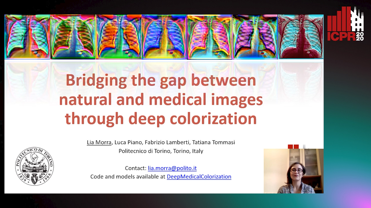
Auto-TLDR; Transfer Learning for Diagnosis on X-ray Images Using Color Adaptation

Slides Poster Similar

Deep learning has thrived by training on large-scale datasets. However, in many applications, as for medical image diagnosis, getting massive amount of data is still prohibitive due to privacy, lack of acquisition homogeneity and annotation cost. In this scenario transfer learning from natural image collections is a standard practice that attempts to tackle shape, texture and color discrepancy all at once through pretrained model fine-tuning. In this work we propose to disentangle those challenges and design a dedicated network module that focuses on color adaptation. We combine learning from scratch of the color module with transfer learning of different classification backbones obtaining an end-to-end, easy-to-train architecture for diagnostic image recognition on X-ray images. Extensive experiments show how our approach is particularly efficient in case of data scarcity and provides a new path for further transferring the learned color information across multiple medical datasets.

CAggNet: Crossing Aggregation Network for Medical Image Segmentation

Xu Cao, Yanghao Lin

Responsive image

Auto-TLDR; Crossing Aggregation Network for Medical Image Segmentation

Slides Poster Similar

In this paper, we present Crossing Aggregation Network (CAggNet), a novel densely connected semantic segmentation method for medical image analysis. The crossing aggregation network absorbs the idea of deep layer aggregation and makes significant innovations in layer connection and semantic information fusion. In this architecture, the traditional skip-connection structure of general U-Net is replaced by aggregations of multi-level down-sampling and up-sampling layers. This enables the network to fuse information interactively flows at different levels of layers in semantic segmentation. It also introduces weighted aggregation module to aggregate multi-scale output information. We have evaluated and compared our CAggNet with several advanced U-Net based methods in two public medical image datasets, including the 2018 Data Science Bowl nuclei detection dataset and the 2015 MICCAI gland segmentation competition dataset. Experimental results indicate that CAggNet improves medical object recognition and achieves a more accurate and efficient segmentation compared to existing improved U-Net and UNet++ structure.

BCAU-Net: A Novel Architecture with Binary Channel Attention Module for MRI Brain Segmentation

Yongpei Zhu, Zicong Zhou, Guojun Liao, Kehong Yuan

Responsive image

Auto-TLDR; BCAU-Net: Binary Channel Attention U-Net for MRI brain segmentation

Slides Poster Similar

Recently deep learning-based networks have achieved advanced performance in medical image segmentation. However, the development of deep learning is slow in magnetic resonance image (MRI) segmentation of normal brain tissues. In this paper, inspired by channel attention module, we propose a new architecture, Binary Channel Attention U-Net (BCAU-Net), by introducing a novel Binary Channel Attention Module (BCAM) into skip connection of U-Net, which can take full advantages of the channel information extracted from the encoding path and corresponding decoding path. To better aggregate multi-scale spatial information of the feature map, spatial pyramid pooling (SPP) modules with different pooling operations are used in BCAM instead of original average-pooling and max-pooling operations. We verify this model on two datasets including IBSR and MRBrainS18, and obtain better performance on MRI brain segmentation compared with other methods. We believe the proposed method can advance the performance in brain segmentation and clinical diagnosis.

A Multi-Task Contextual Atrous Residual Network for Brain Tumor Detection & Segmentation

Ngan Le, Kashu Yamazaki, Quach Kha Gia, Thanh-Dat Truong, Marios Savvides

Responsive image

Auto-TLDR; Contextual Brain Tumor Segmentation Using 3D atrous Residual Networks and Cascaded Structures

Poster Similar

In recent years, deep neural networks have achieved state-of-the-art performance in a variety of recognition and segmentation tasks in medical imaging including brain tumor segmentation. We investigate that segmenting brain tumor is facing to the imbalanced data problem where the number of pixels belonging to background class (non tumor pixel) is much larger than the number of pixels belonging to foreground class (tumor pixel). To address this problem, we propose a multi-task network which is formed as a cascaded structure and designed to share the feature maps. Our model consists of two targets, i.e., (i) effectively differentiating brain tumor regions and (ii) estimating brain tumor masks. The first task is performed by our proposed contextual brain tumor detection network, which plays the role of an attention gate and focuses on the region around brain tumor only while ignore the background (non tumor area). Instead of processing every pixel, our contextual brain tumor detection network only processes contextual regions around ground-truth instances and this strategy helps to produce meaningful regions proposals. The second task is built upon a 3D atrous residual network and under an encode-decode network in order to effectively segment both large and small objects (brain tumor). Our 3D atrous residual network is designed with a skip connection to enables the gradient from the deep layers to be directly propagated to shallow layers, thus, features of different depths are preserved and used for refining each other. In order to incorporate larger contextual information in volume MRI data, our network is designed by 3D atrous convolution with various kernel sizes, which enlarges the receptive field of filters. Our proposed network has been evaluated on various datasets including BRATS2015, BRATS2017 and BRATS2018 datasets with both validation set and testing set. Our performance has been benchmarked by both region-based metrics and surface-based metrics. We also have conducted comparisons against state-of-the-art approaches.

DARN: Deep Attentive Refinement Network for Liver Tumor Segmentation from 3D CT Volume

Yao Zhang, Jiang Tian, Cheng Zhong, Yang Zhang, Zhongchao Shi, Zhiqiang He

Responsive image

Auto-TLDR; Deep Attentive Refinement Network for Liver Tumor Segmentation from 3D Computed Tomography Using Multi-Level Features

Slides Poster Similar

Automatic liver tumor segmentation from 3D Computed Tomography (CT) is a necessary prerequisite in the interventions of hepatic abnormalities and surgery planning. However, accurate liver tumor segmentation remains challenging due to the large variability of tumor sizes and inhomogeneous texture. Recent advances based on Fully Convolutional Network (FCN) in liver tumor segmentation draw on success of learning discriminative multi-level features. In this paper, we propose a Deep Attentive Refinement Network (DARN) for improved liver tumor segmentation from CT volumes by fully exploiting both low and high level features embedded in different layers of FCN. Different from existing works, we exploit attention mechanism to leverage the relation of different levels of features encoded in different layers of FCN. Specifically, we introduce a Semantic Attention Refinement (SemRef) module to selectively emphasize global semantic information in low level features with the guidance of high level ones, and a Spatial Attention Refinement (SpaRef) module to adaptively enhance spatial details in high level features with the guidance of low level ones. We evaluate our network on the public MICCAI 2017 Liver Tumor Segmentation Challenge dataset (LiTS dataset) and it achieves state-of-the-art performance. The proposed refinement modules are an effective strategy to exploit multi-level features and has great potential to generalize to other medical image segmentation tasks.

RescueNet: Joint Building Segmentation and Damage Assessment from Satellite Imagery

Rohit Gupta, Mubarak Shah

Responsive image

Auto-TLDR; RescueNet: End-to-End Building Segmentation and Damage Classification for Humanitarian Aid and Disaster Response

Slides Poster Similar

Accurate and fine-grained information about the extent of damage to buildings is essential for directing Humanitarian Aid and Disaster Response (HADR) operations in the immediate aftermath of any natural calamity. In recent years, satellite and UAV (drone) imagery has been used for this purpose, sometimes aided by computer vision algorithms. Existing Computer Vision approaches for building damage assessment typically rely on a two stage approach, consisting of building detection using an object detection model, followed by damage assessment through classification of the detected building tiles. These multi-stage methods are not end-to-end trainable, and suffer from poor overall results. We propose RescueNet, a unified model that can simultaneously segment buildings and assess the damage levels to individual buildings and can be trained end-to end. In order to to model the composite nature of this problem, we propose a novel localization aware loss function, which consists of a Binary Cross Entropy loss for building segmentation, and a foreground only selective Categorical Cross-Entropy loss for damage classification, and show significant improvement over the widely used Cross-Entropy loss. RescueNet is tested on the large scale and diverse xBD dataset and achieves significantly better building segmentation and damage classification performance than previous methods and achieves generalization across varied geographical regions and disaster types.

Encoder-Decoder Based Convolutional Neural Networks with Multi-Scale-Aware Modules for Crowd Counting

Pongpisit Thanasutives, Ken-Ichi Fukui, Masayuki Numao, Boonserm Kijsirikul

Responsive image

Auto-TLDR; M-SFANet and M-SegNet for Crowd Counting Using Multi-Scale Fusion Networks

Slides Poster Similar

In this paper, we proposed two modified neural networks based on dual path multi-scale fusion networks (SFANet) and SegNet for accurate and efficient crowd counting. Inspired by SFANet, the first model, which is named M-SFANet, is attached with atrous spatial pyramid pooling (ASPP) and context-aware module (CAN). The encoder of M-SFANet is enhanced with ASPP containing parallel atrous convolutional layers with different sampling rates and hence able to extract multi-scale features of the target object and incorporate larger context. To further deal with scale variation throughout an input image, we leverage the CAN module which adaptively encodes the scales of the contextual information. The combination yields an effective model for counting in both dense and sparse crowd scenes. Based on the SFANet decoder structure, M-SFANet's decoder has dual paths, for density map and attention map generation. The second model is called M-SegNet, which is produced by replacing the bilinear upsampling in SFANet with max unpooling that is used in SegNet. This change provides a faster model while providing competitive counting performance. Designed for high-speed surveillance applications, M-SegNet has no additional multi-scale-aware module in order to not increase the complexity. Both models are encoder-decoder based architectures and are end-to-end trainable. We conduct extensive experiments on five crowd counting datasets and one vehicle counting dataset to show that these modifications yield algorithms that could improve state-of-the-art crowd counting methods.

DE-Net: Dilated Encoder Network for Automated Tongue Segmentation

Hui Tang, Bin Wang, Jun Zhou, Yongsheng Gao

Responsive image

Auto-TLDR; Automated Tongue Image Segmentation using De-Net

Slides Poster Similar

Automated tongue recognition is a growing research field due to global demand for personal health care. Using mobile devices to take tongue pictures is convenient and of low cost for tongue recognition. It is particularly suitable for self-health evaluation of the public. However, images taken by mobile devices are easily affected by various imaging environment, which makes fine segmentation a more challenging task compared with those taken by specialized acquisition devices. Deep learning approaches are promising for tongue image segmentation because they have powerful feature learning and representation capability. However, the successive pooling operations in these methods lead to loss of information on image details, making them fail when segmenting low-quality images captured by mobile devices. To address this issue, we propose a dilated encoder network (DE-Net) to capture more high-level features and get high-resolution output for automated tongue image segmentation. In addition, we construct two tongue image datasets which contain images taken by specialized devices and mobile devices, respectively, to verify the effectiveness of the proposed method. Experimental results on both datasets demonstrate that the proposed method outperforms the state-of-the-art methods in tongue image segmentation.